User login
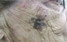
Q: Which is the best diagnosis?
- Seborrheic keratosis
- Pigmented basal cell carcinoma
- Congenital nevus
- Lentigo maligna melanoma
- Actinic purpura
A: The answer is lentigo maligna melanoma. Biopsy revealed the spread of atypical melanocytes along the dermal-epidermal junction and in irregularly shaped and confluent nests. Focal invasion into the papillary dermis was noted when definitive excision was performed.
Lentigo maligna melanoma develops on sun-damaged skin.1 Although most common in older people, lentigo maligna melanoma is now sometimes encountered in younger people. In either case, it is identified using the ABCDE rule of asymmetry, irregular border, color variations, diameter larger than 6 mm, and evolution.2
Seborrheic keratosis often has a rough surface and looks “stuck on.” Basal cell carcinoma usually has a waxy texture, may have a “rolled” border, and usually has telangiectatic vessels at the margins. A congenital nevus is typically stable and long-standing. Many nevi have more uniform pigmentation and lack the different colors and hues typical of melanoma. Purpura usually resolves after a few weeks.
An adequate biopsy is essential for diagnosis and excludes the other entities mentioned in the differential diagnosis.
Surgical treatment with adequate margin control is the cornerstone of therapy.3,4 Topical imiquimod cream (Aldara) may be of value for precursor lentigo maligna lesions proven to be in situ,5 but this is not advocated for invasive disease. Since treatment of smaller lesions is much less difficult and disfiguring, clinicians should be suspicious of any persistent, evolving pigmentary abnormality on sun-damaged skin. Biopsy clarifies the diagnosis.
SUGGESTED READING
- Cohen PJ, Lambert WC, Hill GJ, Schwartz RA. Melanoma. In:Schwartz RA, editor. Skin Cancer: Recognition and Management. New York: Springer-Verlag; 1988:104–105.
- Brodell RT, Helms SE. The changing mole. Additional warning signs of malignant melanoma. Postgrad Med 1998; 104:145–148.
- Zitelli JA, Brown CD, Hanusa BH. Surgical margins for excision of primary cutaneous melanoma. J Am Acad Dermatol 1997; 37:422–429.
- Bub JL, Berg D, Slee A, Odland B. Management of lentigo maligna and lentigo maligna melanoma with staged excision: a 5-year follow-up. Arch Dermatol 2004; 140:552–558.
- Hopson B, richey D, Sajben FP. Treatment of lentigo maligna with imiquimod 5% cream. J Drugs Dermatol 2007; 6:1037–1040.
Q: Which is the best diagnosis?
- Seborrheic keratosis
- Pigmented basal cell carcinoma
- Congenital nevus
- Lentigo maligna melanoma
- Actinic purpura
A: The answer is lentigo maligna melanoma. Biopsy revealed the spread of atypical melanocytes along the dermal-epidermal junction and in irregularly shaped and confluent nests. Focal invasion into the papillary dermis was noted when definitive excision was performed.
Lentigo maligna melanoma develops on sun-damaged skin.1 Although most common in older people, lentigo maligna melanoma is now sometimes encountered in younger people. In either case, it is identified using the ABCDE rule of asymmetry, irregular border, color variations, diameter larger than 6 mm, and evolution.2
Seborrheic keratosis often has a rough surface and looks “stuck on.” Basal cell carcinoma usually has a waxy texture, may have a “rolled” border, and usually has telangiectatic vessels at the margins. A congenital nevus is typically stable and long-standing. Many nevi have more uniform pigmentation and lack the different colors and hues typical of melanoma. Purpura usually resolves after a few weeks.
An adequate biopsy is essential for diagnosis and excludes the other entities mentioned in the differential diagnosis.
Surgical treatment with adequate margin control is the cornerstone of therapy.3,4 Topical imiquimod cream (Aldara) may be of value for precursor lentigo maligna lesions proven to be in situ,5 but this is not advocated for invasive disease. Since treatment of smaller lesions is much less difficult and disfiguring, clinicians should be suspicious of any persistent, evolving pigmentary abnormality on sun-damaged skin. Biopsy clarifies the diagnosis.
SUGGESTED READING
- Cohen PJ, Lambert WC, Hill GJ, Schwartz RA. Melanoma. In:Schwartz RA, editor. Skin Cancer: Recognition and Management. New York: Springer-Verlag; 1988:104–105.
- Brodell RT, Helms SE. The changing mole. Additional warning signs of malignant melanoma. Postgrad Med 1998; 104:145–148.
- Zitelli JA, Brown CD, Hanusa BH. Surgical margins for excision of primary cutaneous melanoma. J Am Acad Dermatol 1997; 37:422–429.
- Bub JL, Berg D, Slee A, Odland B. Management of lentigo maligna and lentigo maligna melanoma with staged excision: a 5-year follow-up. Arch Dermatol 2004; 140:552–558.
- Hopson B, richey D, Sajben FP. Treatment of lentigo maligna with imiquimod 5% cream. J Drugs Dermatol 2007; 6:1037–1040.
