User login
A WELL-DEVELOPED AND PREVIOUSLY HEALTHY INFANT was brought to our emergency department (ED) by her parents, who said that their child was experiencing significant toe pain. Two reddened, swollen toes (FIGURE) were immediately identifiable on the left foot. The patient’s parents denied any trauma or unusual activity involving the infant.
FIGURE
Two reddened and swollen toes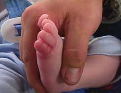
WHAT IS YOUR DIAGNOSIS?
HOW WOULD YOU TREAT THIS PATIENT?
Diagnosis: Hair tourniquet
Upon close inspection, we discovered hair thread tourniquets constricting the fourth and fifth toes on the baby’s left foot.
Hair tourniquet syndrome primarily affects infants in the first few months of life. The average age of occurrence is 4 months, a time when maternal postpartum hair loss (telogen effluvium) is at its maximum.1,2 It is worth noting, however, that this syndrome has also been observed in toddlers and adolescents.3
Toes are most frequently involved, although cases of hair tourniquets affecting the fingers, penis, labia, clitoris and uvula have been reported.2-7 Infants may be brought to the office or ED with irritability or crying, or with an erythematous extremity.
Why tourniquets are overlooked
Infantile tourniquets may be overlooked because of the fine nature of human hair, the swelling of the involved appendage (which can hide the tourniquet itself), and the presence of baby booties, footed pajamas, and mittens that may obscure injured digits from view.
These tourniquets can cause significant morbidity if they are not quickly identified and removed. The tensile strength of human hair is quite strong, allowing for strangulation and even amputation of appendages. The repetitive motion of hands and feet inside booties or mittens allows constriction to increase.
As this tightening occurs, the tourniquet may cause constrictive lymphatic obstruction, edema of the involved soft tissues, and secondary vascular obstruction of venous outflow and arterial perfusion.5,8,9 Tourniquets can also cut through skin, injuring deeper tissues.
In most instances, the damaged tissue will rapidly reperfuse once the offending tourniquet is unraveled or removed. However, some cases may result in epithelialization over the tourniquet, ischemia, necrosis, and gangrene leading to amputation. Quick recognition of the condition and immediate removal of the constricting tourniquet are key to saving the injured appendage.
How best to remove the tourniquet
Methods of tourniquet removal include unwrapping, cutting, or dissolving the hair with commercial hair removal agents such as Nair (Church & Dwight Co, Inc, Princeton, NJ).2,10-12
Unwrapping. In cases where the tourniquet is easily visualized and minimal edema is present, simply unwrapping the constricting hair may be successful. This can be accomplished by identifying a loose end of the hair, grasping the free end with a pinching instrument such as a hemostat or forceps, and carefully unwrapping the hair from the appendage1,11 (strength of recommendation [SOR]: B).
Cutting. If cutting the hair is necessary due to the presence of mild to moderate edema or failure of the unwrapping technique, a blunt probe may be inserted between the hair and the appendage to protect soft tissues from the cutting implement. Once the probe has been inserted, the tourniquet may be cut using scissors or a #11 scalpel blade applied to the surface of the blunt instrument1,11 (SOR: C). Alternative instrumentation, including a #12 Bard Parker curved scalpel blade and a Littauer suture-removal scissor, may be useful when the tourniquet is too tightly wound to allow for insertion of a blunt probe instrument (SOR: C).
Dissolving. A commercially available depilatory agent, such as Nair, may be useful for mild cases, but would not be appropriate when a tourniquet has cut into the skin. Calcium thioglycolate, a common depilatory active ingredient, breaks down disulfide bonds in keratin, thereby weakening hair strands. Chemical agents containing calcium thioglycolate should be used with caution, as keratin is also present in the epidermis and use of these agents may cause irritation to the skin.
When an incisional approach is needed
At times, epithelialization over the tourniquet or severe swelling of a digit may necessitate an incisional approach. If there is any doubt about whether you can completely remove all of the strands of the tourniquet, an incision into the digit itself must be made to disrupt constriction. Historically, a digital nerve block has been the preferred mode of analgesia; however, recent evidence suggests that less invasive pain management strategies, such as a sucrose pacifier, EMLA cream, or ZAP topical analgesia gel may be effective13 (SOR: A).
If you must use this approach, you’ll need to consider the placement of the digital neurovascular bundles of the fingers and toes, located at approximately the 2, 4, 8, and 10 o’clock positions. Following sterile preparation and draping, a longitudinal incision should be made at either the 3 or 9 o’clock position, thus locating it between neurovascular bundles1,11 (SOR: B).
Alternatively, a longitudinal incision can be made directly over the extensor tendon, located dorsally at 12 o’clock. Any resulting tendon laceration would be parallel to the tendon fibers, and could be expected to heal with splinting and wound care1,11,14 (SOR: B).
Prior to initiating treatment, parents or caregivers should be warned about the potential for bleeding and pain during the procedure.
Extreme cases may require surgery
Surgical consultation may be required in extreme cases of edema, neurovascular compromise, necrosis, amputation, or failure to completely remove the tourniquet.2
Other factors to keep in mind. While classically described as consisting of a single hair, tourniquets may be comprised of multiple hairs. That’s why it’s important to carefully inspect the area to be sure that all strands have been removed. Thorough treatment should include consideration of child abuse, tetanus immunization, and the need for antibiotics1,2,11 (SOR: B).
Relief for our young patient
To remove our patient’s hair tourniquets, we carefully cut the fibers with hooked Littauer suture-removal scissors and unwrapped the hair. Damage to the plantar aspect of both toes was significant enough that we had to cut through the soft tissue and into the flexor tendon to completely remove the hair. No sutures were required as the wound edges were well approximated without closure.
We cleaned and dressed the injured toes and arranged for close follow-up. The patient’s recovery was uneventful.
To avoid hair tourniquet syndrome, counsel parents to turn mittens and booties inside out to check for loose hairs. Also, advise parents to make sure there aren’t any hairs wrapped around their baby’s fingers or toes. Vigilance on the part of health care providers can provide quick recognition and appropriate treatment of this condition.
CORRESPONDENCE
Tania D. Strout, PhD, RN, MS, Director of Research, Department of Emergency Medicine, Maine Medical Center, 47 Bramhall Street, Portland, ME 04102; [email protected]
1. Loiselle JM, Cronan KM. Hair tourniquet removal. In: King C, Henretig FM, eds. Textbook of Pediatric Emergency Procedures. 2nd ed. Philadelphia, Pa: Lippincott Williams & Wilkins; 2008:1065–1069.
2. Strahlman RS. Toe tourniquet syndrome in association with maternal hair loss. Pediatrics. 2003;111:685-687.
3. Bacon JL, Burgis JT. Hair thread tourniquet syndrome in adolescents: a presentation and review of the literature. J Pediatr Adolesc Gynecol. 2005;18:155-156.
4. Badawy H, Soliman A, Ouf A, et al. Progressive hair coil penile tourniquet syndrome: multicenter experience with 25 cases. J Pediatr Surg. 2010;45:1514-1518.
5. Kuo JH, Smith LM, Berkowitz CD. A hair tourniquet resulting in strangulation and amputation of the clitoris. Obstet Gynecol. 2002;99:939-941.
6. McNeal RM, Cruickshank JC. Strangulation of the uvula by hair wrapping. Clin Pediatr (Phila). 1987;26:599-600.
7. Krishna S, Paul RI. Hair tourniquet of the uvula. J Emerg Med. 2003;24:325-326.
8. Rich MA, Keating MA. Hair tourniquet syndrome of the clitoris. J Urol. 1999;162:190-191.
9. Sylwestrzak MS, Fischer BF, Fischer H. Recurrent clitoral tourniquet syndrome. Pediatrics. 2000;105:866-867.
10. Douglass DD. Dissolving hair wrapped around an infant’s digit. J Pediatr. 1977;91:162.-
11. Cardriche D. Hair tourniquet removal. Available at: http://emedicine.medscape.com/article/1348969-overview. Updated January 30, 2012. Accessed October 3, 2012.
12. Peckler B, Hsu CK. Tourniquet syndrome: a review of constricting band removal. J Emerg Med. 2001;20:253-262.
13. Stevens B, Yamada J, Ohlsson A. Sucrose for analgesia in newborn infants undergoing painful procedures. Cochrane Database Syst Rev. 2010;(1):CD001069.-
14. Barton DJ, Sloan GM, Nichter LS, et al. Hair-thread tourniquet syndrome. Pediatrics. 1988;82:925-928.
A WELL-DEVELOPED AND PREVIOUSLY HEALTHY INFANT was brought to our emergency department (ED) by her parents, who said that their child was experiencing significant toe pain. Two reddened, swollen toes (FIGURE) were immediately identifiable on the left foot. The patient’s parents denied any trauma or unusual activity involving the infant.
FIGURE
Two reddened and swollen toes
WHAT IS YOUR DIAGNOSIS?
HOW WOULD YOU TREAT THIS PATIENT?
Diagnosis: Hair tourniquet
Upon close inspection, we discovered hair thread tourniquets constricting the fourth and fifth toes on the baby’s left foot.
Hair tourniquet syndrome primarily affects infants in the first few months of life. The average age of occurrence is 4 months, a time when maternal postpartum hair loss (telogen effluvium) is at its maximum.1,2 It is worth noting, however, that this syndrome has also been observed in toddlers and adolescents.3
Toes are most frequently involved, although cases of hair tourniquets affecting the fingers, penis, labia, clitoris and uvula have been reported.2-7 Infants may be brought to the office or ED with irritability or crying, or with an erythematous extremity.
Why tourniquets are overlooked
Infantile tourniquets may be overlooked because of the fine nature of human hair, the swelling of the involved appendage (which can hide the tourniquet itself), and the presence of baby booties, footed pajamas, and mittens that may obscure injured digits from view.
These tourniquets can cause significant morbidity if they are not quickly identified and removed. The tensile strength of human hair is quite strong, allowing for strangulation and even amputation of appendages. The repetitive motion of hands and feet inside booties or mittens allows constriction to increase.
As this tightening occurs, the tourniquet may cause constrictive lymphatic obstruction, edema of the involved soft tissues, and secondary vascular obstruction of venous outflow and arterial perfusion.5,8,9 Tourniquets can also cut through skin, injuring deeper tissues.
In most instances, the damaged tissue will rapidly reperfuse once the offending tourniquet is unraveled or removed. However, some cases may result in epithelialization over the tourniquet, ischemia, necrosis, and gangrene leading to amputation. Quick recognition of the condition and immediate removal of the constricting tourniquet are key to saving the injured appendage.
How best to remove the tourniquet
Methods of tourniquet removal include unwrapping, cutting, or dissolving the hair with commercial hair removal agents such as Nair (Church & Dwight Co, Inc, Princeton, NJ).2,10-12
Unwrapping. In cases where the tourniquet is easily visualized and minimal edema is present, simply unwrapping the constricting hair may be successful. This can be accomplished by identifying a loose end of the hair, grasping the free end with a pinching instrument such as a hemostat or forceps, and carefully unwrapping the hair from the appendage1,11 (strength of recommendation [SOR]: B).
Cutting. If cutting the hair is necessary due to the presence of mild to moderate edema or failure of the unwrapping technique, a blunt probe may be inserted between the hair and the appendage to protect soft tissues from the cutting implement. Once the probe has been inserted, the tourniquet may be cut using scissors or a #11 scalpel blade applied to the surface of the blunt instrument1,11 (SOR: C). Alternative instrumentation, including a #12 Bard Parker curved scalpel blade and a Littauer suture-removal scissor, may be useful when the tourniquet is too tightly wound to allow for insertion of a blunt probe instrument (SOR: C).
Dissolving. A commercially available depilatory agent, such as Nair, may be useful for mild cases, but would not be appropriate when a tourniquet has cut into the skin. Calcium thioglycolate, a common depilatory active ingredient, breaks down disulfide bonds in keratin, thereby weakening hair strands. Chemical agents containing calcium thioglycolate should be used with caution, as keratin is also present in the epidermis and use of these agents may cause irritation to the skin.
When an incisional approach is needed
At times, epithelialization over the tourniquet or severe swelling of a digit may necessitate an incisional approach. If there is any doubt about whether you can completely remove all of the strands of the tourniquet, an incision into the digit itself must be made to disrupt constriction. Historically, a digital nerve block has been the preferred mode of analgesia; however, recent evidence suggests that less invasive pain management strategies, such as a sucrose pacifier, EMLA cream, or ZAP topical analgesia gel may be effective13 (SOR: A).
If you must use this approach, you’ll need to consider the placement of the digital neurovascular bundles of the fingers and toes, located at approximately the 2, 4, 8, and 10 o’clock positions. Following sterile preparation and draping, a longitudinal incision should be made at either the 3 or 9 o’clock position, thus locating it between neurovascular bundles1,11 (SOR: B).
Alternatively, a longitudinal incision can be made directly over the extensor tendon, located dorsally at 12 o’clock. Any resulting tendon laceration would be parallel to the tendon fibers, and could be expected to heal with splinting and wound care1,11,14 (SOR: B).
Prior to initiating treatment, parents or caregivers should be warned about the potential for bleeding and pain during the procedure.
Extreme cases may require surgery
Surgical consultation may be required in extreme cases of edema, neurovascular compromise, necrosis, amputation, or failure to completely remove the tourniquet.2
Other factors to keep in mind. While classically described as consisting of a single hair, tourniquets may be comprised of multiple hairs. That’s why it’s important to carefully inspect the area to be sure that all strands have been removed. Thorough treatment should include consideration of child abuse, tetanus immunization, and the need for antibiotics1,2,11 (SOR: B).
Relief for our young patient
To remove our patient’s hair tourniquets, we carefully cut the fibers with hooked Littauer suture-removal scissors and unwrapped the hair. Damage to the plantar aspect of both toes was significant enough that we had to cut through the soft tissue and into the flexor tendon to completely remove the hair. No sutures were required as the wound edges were well approximated without closure.
We cleaned and dressed the injured toes and arranged for close follow-up. The patient’s recovery was uneventful.
To avoid hair tourniquet syndrome, counsel parents to turn mittens and booties inside out to check for loose hairs. Also, advise parents to make sure there aren’t any hairs wrapped around their baby’s fingers or toes. Vigilance on the part of health care providers can provide quick recognition and appropriate treatment of this condition.
CORRESPONDENCE
Tania D. Strout, PhD, RN, MS, Director of Research, Department of Emergency Medicine, Maine Medical Center, 47 Bramhall Street, Portland, ME 04102; [email protected]
1. Loiselle JM, Cronan KM. Hair tourniquet removal. In: King C, Henretig FM, eds. Textbook of Pediatric Emergency Procedures. 2nd ed. Philadelphia, Pa: Lippincott Williams & Wilkins; 2008:1065–1069.
2. Strahlman RS. Toe tourniquet syndrome in association with maternal hair loss. Pediatrics. 2003;111:685-687.
3. Bacon JL, Burgis JT. Hair thread tourniquet syndrome in adolescents: a presentation and review of the literature. J Pediatr Adolesc Gynecol. 2005;18:155-156.
4. Badawy H, Soliman A, Ouf A, et al. Progressive hair coil penile tourniquet syndrome: multicenter experience with 25 cases. J Pediatr Surg. 2010;45:1514-1518.
5. Kuo JH, Smith LM, Berkowitz CD. A hair tourniquet resulting in strangulation and amputation of the clitoris. Obstet Gynecol. 2002;99:939-941.
6. McNeal RM, Cruickshank JC. Strangulation of the uvula by hair wrapping. Clin Pediatr (Phila). 1987;26:599-600.
7. Krishna S, Paul RI. Hair tourniquet of the uvula. J Emerg Med. 2003;24:325-326.
8. Rich MA, Keating MA. Hair tourniquet syndrome of the clitoris. J Urol. 1999;162:190-191.
9. Sylwestrzak MS, Fischer BF, Fischer H. Recurrent clitoral tourniquet syndrome. Pediatrics. 2000;105:866-867.
10. Douglass DD. Dissolving hair wrapped around an infant’s digit. J Pediatr. 1977;91:162.-
11. Cardriche D. Hair tourniquet removal. Available at: http://emedicine.medscape.com/article/1348969-overview. Updated January 30, 2012. Accessed October 3, 2012.
12. Peckler B, Hsu CK. Tourniquet syndrome: a review of constricting band removal. J Emerg Med. 2001;20:253-262.
13. Stevens B, Yamada J, Ohlsson A. Sucrose for analgesia in newborn infants undergoing painful procedures. Cochrane Database Syst Rev. 2010;(1):CD001069.-
14. Barton DJ, Sloan GM, Nichter LS, et al. Hair-thread tourniquet syndrome. Pediatrics. 1988;82:925-928.