User login
RIO GRANDE, P.R. – Langerhans cell histiocytosis can be benign and self-limited, or a disseminated disease with the potential for significant morbidity and death, said Dr. Anthony J. Mancini.
The mortality risk depends on whether high-risk organs such as the liver or spleen are involved, as well as the early response to treatment, said Dr. Mancini, professor of pediatrics and dermatology at Northwestern University and Children’s Memorial Hospital in Chicago.
Langerhans cell histiocytosis (LCH) was one of five pediatric dermatology diagnoses Dr. Mancini urged dermatologists at the annual Caribbean Dermatology Symposium not to miss. Prompt identification of these conditions can improve young patients’ outcomes and reduce the risk for complications later, he said.
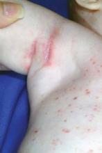
The clinical diagnosis of LCH in infants and children includes scaly erythematous seborrheic-like dermatitis, brown or red papules that may be lichenoid, and erythema in skin fold areas including the groin and the anterior neck folds, often with punctate erosions, he noted. Petechiae may be present, and crusted papules on the palms and soles are occasionally seen in young children.
Histopathology of LCH shows a granulomatous infiltrate that is epidermotropic, and the presence of positive CD1a or CD207 staining confirms the diagnosis, he said. An important distinction between LCH and streptococcal intertrigo: The strep responds rapidly to antibiotics and the erosive changes are more diffuse rather than focal.
Kawasaki Disease
"The most feared complication of Kawasaki disease (KD) is that of coronary artery aneurysm," said Dr. Mancini. "So it is very important to think about this disorder and get a rapid diagnosis," he said. The diagnosis is purely clinical, he noted, without the availability of a confirmatory diagnostic test at this time. Therefore, "have a high index of suspicion" for this disorder, he said.
Clinical criteria for KD include oropharyngeal changes, extremity changes, and a polymorphous skin eruption. Common prodromal symptoms may include irritability, vomiting, anorexia, cough, diarrhea, abdominal pain, and joint pain. "These kids appear sick," Dr. Mancini said.
Skin findings in KD may take on several morphologies, including morbilliform, urticarial, serum sickness-like, or even pustular.
One key finding that is suggestive of KD: Accentuation of erythema in skin folds, especially in the form of perineal desquamation, he said. However, blisters and purpura are not typically associated with the disorder.
Congenital Immunodeficiency
"In many patients, skin manifestations may be the presenting finding" of congenital immunodeficiency, Dr. Mancini said. In a recent study of 128 patients with primary immunodeficiency, 48% had skin manifestations, and these were the presenting feature in 39% with total PID and 82% of those with skin lesions (Pediatr. Dermatol. 2011;28;494-501).
Consider congenital immunodeficiency (CID) in cases of severe atopic or seborrheic dermatitis, intertrigo, or erythroderma that are resistant to therapy, said Dr. Mancini. Growth failure, alopecia, and recurrent infections (especially with unusual organisms) also can be signs of CID.
Graft vs. host disease (GVHD) in a skin biopsy increases suspicion for CID, he added.
An initial evaluation for suspected CID should include a complete blood count, quantitative immunoglobulins, mitogen stimulation assay, tetanus titer, T- and B-cell flow cytometry, and nitroblue tetrazolium or chronic granulomatous disease flow assay, said Dr. Mancini.
There are many congenital immunodeficiencies that may present with skin findings, including Wiskott-Aldrich syndrome, chronic mucocutaneous candidiasis, severe combined immunodeficiency, and leukocyte adhesion deficiency.
Drug Hypersensitivity Syndrome (DRESS)
Also known as Drug Reaction With Eosinophilia and Systemic Symptoms (DRESS), drug hypersensitivity tends to occur from 3-8 weeks after starting a particular medication, said Dr. Mancini. A skin rash is one of the most common findings, occurring in 77% to 100% of patients, he said. Facial edema is common, especially in the periorbital area. DRESS also can include fever, lymph node enlargement, and internal organ involvement.
Other clinical symptoms include conjunctivitis, pharyngitis, vomiting, and diarrhea. Telltale hematology findings indicative of DRESS include atypical lymphocytes and eosinophilia; pancytopenia occasionally appears.
The first line of defense is recognition and withdrawal from the drug. Serial monitoring of liver function studies is vital, given the fulminant hepatitis that can ensue, Dr. Mancini said. Treatment with systemic steroids and/or intravenous immunoglobulin is occasionally used, albeit controversially, he noted. Complete resolution of symptoms may take as long as 6 months.
Neonatal Herpes
The risk for neonatal infection with herpes simplex virus (HSV) is highest when the mother has active genital herpes at the time of delivery, Dr. Mancini said. If left untreated, the mortality rate for central nervous system or disseminated neonatal herpes is estimated at 50%-90%, so rapid recognition and diagnosis is vital, he said. The central nervous system, liver, lungs, adrenal glands, and bone marrow are important sites of potential involvement when the process disseminates, he added.
The classic skin presentation of neonatal HSV infection is vesicles and vesiculopustules that may be grouped on a red base or may become hemorrhagic. Bullae, widespread skin erosions, and polycyclic patches occasionally may be seen, and scarring may be present. Prompt evaluation is important, and intravenous acyclovir should be immediately instituted if the diagnosis of neonatal HSV is being considered.
Dr. Mancini said he had no financial conflicts to disclose.
EXPERT ANALYSIS FROM THE ANNUAL CARIBBEAN DERMATOLOGY SYMPOSIUM

