User login

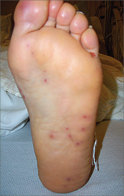
The FP hospitalized the patient for suspected acute bacterial endocarditis and ordered an echocardiogram, which showed vegetations on the tricuspid valve. He realized that the non-painful lesions on the palms and soles were Janeway lesions. The one painful lesion within the pulp of the big toe was an Osler node. (Remember “O” for “ouch” and Osler.) Janeway lesions are caused by septic emboli that form microabscesses in the dermis (without epidermal involvement). Osler nodes are tender and the result of the deposition of immune complexes.
Other clinical features of endocarditis include:
- fever, seen in 85% to 99% of patients. It is typically low-grade—less than 39°C (102.2°F).
- new or changing heart murmur
- septic emboli, seen in up to 60% of patients
- intracranial hemorrhages, seen in up to 40% of patients
- mycotic aneurysms
- splinter hemorrhages
- glomerulonephritis
- Roth spots—that is, retinal hemorrhages from microemboli
- positive rheumatoid factor.
Treatment begins with empiric antibiotics after the initial blood cultures are drawn. It is important to cover Staphylococcus aureus in injection drug users.
In this case, the FP started the patient on nafcillin 2 g every 4 hours and gentamicin 1 mg/kg every 8 hours. Vancomycin is used instead of nafcillin when there is a concern about methicillin-resistant Staphylococcus aureus (MRSA) from a prior history of MRSA infection.
This patient’s blood cultures grew out methicillin sensitive Staphylococcus aureus, so her antibiotic regimen was continued for 6 weeks with full clearance of the endocarditis. The patient was also willing to enter a drug rehabilitation program.
Photos courtesy of David A. Kasper DO. Text for Photo Rounds Friday courtesy of Richard P. Usatine, MD. This case was adapted from: Chumley H. Endocarditis. In: Usatine R, Smith M, Mayeaux EJ, et al, eds. Color Atlas of Family Medicine. 2nd ed. New York, NY: McGraw-Hill; 2013:287-291.
To learn more about the Color Atlas of Family Medicine, see: http://www.amazon.com/Color-Family-Medicine-Richard-Usatine/dp/0071769641/
You can now get the second edition of the Color Atlas of Family Medicine as an app by clicking this link: http://usatinemedia.com/