User login

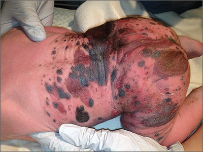
Giant congenital nevus was diagnosed in this patient. Congenital melanocytic nevi (CMN) are pigmented lesions that are present at birth and created by the abnormal migration of neural crest cells during embryogenesis. Nevi are categorized by size as small (<1.5 cm), medium (1.5-20 cm), large (>20 cm), or giant (>40 cm).
Congenital nevi tend to start out flat, with uniform pigmentation, but can become more variegated in texture and color as normal growth and development continue. Giant congenital nevi, which are rare, are likely to thicken, darken, and enlarge as the patient grows. Some nevi may develop very coarse or dark hair. CMN can cover any part of the body and occur independent of skin color and other ethnic factors.
CMN may present in almost any location and may be brown, black, pink, or purple in color. Café au lait macules, blue-gray spots, nevus of Ota, nevus spilus, and vascular malformations are part of the differential diagnosis for CMN and have individual location and color characteristics that set them apart clinically.
Patients with CMN are at increased risk for neurocutaneous melanosis (NCM; a melanocyte proliferation in the central nervous system) and melanoma. Magnetic resonance imaging (MRI) is helpful to exclude NCM. Treatment options for patients with large and giant CMN include early curettage, local excision, dermabrasion, and laser therapy. (There is, however, considerable debate about the value of surgery.) The newborn in the case underwent an MRI and the results were normal. At 4 months of age, he hadn’t developed any neurologic symptoms. The child’s nevi continue to grow and he sees his family physician for routine well-child visits and a dermatologist annually to monitor the nevi.
Adapted from: Karnes J, Griffin C. Large plaques on a baby boy. J Fam Pract. 2016;65:407-409.
