User login

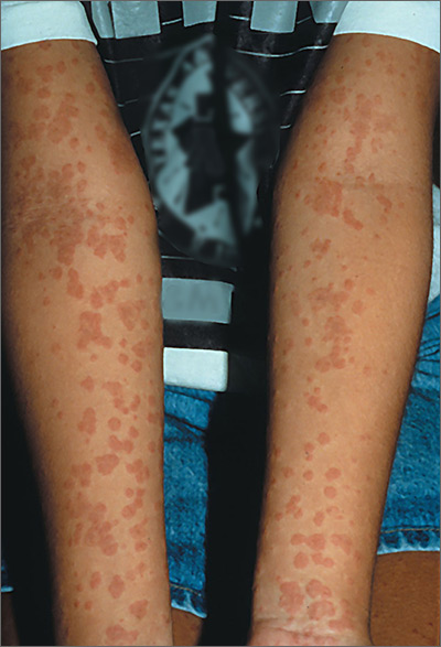
Based on the results of the punch biopsy and the slight scale seen on the periphery of the lesions (a collarette scale pattern), the FP made a diagnosis of pityriasis rosea.
The distribution of the lesions in this case was not typical for pityriasis rosea; lesions are typically found on the trunk (not the arms) and may start with a herald patch. Given the distribution of the lesions in this case, the more precise diagnosis was inverse pityriasis rosea.
The physician explained to the patient and her mother that the rash would resolve spontaneously and was unlikely to leave any scarring. Six months later, the FP saw the mother for an unrelated issue and she said her daughter’s rash had gotten better within a month of her daughter’s visit, and there had been no scarring.
Photos and text for Photo Rounds Friday courtesy of Richard P. Usatine, MD. This case was adapted from: Henderson D, Usatine R. Pityriasis rosea. In: Usatine R, Smith M, Mayeaux EJ, et al, eds. Color Atlas of Family Medicine. 2nd ed. New York, NY: McGraw-Hill; 2013: 896-900.
To learn more about the Color Atlas of Family Medicine, see: www.amazon.com/Color-Family-Medicine-Richard-Usatine/dp/0071769641/
You can now get the second edition of the Color Atlas of Family Medicine as an app by clicking on this link: usatinemedia.com
