User login
A mother brings her 5-year-old boy in to your office because she is concerned about a rash on his legs that seems to be worsening. She tells you that he had a runny nose and a mild cough a week earlier, but that those symptoms resolved before the rash developed. He has also complained of “belly pain.”
The boy’s mother says he’s been less active and more irritable since the onset of the rash, and that he is hardly eating. She also tells you that earlier in the day, her son told her that it hurts to walk.
You dig deeper…
A complete review of systems is otherwise negative. The 5-year-old was born at term without complication. He has met all developmental milestones and his immunizations are up to date. He takes no medications.
The boy’s vital signs are normal. He has an erythematous maculopapular rash distributed on his legs symmetrically; it is palpable, nontender, and nonblanching. You detect no abnormalities in abdominal, neurologic, or musculoskeletal examinations.
A complete blood count (CBC) and basic metabolic panel (BMP) reveal mild leukocytosis with a normal differential. Urinalysis shows moderate blood and trace protein. Laboratory results are otherwise normal.
WHAT IS YOUR DIAGNOSIS?
A classic presentation revealed: Henoch-Schönlein purpura
The combination of rash (FIGURE), abdominal pain, arthralgia, and evidence of renal involvement are the classic symptoms for Henoch-Schönlein purpura (HSP),1 the most common systemic vasculitis of childhood.2 Fever may also be present. Boys are affected nearly twice as often as girls. HSP occurs only rarely among adults, and men and women are affected equally. The estimated annual incidence in children is 20 cases per 100,000, with most cases occurring in those between the ages of 4 and 6 years.3
Although the exact cause of HSP is unknown, 75% of cases follow an upper respiratory infection. Streptococcus, Mycoplasma, adenovirus, parvovirus, Epstein-Barr virus, and varicella have all been implicated as offending pathogens. A number of case reports have also described the disease after the use of certain drugs, including penicillin, ampicillin, and quinine; and after the administration of vaccines, including those for typhoid, measles, yellow fever, and cholera. In all cases, the underlying mechanism is thought to be a systemic rise in immunoglobulin A (IgA), which forms immune complexes deposited in arterioles, capillaries, and venules.4 The precise interactions in the mechanism of disease are yet to be determined.
FIGURE
Rash and leg pain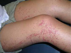
Like the patient described in the text, this 11-year-old girl had a similar rash on her legs, as well as leg and abdominal pain.
HSP is usually benign, self-limited
HSP signs and symptoms may occur in any order over a period of days to weeks. Symptoms tend to be less severe in younger children than in older children and adults. Besides abdominal and renal systems, the lungs and central nervous system (CNS) may be affected, although rarely in children. Pulmonary involvement, if it does occur, usually manifests as diffuse alveolar hemorrhage or interstitial pneumonia or fibrosis.5 CNS vasculitis may lead to cerebral hemorrhage.6
HSP usually is a benign, self-limited disorder. The average disease course is 4 weeks, although it may be as brief as 3 days or as long as 2 years. Up to 94% of children (and 89% of adults) recover fully.7 However, the potential for chronic renal disease does exist.8
The recurrence rate of HSP is generally reported as 40%;8 patients who develop nephritis have higher recurrence rates.9
Rash is almost always present
Nearly all of those affected with HSP exhibit a rash. In children, it may begin as urticarial or maculopapular skin lesions. The rash typically is a palpable purpura, with lesions measuring 2 to 10 mm in diameter. Classically, the rash appears symmetrically on the extensor surfaces of the arms, legs, and buttocks. Lesions may also occur on the face and ears; however, the rash usually spares the trunk.2 The rash generally fades over several days and gives way to darkened pigment. The purpuric lesions resolve more quickly with bed rest and tend to reappear when the patient resumes activity.
Bullous or necrotic lesions—although reported to occur in up to 60% of affected adults—are uncommon in children. If you had noted such lesions in this case, you would have had to rule out toxic vasculitides and meningococcal septicemia or other septic emboli.10
Arthralgia affects lower extremities
Arthralgia is the second most common clinical manifestation of HSP, occurring in approximately 82% of patients. Joint pain associated with HSP is likely due to periarticular soft tissue edema, and most commonly occurs in the hips, knees, or ankles.1,11 The arthralgia of HSP is transient and self-limited and does not cause permanent damage to the joints. Like the rash, joint pain tends to decrease with bed rest and increase with activity.11
Gastrointestinal involvement is rarely serious
Gastrointestinal involvement has been reported in 63% of patients with HSP. Symptoms typically include a colicky abdominal pain, associated with nausea and vomiting. These symptoms probably arise secondary to edema of the bowel wall and vasculitis of the gastrointestinal tract. The pain most often develops within 8 days of the appearance of the rash, but it has been reported to appear within weeks to months of cutaneous changes.12
Intussusception is the most common gastrointestinal complication in patients with HSP, with a reported overall incidence of 3.5% in hospitalized patients with severe abdominal pain. Gastrointestinal bleeding, presenting as either melena or hematochezia, has been reported in 25% of patients with HSP. Occult bleeding may occur in up to 50% of patients.12
More serious complications are rare, and they include bowel infarction and perforation, usually in the jejunum or ileum, and pancreatitis, cholecystitis, or protein-losing enteropathy secondary to HSP.
Renal involvement may require close follow-up
Renal disease has been reported to affect 30% to 70% of all HSP patients.13 Onset occurs within weeks to months after other symptoms of HSP. Most patients with renal involvement have only mild disease, such as asymptomatic hematuria and proteinuria.1,7,13 HSP is thought to account for approximately 15% of all glomerulopathies in childhood.13
Although renal disease in HSP is generally benign, such complications as nephrotic syndrome, hypertension, and acute and chronic renal failure may occur. Adults are much more susceptible to the latter complication.
When the diagnosis is unclear, renal biopsy may help confirm the presence of disease. Evidence of HSP is identical to that seen with IgA nephropathy.11,13 The percentage of glomeruli showing crescents on renal biopsy seems to be the most important prognostic finding, with crescent formation involving more than 50% of the glomeruli carrying a poor prognosis.14
Diagnosis is mainly clinical
Clinical diagnosis is not difficult with the classic 4 symptoms present (rash, abdominal pain, arthralgia, and evidence of renal involvement). However, when the presentation is less straightforward, confirmation of the diagnosis may depend on biopsy of the affected organ (eg, skin, kidney) demonstrating leukocytoclastic vasculitis with IgA deposition.
The differential diagnosis of HSP is large, and includes acute abdomen, meningococcal meningitis or septicemia, rheumatoid arthritis, idiopathic thrombocytopenic purpura, and systemic lupus erythematosus.15
Lab tests are minimally helpful. No lab test or imaging study is sensitive or specific for HSP. An elevated serum IgA level suggests the disease. A CBC may show leukocytosis and thrombocytosis, but test results may also be normal. The erythrocyte sedimentation rate may be elevated. On a BMP, blood urea nitrogen and creatinine levels may be elevated secondary to renal involvement or dehydration associated with HSP. Finally, hematuria or proteinuria show up on urinalysis in 30% to 70% of patients with HSP.2,12,13
What treatment is indicated?
Most patients with HSP recover completely without any specific intervention other than reassurance, bed rest, and supportive care. Arthralgia usually responds to nonsteroidal anti-inflammatory drugs and corticosteroids. Hospitalization is warranted when patients have a depleted volume status or inadequately controlled pain. With an otherwise un-complicated illness, watchful management suffices. Gastrointestinal and renal complications may require more aggressive therapy.
Gastrointestinal complications. As noted earlier, the most common gastrointestinal complication is intussusception. No definite measures for preventing intussusception appear in the literature. Some evidence supports the use of corticosteroids for severe abdominal symptoms,16,17 but in general, corticosteroids are not indicated for extrarenal manifestations. More serious complications such as bowel infarction or perforation, pancreatitis, or cholecystitis are rare.
Renal disease. Much research has focused on treatment options for patients with renal complications, due to the possibility of long-term debilitating effects. Although no evidence supports corticosteroid use for patients with mild renal involvement,16 for patients with severe renal disease—defined as crescenteric nephritis on biopsy, usually complicated by oliguria and hypertension—corticosteroids may help prevent irreversible glomerular injury.
Other agents that have been used for severe renal disease include azathioprine, cyclophosphamide, and dipyridamole. These drugs have shown some success in resolving symptoms, but their use remains controversial.16,18 Finally, plasmapheresis has led to significant clinical improvement for a small number of patients with severe, rapidly progressing HSP.
Caveat. The effectiveness of all treatments for HSP remains in question, given that patients’ symptoms may simply resolve spontaneously. A clear answer will depend on further research.
Rest, fluids, and ibuprofen for your patient
You send your patient home with his mother, and advise her to provide him with supportive care, including oral hydration. You tell her to encourage rest and to give symptomatic pain relief with an over-the-counter medication such as ibuprofen.
You see the child again in 3 days, by which time his arthralgias are improving. Repeat urinalysis shows continuing moderate blood and trace protein. You follow him biweekly for 2 weeks, and then weekly for 4 more weeks. Six weeks after his initial presentation, his symptoms completely resolve, and urinalysis shows no evidence of hematuria or proteinuria. His blood pressure is normal at all visits.
You see the patient monthly for the next 5 months. Symptoms do not recur, and urinalysis results are normal. You counsel the boy’s mother regarding the risk of recurrence, and advise her to contact the office immediately if symptoms return.
CORRESPONDENCE Shailendra K. Saxena, MD, PhD, Department of Family Medicine, 10828 John Galt Boulevard, Omaha, NE 68137; [email protected]
1. Roberts PF, Waller TA, Brinker TM, et al. Henoch-Schönlein purpura: a review article. South Med J. 2007;100:821-824.
2. Tizard EJ. Henoch-Schönlein purpura. Arch Dis Child. 1999;80:380-383.
3. Gardner-Medwin JM, Dolezalova P, Cummins C, et al. Incidence of Henoch-Schönlein purpura, Kawasaki disease, and rare vasculitides in children of different ethnic origins. Lancet. 2002;360:1197-1202.
4. Yang YH, Chuang YH, Wang LC, et al. The immunology of Henoch-Schönlein purpura. Autoimmun Rev. 2008;7:179-184.
5. Nadrous HF, Yu AC, Specks U, et al. Pulmonary involvement in Henoch-Schönlein purpura. Mayo Clin Proc. 2004;79:1151-1157.
6. Elinson P, Foster KW, Kaufman DB. Magnetic resonance imaging of central nervous system vasculitis: a case report of Henoch-Schönlein purpura. Acta Pediatr. 2008;79:710-713.
7. Blanco R, Martínez-Taboada VM, Rodríguez-Valverde V, et al. Henoch-Schönlein purpura in adulthood and childhood: two different expressions of the same syndrome. Arthritis Rheum. 1997;40:859-864.
8. Dillon MJ. Henoch-Schönlein purpura (treatment and outcome). Cleve Clin J Med. 2002;69(suppl):S121-S123.
9. Alfredo CS, Nunes NA, Len CA, et al. Henoch-Schönlein purpura: recurrence and chronicity. J Pediatr (Rio J). 2007;83:177-180.
10. Morelli JG. Vascular disorders. In: Kliegman RM, Behrman RE, Jensen HB, eds. Nelson Textbook of Pediatrics. 18th ed. Philadelphia, Pa: Saunders Elsevier; 2007:2667–2673.
11. Trapani S, Micheli A, Grisolia F, et al. Henoch-Schönlein purpura in childhood: epidemiological and clinical analysis of 150 cases over a 5-year period and review of literature. Semin Arthritis Rheum. 2005;35:143-153.
12. Chang WL, Yang YH, Lin YT, et al. Gastrointestinal manifestations in Henoch-Schönlein purpura: a review of 261 patients. Acta Pediatr. 2004;93:1427-1431.
13. Chang WL, Yang YH, Wang LC, et al. Renal manifestations in Henoch-Schönlein purpura: a 10-year clinical study. Pediatr Nephrol. 2005;20:1269-1272.
14. Bogdanovic R. Henoch-Schönlein purpura nephritis in children: risk factors, prevention and treatment. Acta Paediatr. 2009;98:1882-1889.
15. Kraft DM, Mckee D, Scott C. Henoch-Schönlein purpura: a review. Am Fam Phys. 1998;58:405-408,411.
16. Szer IS. Gastrointestinal and renal involvement in vasculitis: management strategies in Henoch-Schönlein purpura. Cleve Clin J Med. 1999;66:312-317.
17. Leung SP. Use of intravenous hydrocortisone in Henoch-Schönlein purpura. J Paediatr Child Health. 2001;37:309-310.
18. Zaffanello M, Brugnara M, Franchini M, et al. Therapy for children with Henoch-Schönlein purpura nephritis: a systematic review. ScientificWorldJournal. 2007;7:20-30.
A mother brings her 5-year-old boy in to your office because she is concerned about a rash on his legs that seems to be worsening. She tells you that he had a runny nose and a mild cough a week earlier, but that those symptoms resolved before the rash developed. He has also complained of “belly pain.”
The boy’s mother says he’s been less active and more irritable since the onset of the rash, and that he is hardly eating. She also tells you that earlier in the day, her son told her that it hurts to walk.
You dig deeper…
A complete review of systems is otherwise negative. The 5-year-old was born at term without complication. He has met all developmental milestones and his immunizations are up to date. He takes no medications.
The boy’s vital signs are normal. He has an erythematous maculopapular rash distributed on his legs symmetrically; it is palpable, nontender, and nonblanching. You detect no abnormalities in abdominal, neurologic, or musculoskeletal examinations.
A complete blood count (CBC) and basic metabolic panel (BMP) reveal mild leukocytosis with a normal differential. Urinalysis shows moderate blood and trace protein. Laboratory results are otherwise normal.
WHAT IS YOUR DIAGNOSIS?
A classic presentation revealed: Henoch-Schönlein purpura
The combination of rash (FIGURE), abdominal pain, arthralgia, and evidence of renal involvement are the classic symptoms for Henoch-Schönlein purpura (HSP),1 the most common systemic vasculitis of childhood.2 Fever may also be present. Boys are affected nearly twice as often as girls. HSP occurs only rarely among adults, and men and women are affected equally. The estimated annual incidence in children is 20 cases per 100,000, with most cases occurring in those between the ages of 4 and 6 years.3
Although the exact cause of HSP is unknown, 75% of cases follow an upper respiratory infection. Streptococcus, Mycoplasma, adenovirus, parvovirus, Epstein-Barr virus, and varicella have all been implicated as offending pathogens. A number of case reports have also described the disease after the use of certain drugs, including penicillin, ampicillin, and quinine; and after the administration of vaccines, including those for typhoid, measles, yellow fever, and cholera. In all cases, the underlying mechanism is thought to be a systemic rise in immunoglobulin A (IgA), which forms immune complexes deposited in arterioles, capillaries, and venules.4 The precise interactions in the mechanism of disease are yet to be determined.
FIGURE
Rash and leg pain
Like the patient described in the text, this 11-year-old girl had a similar rash on her legs, as well as leg and abdominal pain.
HSP is usually benign, self-limited
HSP signs and symptoms may occur in any order over a period of days to weeks. Symptoms tend to be less severe in younger children than in older children and adults. Besides abdominal and renal systems, the lungs and central nervous system (CNS) may be affected, although rarely in children. Pulmonary involvement, if it does occur, usually manifests as diffuse alveolar hemorrhage or interstitial pneumonia or fibrosis.5 CNS vasculitis may lead to cerebral hemorrhage.6
HSP usually is a benign, self-limited disorder. The average disease course is 4 weeks, although it may be as brief as 3 days or as long as 2 years. Up to 94% of children (and 89% of adults) recover fully.7 However, the potential for chronic renal disease does exist.8
The recurrence rate of HSP is generally reported as 40%;8 patients who develop nephritis have higher recurrence rates.9
Rash is almost always present
Nearly all of those affected with HSP exhibit a rash. In children, it may begin as urticarial or maculopapular skin lesions. The rash typically is a palpable purpura, with lesions measuring 2 to 10 mm in diameter. Classically, the rash appears symmetrically on the extensor surfaces of the arms, legs, and buttocks. Lesions may also occur on the face and ears; however, the rash usually spares the trunk.2 The rash generally fades over several days and gives way to darkened pigment. The purpuric lesions resolve more quickly with bed rest and tend to reappear when the patient resumes activity.
Bullous or necrotic lesions—although reported to occur in up to 60% of affected adults—are uncommon in children. If you had noted such lesions in this case, you would have had to rule out toxic vasculitides and meningococcal septicemia or other septic emboli.10
Arthralgia affects lower extremities
Arthralgia is the second most common clinical manifestation of HSP, occurring in approximately 82% of patients. Joint pain associated with HSP is likely due to periarticular soft tissue edema, and most commonly occurs in the hips, knees, or ankles.1,11 The arthralgia of HSP is transient and self-limited and does not cause permanent damage to the joints. Like the rash, joint pain tends to decrease with bed rest and increase with activity.11
Gastrointestinal involvement is rarely serious
Gastrointestinal involvement has been reported in 63% of patients with HSP. Symptoms typically include a colicky abdominal pain, associated with nausea and vomiting. These symptoms probably arise secondary to edema of the bowel wall and vasculitis of the gastrointestinal tract. The pain most often develops within 8 days of the appearance of the rash, but it has been reported to appear within weeks to months of cutaneous changes.12
Intussusception is the most common gastrointestinal complication in patients with HSP, with a reported overall incidence of 3.5% in hospitalized patients with severe abdominal pain. Gastrointestinal bleeding, presenting as either melena or hematochezia, has been reported in 25% of patients with HSP. Occult bleeding may occur in up to 50% of patients.12
More serious complications are rare, and they include bowel infarction and perforation, usually in the jejunum or ileum, and pancreatitis, cholecystitis, or protein-losing enteropathy secondary to HSP.
Renal involvement may require close follow-up
Renal disease has been reported to affect 30% to 70% of all HSP patients.13 Onset occurs within weeks to months after other symptoms of HSP. Most patients with renal involvement have only mild disease, such as asymptomatic hematuria and proteinuria.1,7,13 HSP is thought to account for approximately 15% of all glomerulopathies in childhood.13
Although renal disease in HSP is generally benign, such complications as nephrotic syndrome, hypertension, and acute and chronic renal failure may occur. Adults are much more susceptible to the latter complication.
When the diagnosis is unclear, renal biopsy may help confirm the presence of disease. Evidence of HSP is identical to that seen with IgA nephropathy.11,13 The percentage of glomeruli showing crescents on renal biopsy seems to be the most important prognostic finding, with crescent formation involving more than 50% of the glomeruli carrying a poor prognosis.14
Diagnosis is mainly clinical
Clinical diagnosis is not difficult with the classic 4 symptoms present (rash, abdominal pain, arthralgia, and evidence of renal involvement). However, when the presentation is less straightforward, confirmation of the diagnosis may depend on biopsy of the affected organ (eg, skin, kidney) demonstrating leukocytoclastic vasculitis with IgA deposition.
The differential diagnosis of HSP is large, and includes acute abdomen, meningococcal meningitis or septicemia, rheumatoid arthritis, idiopathic thrombocytopenic purpura, and systemic lupus erythematosus.15
Lab tests are minimally helpful. No lab test or imaging study is sensitive or specific for HSP. An elevated serum IgA level suggests the disease. A CBC may show leukocytosis and thrombocytosis, but test results may also be normal. The erythrocyte sedimentation rate may be elevated. On a BMP, blood urea nitrogen and creatinine levels may be elevated secondary to renal involvement or dehydration associated with HSP. Finally, hematuria or proteinuria show up on urinalysis in 30% to 70% of patients with HSP.2,12,13
What treatment is indicated?
Most patients with HSP recover completely without any specific intervention other than reassurance, bed rest, and supportive care. Arthralgia usually responds to nonsteroidal anti-inflammatory drugs and corticosteroids. Hospitalization is warranted when patients have a depleted volume status or inadequately controlled pain. With an otherwise un-complicated illness, watchful management suffices. Gastrointestinal and renal complications may require more aggressive therapy.
Gastrointestinal complications. As noted earlier, the most common gastrointestinal complication is intussusception. No definite measures for preventing intussusception appear in the literature. Some evidence supports the use of corticosteroids for severe abdominal symptoms,16,17 but in general, corticosteroids are not indicated for extrarenal manifestations. More serious complications such as bowel infarction or perforation, pancreatitis, or cholecystitis are rare.
Renal disease. Much research has focused on treatment options for patients with renal complications, due to the possibility of long-term debilitating effects. Although no evidence supports corticosteroid use for patients with mild renal involvement,16 for patients with severe renal disease—defined as crescenteric nephritis on biopsy, usually complicated by oliguria and hypertension—corticosteroids may help prevent irreversible glomerular injury.
Other agents that have been used for severe renal disease include azathioprine, cyclophosphamide, and dipyridamole. These drugs have shown some success in resolving symptoms, but their use remains controversial.16,18 Finally, plasmapheresis has led to significant clinical improvement for a small number of patients with severe, rapidly progressing HSP.
Caveat. The effectiveness of all treatments for HSP remains in question, given that patients’ symptoms may simply resolve spontaneously. A clear answer will depend on further research.
Rest, fluids, and ibuprofen for your patient
You send your patient home with his mother, and advise her to provide him with supportive care, including oral hydration. You tell her to encourage rest and to give symptomatic pain relief with an over-the-counter medication such as ibuprofen.
You see the child again in 3 days, by which time his arthralgias are improving. Repeat urinalysis shows continuing moderate blood and trace protein. You follow him biweekly for 2 weeks, and then weekly for 4 more weeks. Six weeks after his initial presentation, his symptoms completely resolve, and urinalysis shows no evidence of hematuria or proteinuria. His blood pressure is normal at all visits.
You see the patient monthly for the next 5 months. Symptoms do not recur, and urinalysis results are normal. You counsel the boy’s mother regarding the risk of recurrence, and advise her to contact the office immediately if symptoms return.
CORRESPONDENCE Shailendra K. Saxena, MD, PhD, Department of Family Medicine, 10828 John Galt Boulevard, Omaha, NE 68137; [email protected]
1. Roberts PF, Waller TA, Brinker TM, et al. Henoch-Schönlein purpura: a review article. South Med J. 2007;100:821-824.
2. Tizard EJ. Henoch-Schönlein purpura. Arch Dis Child. 1999;80:380-383.
3. Gardner-Medwin JM, Dolezalova P, Cummins C, et al. Incidence of Henoch-Schönlein purpura, Kawasaki disease, and rare vasculitides in children of different ethnic origins. Lancet. 2002;360:1197-1202.
4. Yang YH, Chuang YH, Wang LC, et al. The immunology of Henoch-Schönlein purpura. Autoimmun Rev. 2008;7:179-184.
5. Nadrous HF, Yu AC, Specks U, et al. Pulmonary involvement in Henoch-Schönlein purpura. Mayo Clin Proc. 2004;79:1151-1157.
6. Elinson P, Foster KW, Kaufman DB. Magnetic resonance imaging of central nervous system vasculitis: a case report of Henoch-Schönlein purpura. Acta Pediatr. 2008;79:710-713.
7. Blanco R, Martínez-Taboada VM, Rodríguez-Valverde V, et al. Henoch-Schönlein purpura in adulthood and childhood: two different expressions of the same syndrome. Arthritis Rheum. 1997;40:859-864.
8. Dillon MJ. Henoch-Schönlein purpura (treatment and outcome). Cleve Clin J Med. 2002;69(suppl):S121-S123.
9. Alfredo CS, Nunes NA, Len CA, et al. Henoch-Schönlein purpura: recurrence and chronicity. J Pediatr (Rio J). 2007;83:177-180.
10. Morelli JG. Vascular disorders. In: Kliegman RM, Behrman RE, Jensen HB, eds. Nelson Textbook of Pediatrics. 18th ed. Philadelphia, Pa: Saunders Elsevier; 2007:2667–2673.
11. Trapani S, Micheli A, Grisolia F, et al. Henoch-Schönlein purpura in childhood: epidemiological and clinical analysis of 150 cases over a 5-year period and review of literature. Semin Arthritis Rheum. 2005;35:143-153.
12. Chang WL, Yang YH, Lin YT, et al. Gastrointestinal manifestations in Henoch-Schönlein purpura: a review of 261 patients. Acta Pediatr. 2004;93:1427-1431.
13. Chang WL, Yang YH, Wang LC, et al. Renal manifestations in Henoch-Schönlein purpura: a 10-year clinical study. Pediatr Nephrol. 2005;20:1269-1272.
14. Bogdanovic R. Henoch-Schönlein purpura nephritis in children: risk factors, prevention and treatment. Acta Paediatr. 2009;98:1882-1889.
15. Kraft DM, Mckee D, Scott C. Henoch-Schönlein purpura: a review. Am Fam Phys. 1998;58:405-408,411.
16. Szer IS. Gastrointestinal and renal involvement in vasculitis: management strategies in Henoch-Schönlein purpura. Cleve Clin J Med. 1999;66:312-317.
17. Leung SP. Use of intravenous hydrocortisone in Henoch-Schönlein purpura. J Paediatr Child Health. 2001;37:309-310.
18. Zaffanello M, Brugnara M, Franchini M, et al. Therapy for children with Henoch-Schönlein purpura nephritis: a systematic review. ScientificWorldJournal. 2007;7:20-30.