User login
Official news magazine of the Society of Hospital Medicine
Copyright by Society of Hospital Medicine or related companies. All rights reserved. ISSN 1553-085X
nav[contains(@class, 'nav-ce-stack nav-ce-stack__large-screen')]
header[@id='header']
div[contains(@class, 'header__large-screen')]
div[contains(@class, 'read-next-article')]
div[contains(@class, 'main-prefix')]
div[contains(@class, 'nav-primary')]
nav[contains(@class, 'nav-primary')]
section[contains(@class, 'footer-nav-section-wrapper')]
footer[@id='footer']
section[contains(@class, 'nav-hidden')]
div[contains(@class, 'ce-card-content')]
nav[contains(@class, 'nav-ce-stack')]
div[contains(@class, 'view-medstat-quiz-listing-panes')]
div[contains(@class, 'pane-article-sidebar-latest-news')]
div[contains(@class, 'pane-pub-article-hospitalist')]

What Is the Best Management Strategy for Postoperative Atrial Fibrillation?
Clinical question: What is the best management strategy for postoperative atrial fibrillation?
Bottom line: For new-onset atrial fibrillation (AF) following cardiac surgery, both rate control and rhythm control are reasonable strategies. There is no a clear advantage of one over the other. (LOE = 1b)
Reference: Gillinov AM, Bagiella E, Moskowitz AJ, et al. Rate control versus rhythm control for atrial fibrillation after cardiac surgery. N Engl J Med 2016;374(20):1911–1921.
Study design: Randomized controlled trial (nonblinded)
Funding source: Government
Allocation: Concealed
Setting: Inpatient (any location) with outpatient follow-up
Synopsis
Postoperative AF is a common complication of cardiac surgery. In this trial, investigators identified more than 2000 patients who were undergoing coronary-artery bypass grafting and/or cardiac valve surgery. Of these patients, one-third developed new-onset AF and were randomized to receive either rate control or rhythm control.
In the rate-control group, patients received medications to slow heart rate to less than 100 beats per minute. If sinus rhythm was not achieved, these patients could then receive rhythm control per their physician's discretion. In the rhythm-control group, patients received amiodarone with or without rate-lowering medication, followed by cardioversion if AF persisted for 24 to 48 hours. The crossover rate in both groups was approximately 25% due to either drug ineffectiveness in the rate-control group or drug side effects in the rhythm-control group. All patients who remained in AF after 48 hours received anticoagulation.
The 2 groups were similar at baseline: mean age was 69 years, 75% were male, and 94% were white. Intention-to-treat analysis was used to test the primary endpoint of number of days in the emergency department or hospital within 60 days after randomization. There was no significant difference detected in this outcome between the 2 groups, even when the initial length of stay was adjusted for discharge readiness from an AF perspective. A sensitivity analysis accounting for the large number of crossovers also confirmed this finding. More than 90% of patients in both groups had a stable heart rhythm at the 60-day follow-up. Complication rates and 30-day readmission rates were also similar in the 2 groups.
Dr. Kulkarni is an assistant professor of hospital medicine at Northwestern University in Chicago.
Early Initiation of Renal Replacement Therapy Improves Mortality in Critically Ill Patients with Acute Kidney Injury
Clinical question: For critically ill patients with acute kidney injury, does early initiation of renal replacement therapy improve mortality?
Bottom line: In this single-center study, early initiation of renal replacement therapy (RRT) in critically ill patients with acute kidney injury (AKI) decreased the number of deaths at 90 days. Larger studies are required to confirm this finding. Although some patients may prefer to avoid dialysis and its inherent risks, this preference must be balanced with the greater risk of mortality that may occur by not undergoing this treatment early on. (LOE = 1b)
Reference: Zarbock A, Kellum JA, Schmidt C, et al. Effect of early vs delayed initiation of renal replacement therapy on mortality in critically ill patients with acute kidney injury. JAMA 2016;315(20):2190–2199.
Study design: Randomized controlled trial (nonblinded)
Funding source: Foundation
Allocation: Concealed
Setting: Inpatient (ICU only)
Synopsis
To study the optimal time for initiation of RRT for critically ill patients with AKI, these authors recruited patients with severe sepsis, pressor requirements, refractory fluid overload, or nonrenal organ dysfunction who developed stage 2 AKI (urine output < 0.5 mL/kg/h for more than 12 hours or a 2-fold increase in serum creatinine from baseline). Patients with chronic kidney disease, glomerulonephritis, interstitial nephritis, vasculitis, and postrenal obstruction were excluded, among others.
Overall, 231 patients were randomized to receive either early RRT or delayed RRT. RRT was delivered initially as continuous venovenous hemodiafiltration and could be changed to an intermittent procedure such as intermittent hemodialysis or sustained low-efficiency daily dialysis if renal recovery did not occur after 7 days. Early RRT was initiated within 8 hours of diagnosis of stage 2 AKI while delayed RRT was initiated within 12 hours after patients had developed stage 3 AKI (urine output < 0.3mL/kg/h for more than 24 hours or a 3-fold increase in serum creatinine from baseline) or if patients had an absolute indication for RRT. Patients in the 2 groups had similar baseline Sequential Organ Failure Assessment scores and almost all were surgical patients. Although all patients in the early group received RRT, 9% of patients in the delayed group did not, mostly because they did not progress to stage 3 AKI.
Early RRT resulted in a significantly decreased 90-day mortality rate as compared with delayed RRT (39% vs 55%; P = .03). Patients in the early group also had a decreased duration of RRT (9 days vs 25 days; P = .04), decreased length of hospital stay (51 days vs 82 days; P < .001), and greater recovery of renal function at 90 days (54% vs 39%; P = .02). The authors postulate that initiating early RRT may prevent further injury to the kidneys and other organs by reducing systemic inflammation.
Dr. Kulkarni is an assistant professor of hospital medicine at Northwestern University in Chicago.
Persistent Fatty Liver Increases Risk of Carotid Atherosclerosis
NEW YORK - Patients with persistent nonalcoholic fatty liver disease (NAFLD) face a significantly elevated risk of carotid atherosclerosis, according to a new study of Korean men.
"The most interesting finding of our research is that regression of fatty liver is associated with reduced risk of subclinical carotid atherosclerosis compared to persistent fatty liver," said Dr. Geum-Youn Gwak from Sungkyunkwan University School of Medicine in Seoul.
"So, if somebody has fatty liver at one point, he or she should try hard to resolve fatty liver. Otherwise, it is highly likely that he or she would get cardiovascular disease one day," Dr. Gwak told Reuters Health by email.
NAFLD is associated with metabolic syndrome, diabetes, and cardiovascular disease morbidity and mortality, and several studies have shown that fatty liver is associated with markers of subclinical atherosclerosis.
Dr. Gwak's team conducted a retrospective longitudinal study of 8,020 men to assess the independent association of NAFLD with the development of subclinical carotid atherosclerosis identified by ultrasound, as defined by the development of an abnormally increased carotid intima-media thickness (CIMT) or of carotid plaque.
At baseline, 39.7% of the men had NAFLD, and 17.6% of these showed regression of NAFLD during follow-up. Nearly a quarter of men (23.1%) without NAFLD at baseline had developed it by the end of follow-up, which lasted a median of 3.3 years.
The three-year cumulative incidence of subclinical carotid atherosclerosis was 14.3%, the researchers report in Gastroenterology, online June 6.
The risk of developing carotid atherosclerosis was 23% higher among men with persistent NAFLD than among those without the condition (p<0.001). This association persisted after adjusting for smoking, alcohol use, body mass index, and weight change but disappeared after adjustment for metabolic variables.
This suggests that metabolic factors mediate the association between NAFLD and the development of carotid atherosclerosis, the researchers note.
Men whose NAFLD regressed had an 18% lower risk of carotid atherosclerosis, compared with men who had persistent NAFLD (p<0.013).
Other factors associated with the development of carotid atherosclerosis included higher baseline NAFLD fibrosis score and baseline or persistent elevations of alanine aminotransferase (ALT) or gamma-glutamyltransferase (GGT).
"Once fatty liver is successfully resolved, the cardiovascular disease risk becomes similar to those without fatty liver at baseline," Dr. Gwak concluded. "That's the key message that physicians should deliver to their fatty liver patients."
SOURCE: http://bit.ly/28ODruY
Gastroenterology 2016.
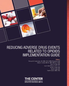
Toolkit Can Help Reduce Opioid-Related Adverse Events

The RADEO toolkit also provides strategies for facilitating policy formation, evaluating current processes, tracking progress against implementation goals, and identifying best practices. Although the RADEO toolkit is designed for the inpatient setting, it also discusses care transitions for patients on opioid therapy to the outpatient setting.
Download the toolkit today at www.hospitalmedicine.org/RADEO. Check out all available quality improvement and patient safety toolkits at www.hospitalmedicine.org/qi.
Submit Your Case Study on Promoting Antimicrobial Stewardship and “Fight the Resistance”


CT Scans Reliable Determinants of Blunt Trauma
NEW YORK - CT scans identify all clinically significant cervical spine injuries in intoxicated patients with blunt trauma, according to a new study.
"I don't think any of the results were particularly surprising to any of us who regularly do trauma care, but what I do think is remarkable about them is that they dispel several long-held myths about the c-spine, intoxicated patients, and the clearance process," Dr. Matthew J. Martin from Legacy Emanuel Medical Center, Portland, Oregon told Reuters Health.
"I think it again confirms that modern CT scan is highly reliable for identifying significant c-spine injuries, but also that the majority of so called 'intoxicated' patients are examinable enough to determine whether the collar can be removed (when combined with the CT scan)," he said.
Up to half of trauma patients are intoxicated, making clearance of the cervical spine a commonly encountered dilemma with both medical and medicolegal implications. Most guidelines indicate that the cervical spine should not be cleared in such patients, resulting in prolonged immobilization or additional imaging even in the face of a normal CT scan.
Dr. Martin's team examined cervical spine clearance practices for intoxicated trauma patients, examined the reliability of cervical spine CT scans for identifying clinically significant injuries (CSIs), and looked for CSIs that might have been missed by CT scans.
Among 1,429 patients who had an alcohol or drug screen performed, 44.2% were intoxicated, the researchers report in JAMA Surgery, online June 15.
Cervical spine injuries were identified in 11.3% of the sober group, 8.1% of the alcohol-intoxicated group, and 12.0% of the drug-intoxicated group.
CT scans yielded negative predictive values of 99.2% for all injuries and 99.8% for unstable injuries. There were five false-negative CT scans, including four central cord syndromes without associated fractures and one potentially unstable injury in a drug-intoxicated patient who presented with clear quadriplegia on examination.
Half of the intoxicated patients were admitted with continued cervical spine immobilization only on the basis of their intoxication. There were no missed CSIs in this group, and all patients were discharged without evidence of an injury or neurologic deficit. They underwent cervical spine immobilization for an average of 15.1 hours, about four times the average time to cervical spine clearance among sober patients (3.7 hours).
"The finding of how long we are keeping these patients in a c-collar based solely on intoxication should raise some eyebrows, and identifies an easy target for process improvement," Dr. Martin said.
"Cervical collars and immobilization are not therapeutic for the vast majority of c-spine injuries; they are really only to prevent inadvertent motion of an unstable c-spine injury," Dr. Martin said. "This is exceedingly rare in a patient who presents with no gross motor deficit, and a high quality CT scan will identify these unstable injuries very reliably. In addition, there are multiple adverse effects of prolonged immobilization, and even of getting an MRI."
"When these are factored in, I think the risk:benefit analysis falls squarely on the side of early clearance based on CT scan," he concluded.
"A key point is that this should be done by experts who are familiar with not only the global concept (the collar can be removed with a negative CT scan), but also the finer points where you could potentially cause harm, or where you should not remove the collar," Dr. Martin added. "This is where a very clear written protocol comes into play and reduces variation or errors that could cause patient harm."
"The results of this study suggest that it is unnecessary to delay cervical spine clearance until intoxicated patients are sober or until magnetic resonance imaging is performed," write Dr. Olubode A. Olufajo and Dr. Ali Salim from Brigham and Women's Hospital, Boston, in a related editorial. "However, caution must be taken in making conclusions based on these data."
"Although the authors conducted the study at an institution with high-quality CT technology and well-trained radiologists, they still recorded a false-negative CT report consistent with a misread," they note. "With the higher potential for this nature of error in lower-resourced settings, it becomes important to compare the costs and benefits of early removal of cervical collars."
They wonder, "With our knowledge that intoxicated patients form up to half of the population of trauma patients, is it really safe to risk irreversible injuries in 1% of the population to save a few hours in cervical clearance times?"
Dr. Stephen Asha from the University of New South Wales in Sydney, Australia, who has reported on various aspects of cervical spine imaging, told Reuters Health by email, "I think this study confirms what clinical experience as well as much of the more recent studies on cervical spine CT scanning tells us, which is that if there is nothing abnormal detected on a new generation, multi-slice CT, then the neck can be cleared."
"Of course there were a few missed injuries, but this needs to be put into context: no one just does a test in isolation, it is always combined with a clinical assessment, and a consideration the mechanism of injury," said Dr. Asha, who was not involved in the new work. "In this case there were five injuries not apparent on the CT scan, but all had obvious spinal cord injury on clinical examination before the CT was done, so these injuries were never going to be missed in a real clinical setting."
"MRI use should be carefully considered because the problem with MRI is that it can be over-sensitive, demonstrating abnormal signal suggesting ligamentous injury in patient who simply have a ligamentous 'strain,'" Dr. Asha explained. "The false-positive results then lead to further periods of inappropriate immobilization and testing, with the accompanying costs, inconvenience, and complications."
"In patients in whom the clinical assessment raises no concerns for injury, then a normal CT should herald the end of investigations," he said. "MRI should be reserved for those where the clinical assessment is abnormal or where the CT is abnormal and further evaluation for ligamentous or spinal injury is required."
Dr. Asha concluded, "If the clinical exam is not concerning and the CT is normal, then clear the neck."
SOURCE: http://bit.ly/28MxHxA and http://bit.ly/28MxHO5
JAMA Surg 2016.
Education, Networking Opportunities Inspire Arizona Hospitalist Vishal Verma, MD, to Start New SHM Chapter

This month, The Hospitalist spotlights Vishal Verma, MD, medical director of the hospitalist program at 4C Medical Group in Scottsdale, Ariz. In addition to being an active SHM member and regular attendee at SHM meetings, Dr. Verma recently purchased a group membership for his hospitalist team. He also started an Arizona chapter of SHM, based on his positive experiences with SHM. He recently spoke with The Hospitalist to share his path to hospital medicine and his inspiration to expand the society’s reach in Arizona.
Question: How did you arrive at a career in hospital medicine?
Answer: After finishing my medical school training at Kasturba Medical College in Manipal, India, I traveled to the United States to further my education and begin my internship and residency. I started my internship in internal medicine at a downtown Brooklyn, N.Y., hospital in 2006. Internship year, though often considered to be a hectic and laborious year, was when I learned for the first time how to care for hospitalized patients. I was chosen by the chief residents as intern of the month in my first month of training. This honor, and the experience of training in an inner-city hospital, further ignited my passion to practice medicine.
It was during my time as an intern and resident when I fully realized the critical role hospitalists play as the main coordinators of care and witnessed their influence on care outcomes. I later served as chief medical resident and was elected by my fellow residents as president of house staff. I was also elected as vice president of the Committee of Interns and Residents (CIR) and served on CIR’s National Executive Board, where I passionately advocated for my patients and fellow colleagues. Serving in various roles provided me with an in-depth knowledge of hospital medicine and helped me build it as my career.
Q: In your current role, how does your membership with SHM help you improve quality of patient care?
A: Our group consists of 14 hospitalists who serve in two community hospitals. Being a member of SHM for many years has been a rewarding experience as it keeps me informed about changes and advances in hospital medicine. Through the Journal of Hospital Medicine, regular webinars, and SHM conferences and annual meetings, SHM helps us enrich our knowledge base on quality, performance, patient experience, coding, practice management, acute and post-acute care, and other aspects of hospital medicine.
At our recent visit to HM16 in San Diego, a few members of our team attended sessions on post-acute care and value-based reimbursements. At the sessions, we learned of the importance of stressing quality and engaging sub-acute rehab facilities in meaningful ways so as to improve the quality of care in skilled nursing facilities and also help to decrease the length of stay from 30 days to closer to 15 days. 4C Medical Group has implemented many suggestions from these lectures and is in the process of transforming our post-acute-care teams.
I also serve as a member of the board of directors for 4C Medical Group, where my association with SHM has helped me give valuable input while we manage the care of over 20,000 patients in acute, sub-acute, and home-based teams as well as outpatient clinics. SHM provides its members with a platform to sharpen their leadership skills and enables members to build a strong network among fellow leaders, which helps us learn about and share best practices, which translates to better quality of care.
For example, a recent presentation by SHM member Dr. Jesse Theisen-Toupal on inpatient management of opioid use disorder was an eye-opener. Learning about harm-reduction strategies for opioid misuse during the presentation was very helpful to us, and we shared the suggestions with our hospitalist team.
Q: What inspired you to start an Arizona chapter in Scottsdale and purchase a group membership for your team?
A: At HM16, I met Debra Beach, manager of membership and outreach programs at SHM, and we discussed how our company can align with SHM and bring our hospitalists on board as members to provide them with a greater network of resources. I was surprised that Arizona did not have a dedicated SHM chapter. Phoenix, one of the U.S.’s largest metropolitan areas, has many large hospital systems employing and contracting thousands of hospitalists. I saw an opening for a great opportunity to take the lead on developing an SHM chapter in Arizona with the support of my 4C colleagues. After discussing this opportunity with other hospitalist groups in Arizona, we came to the conclusion that it would benefit not only our team at 4C but hospitalists statewide.
I am confident that the Arizona chapter of SHM will not only be successful but soon will be contributing nationally to the hospitalist movement. Moreover, SHM will help keep our members educated and informed about the upcoming changes as we transition to a pay-for-performance model of reimbursement and any other healthcare system changes still to come.
I believe in the famous Chinese proverb, “A journey of a thousand miles begins with a single step.” We have taken the first step in our hospitalist group, and without a doubt, SHM’s journey in the state of Arizona shall be a success story. All our members are excited with this new beginning.
Q: How do you see hospital medicine evolving over the next 20 years?
A: 2016 has already been designated as the “Year of the Hospitalist.” I will take it a step further and predict that the next decade will be a decade of hospital medicine. Inpatient care is transforming at a rapid pace, and we need a dedicated and well-trained stream of doctors who are specialists in managing hospitalized patients. Care of hospitalized patients was once fragmented and costly; now with hospitalists as captains of the ship, care can be delivered in more comprehensive, cost-effective ways with better quality and increased performance. The introduction of a separate specialist billing code for hospitalists by the CMS is a step in the right direction.
In the next few years, I would enjoy seeing a separate board for hospitalists with hospital medicine’s own specialty certification. The potential for hospital medicine’s continued growth is tremendous, and I look forward to being a part of its future. TH
Brett Radler is SHM’s communications specialist.

Get up to Speed on the Latest in Pediatric Hospital Medicine

Register, book your hotel, and see the full course schedule at www.phmmeeting.org.
Brett Radler is SHM’s communications coordinator.
Register, book your hotel, and see the full course schedule at www.phmmeeting.org.
Brett Radler is SHM’s communications coordinator.
Register, book your hotel, and see the full course schedule at www.phmmeeting.org.
Brett Radler is SHM’s communications coordinator.
Is It Safe to Discharge a Patient with IDU History, PICC for Outpatient Antimicrobial Therapy?
Case
A 42-year-old female with a history of intravenous (IV) drug use presents with severe neck pain, gait instability, and bilateral C5 motor weakness. A cervical MRI shows inflammation consistent with infection of her cervical spine at C5 and C6 and significant boney destruction. The patient undergoes kyphoplasty and debridement of her cervical spine. Operative cultures are significant for Pseudomonas aeruginosa. Infectious disease consultants recommend parenteral ceftriaxone for six weeks. The patient has no insurance, and efforts to obtain long-term placement are unsuccessful. The patient states that her last use of IV drugs was three months ago, and she insists that she will abstain from illicit IV drug abuse going forward.
Background
Outpatient parenteral antibiotic treatment (OPAT) has proven to be a cost-effective and relatively safe treatment option for most patients.1 For these reasons, it has been encouraged for use among a wide a variety of clinical situations. Intravenous drug users (IDUs) are often underinsured and have few options other than costly treatment in an inpatient acute-care facility.
A history of illicit injection drug use frequently raises questions about the appropriateness of OPAT. Some of our most vulnerable patients are those who abuse illicit drugs. Due to psychiatric, social, and financial factors, their ability to adequately transition to outpatient care may be limited. They are often underinsured, and appropriate options for inpatient post-acute care may not exist. Hospitalists often feel pressure to discharge these patients despite the lack of optimal follow-up care, and they must weigh the risks and benefits in each case.
The enrollment of IDUs into an OPAT service using a peripherally inserted central catheter (PICC) is controversial and often avoided. No clear-cut guidelines concerning the use of OPAT in IDUs by national medical societies exist.2 Consultants are often reluctant to recommend options that deviate from the typical standard of inpatient or directly observed care. The obvious risk is that a PICC line provides easy and tempting access to veins for continued drug abuse. In addition, there is an increased risk of infection and/or thrombosis if the PICC is abused.3
The safety and efficacy of PICC line use for OPAT in IDUs are unknown, and studies addressing these issues are limited. In one study at the National University Hospital of Singapore, 29 IDU patients received OPAT without complications.4 Patients were closely monitored, including by use of a tamper-proof security seal on the PICC. Infective endocarditis was the primary diagnosis in 42% of the cases studied. There were no deaths or cases of PICC abuse reported. In another abstract presentation, 39 IDU patients at Henry Ford Health System in Detroit were discharged to outpatient therapy with a PICC line and demonstrated a high cure rate (73.3%). Nine patients were lost to follow-up.5
No studies have compared OPAT therapy to inpatient therapy in IDU patients.
Back to the Case
Despite multiple attempts and due to financial considerations, no long-term care facility is able to admit the patient for therapy. The frequency of required antibiotics makes outpatient therapy in an infusion center problematic. The primary service is reluctant to discharge the patient home with a PICC line in place due to the potential of abuse and complications. A “Goals of Care” committee, consisting of several physicians from multiple specialties, legal counsel, and case management, is convened to review the case. The committee concludes that, in this particular case, it would be a reasonable option to discharge the patient to home with a PICC line in place to complete OPAT. A patient agreement document is drafted; it describes the complications of PICC line abuse and stipulates that the patient agrees to drug testing throughout the duration of her treatment. A similar agreement is required by the home infusion company. Both documents are signed by the patient, and she is subsequently discharged home.
Bottom Line
Our strategy is to deal with each of these cases as unique situations because no policies, procedures, protocols, or guidelines currently exist. One of the guiding principles should be, despite financial pressures, that the primary focus is on appropriate care of this vulnerable population. A type of “Goals of Care” committee (or organizational equivalent) can be utilized to offer assistance in decision making. Unfortunately, the safety and efficacy of OPAT in IDU patients are uncertain, and there is a lack of studies to support definitive protocols. In select cases, OPAT in IDU patients may be considered, but signed consent of the risks and the patient’s responsibilities concerning OPAT should be clearly documented in the medical record by the discharging team. TH
Dr. Conrad is a hospitalist with Ochsner Health System in New Orleans.
References
- Tice AD, Hoaglund PA, Nolet B, McKinnon PS, Mozaffari E. Cost perspectives for outpatient intravenous antimicrobial therapy. Pharmacotherapy. 2002;22(2, pt 2):63S-70S.
- Tice AD, Rehm SJ, Dalovisio JR, et al. Practice guidelines for outpatient parenteral antimicrobial therapy. IDSA guidelines. Clin Infect Dis. 2004;38(12):1651-1672.
- Chemaly R, de Parres JB, Rehm SJ, et al. Venous thrombosis associated with peripherally inserted central catheters: a retrospective analysis of the Cleveland Clinic experience. Clin Infect Dis. 2002;34(9):1179-1183.
- Ho J, Archuleta S, Sulaiman Z, Fisher D. Safe and successful treatment of intravenous drug users with a peripherally inserted central catheter in an outpatient parenteral antibiotic treatment service. J Antimicrobial Chemotherapy. 2010;65(12):2641-2644.
- Papalekas E, Patel N, Neph A, Moreno D, Zervos M, Reyes K. Outpatient parenteral antimicrobial therapy (OPAT) in intravenous drug users (IVDUs): epidemiology and outcomes. Oral abstract presented at: IDWeek; October 2014; Philadelphia.
LETTER: Point-of-Care Ultrasound: The (Sound) Wave of the Future for Hospitalists
Small devices carried in pockets during rounds can enable hospitalists to make quick decisions at the bedside, enhance and teach physical exam skills, streamline patient flow through the hospital, and potentially avoid the cost and risk of exposure to radiation. Point-of-care (POC) ultrasound enhances both patient satisfaction and the clinician’s professional satisfaction. Hospital medicine will be the next field to rapidly assimilate its use.
POC, or “bedside,” ultrasound has been used by ob-gyns, vascular access, and procedural teams for quite some time. Of late, emergency medicine and critical care physicians have adopted its use. It offers the advantage of gaining immediate information regarding the patient through dynamic imaging and the ability to integrate that information into the clinical picture. This enables providers to make decisions about patient care in real time.
With the advent of affordable handheld devices with quality images, rounding with these devices has become practical for hospitalists. Hospitalists should rapidly embrace this skill set. POC ultrasound can be very useful to quickly improve patient diagnosis, patient satisfaction, patient safety, length of stay, and provider satisfaction.
For example, in patients complaining of dyspnea, for which there is not a clear diagnosis of COPD, congestive heart failure, pulmonary embolism, or pneumonia, a focused cardiac ultrasound can rapidly differentiate between right ventricular dysfunction, left ventricular dysfunction, pericardial effusion, or a hyperdynamic heart. Lung ultrasound with diffuse or focal “B lines,” focal consolidation, and/or pleural effusion can assist in differentiating the cause as well.
POC ultrasound also is a teaching tool that can enhance exam skills. Hospitalists can confirm exam findings and teach as they palpate the liver or percuss the chest. Performing a procedure such as paracentesis or a central line with ultrasound guidance is now considered standard of care in some centers. The literature shows ultrasound guidance is safer even when compared to clinicians skilled in landmark techniques. In addition, many hospitalists and/or trainees will work in areas where 24-7 echo, interventional radiologists, and ultrasound techs are not available. Hospitalists need to know how to use POC ultrasound to serve patients well.
POC ultrasound can also be used in daily care. For heart failure patients, watching the B lines (pulmonary edema), pleural effusions, and inferior vena cava size can avoid over- or under-diuresis and reduce length of stay and cost. The same can be said for patients with percutaneous catheters to ensure proper drainage of the pockets of fluid in the chest or abdomen.
It is important to know the limitations of POC ultrasound. It is best used to answer binary questions (e.g., pericardial effusion present or not). It is a skill to be acquired and honed, and it requires specialized training. There are many one- to two-day courses as well simulators and other means. The basics of image acquisition and interpretation can be found online, and much of it is free. Manufacturers often are willing to provide machines to practice with.
Many patients enjoy seeing the images and having a better understanding of their disease process, which leads to improved patient satisfaction. Overall, there are many benefits for hospitalists.
Gordon Johnson, MD, hospitalist and president, Oregon/Southwest Washington SHM Chapter