User login
American Academy of Neurology (AAN): Annual Meeting
Pathologic Proteins in Alzheimer’s, Parkinson’s Also Collect in Skin Cells
The same dysregulated proteins that form in the brains of patients with Alzheimer’s disease or Parkinson’s disease appear to clump within their skin cells as well, according to new research released Feb. 24 in advance of the annual meeting of the American Academy of Neurology.
In a small prospective study, epidermal samples from patients showed intracellular aggregates of phosphorylated tau, a pathologic hallmark of both Alzheimer’s and Parkinson’s. Patients with Parkinson’s disease also showed a similarly increased expression of alpha-synuclein. But neither protein was present in any of the samples taken from healthy control subjects.
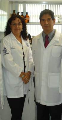
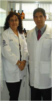
If these findings can be validated in large groups, epidermal protein aggregation might have some potential as a biomarker for both Alzheimer’s and Parkinson’s diseases and perhaps in other proteinopathies, such as frontotemporal dementia, progressive supranuclear palsy, and corticobasal syndrome, according to Dr. Ildefonso Rodríguez-Leyva of the University of San Luis Potosí (Mexico). He and his colleague, Dr. María E. Jiménez-Capdeville, will present their findings at the meeting in April.
Their current work builds on initial data presented at the 2012 annual meeting, and later published (Ann. Clin. Transl. Neurol. 2014;1:471-8). That study showed that about 60% of cells in specific skin structures in patients with Parkinson’s disease had alpha-synuclein inclusions.
The current study included 65 subjects: 20 with Alzheimer’s, 16 with Parkinson’s, 17 with non-neurodegenerative dementia, and 12 age-matched healthy controls. All patients were in the mild-moderate stage of their illnesses. Each underwent a retroauricular punch biopsy. This is an ideal area because the procedure leaves no visible scar and bleeding is easy to control, Dr. Rodríguez-Leyva said in an interview. Samples were analyzed by immunohistochemistry.
Finding these proteins in the skin of patients with neurodegenerative diseases makes sense, Dr. Rodríguez-Leyva said, because neurons and epidermal cells arise from the same fetal lamina – the ectoderm. “Because these cells have the same origin, our thought is that they should have a similar program of protein expression. Therefore, the skin may reflect adverse events taking place in the nervous system.”This initial study didn’t attempt to correlate epidermal protein expression and disease stage. But that study is coming, Dr. Rodríguez-Leyva said. “We can’t be sure at this point if there is any relationship there. We need to test more patients in different points of disease progression to know if the levels of altered proteins correlate with disease evolution.”
Neither Dr. Rodríguez-Leyva nor Dr. Jiménez-Capdeville had any financial disclosures. The study was supported by the National Council of Science and Technology of Mexico.
FROM THE AAN 2015 ANNUAL MEETING
Pathologic proteins in Alzheimer’s, Parkinson’s also collect in skin cells
The same dysregulated proteins that form in the brains of patients with Alzheimer’s disease or Parkinson’s disease appear to clump within their skin cells as well, according to new research released Feb. 24 in advance of the annual meeting of the American Academy of Neurology.
In a small prospective study, epidermal samples from patients showed intracellular aggregates of phosphorylated tau, a pathologic hallmark of both Alzheimer’s and Parkinson’s. Patients with Parkinson’s disease also showed a similarly increased expression of alpha-synuclein. But neither protein was present in any of the samples taken from healthy control subjects.
If these findings can be validated in large groups, epidermal protein aggregation might have some potential as a biomarker for both Alzheimer’s and Parkinson’s diseases and perhaps in other proteinopathies, such as frontotemporal dementia, progressive supranuclear palsy, and corticobasal syndrome, according to Dr. Ildefonso Rodríguez-Leyva of the University of San Luis Potosí (Mexico). He and his colleague, Dr. María E. Jiménez-Capdeville, will present their findings at the meeting in April.
Their current work builds on initial data presented at the 2012 annual meeting, and later published (Ann. Clin. Transl. Neurol. 2014;1:471-8). That study showed that about 60% of cells in specific skin structures in patients with Parkinson’s disease had alpha-synuclein inclusions.
The current study included 65 subjects: 20 with Alzheimer’s, 16 with Parkinson’s, 17 with non-neurodegenerative dementia, and 12 age-matched healthy controls. All patients were in the mild-moderate stage of their illnesses. Each underwent a retroauricular punch biopsy. This is an ideal area because the procedure leaves no visible scar and bleeding is easy to control, Dr. Rodríguez-Leyva said in an interview. Samples were analyzed by immunohistochemistry.
Finding these proteins in the skin of patients with neurodegenerative diseases makes sense, Dr. Rodríguez-Leyva said, because neurons and epidermal cells arise from the same fetal lamina – the ectoderm. “Because these cells have the same origin, our thought is that they should have a similar program of protein expression. Therefore, the skin may reflect adverse events taking place in the nervous system.”This initial study didn’t attempt to correlate epidermal protein expression and disease stage. But that study is coming, Dr. Rodríguez-Leyva said. “We can’t be sure at this point if there is any relationship there. We need to test more patients in different points of disease progression to know if the levels of altered proteins correlate with disease evolution.”
Neither Dr. Rodríguez-Leyva nor Dr. Jiménez-Capdeville had any financial disclosures. The study was supported by the National Council of Science and Technology of Mexico.
On Twitter @alz_gal
FROM THE AAN 2015 ANNUAL MEETING
Key clinical point: The abnormal brain proteins of Alzheimer’s and Parkinson’s may also be expressed in skin cells.
Major finding: Tau and alpha-synuclein were expressed in the epidermal cells of patients, but not in those of healthy controls.
Data source: The prospective study comprising 65 subjects (20 with Alzheimer’s, 16 with Parkinson’s, 17 with non-neurodegenerative dementia, and 12 age-matched healthy controls).
Disclosures: Neither Dr. Rodríguez-Leyva nor Dr. Jiménez-Capdeville had any financial disclosures. The study was supported by the National Council of Science and Technology of Mexico.
Larger and more severe strokes seen with aspirin resistance
Patients with acute ischemic stroke who test positive for aspirin resistance had both larger stroke volume and increased severity, compared with patients without resistance, in an observational study of 311 patients at Korean centers.
Given that previous studies have shown that the use of aspirin is associated with lower stroke severity and decreased infarction growth, the current study’s findings may help to define the effect of aspirin resistance (AR) on stroke severity, since previous studies had provided inconclusive results, Dr. Mi Sun Oh and colleagues at Hallym University Sacred Heart Hospital, Anyang, South Korea, wrote in their abstract. The findings were released Feb. 23 in advance of the annual meeting in April of the American Academy of Neurology.
The investigators enrolled patients with acute ischemic stroke confirmed by diffusion-weighted imaging (DWI) who had received at least 7 days of aspirin therapy before initial stroke symptoms and had been checked for AR within 24 hours of hospital admission. Patients with high prestroke disability scores (modified Rankin Scale score > 2) were excluded, as were those who were taking another antiplatelet or anticoagulant medication concurrently with aspirin on hospital admission.
The abstract did not report detailed patient characteristics or information about type or dose of aspirin; the full results of the study will be presented at the meeting in Washington.
Enrollees were deemed aspirin resistant if a rapid assay detected greater than 550 Aspirin Reaction Units. DWI-observed stroke volume was assessed via a semiautomated threshold technique, and investigators employed the National Institutes of Health Stroke Scale (NIHSS) score to measure initial stroke severity.
Seventy-eight of the 311 patients (25.1%) had AR. Dr. Oh and colleagues reported that median stroke volume was higher for these patients, compared with the aspirin-sensitive group (2.8 cc vs. 1.6 cc), as was least-square mean on multivariate analysis (1.6 cc [95% CI, 1.1-2.1] vs. 1.1 cc [95% CI, 0.7-1.4], P = .036). Median NIHSS scores were also higher for the AR group (4 vs. 3), indicating greater stroke severity, a result that was confirmed by multivariate analysis.
Aspirin resistance is a complicated and heterogeneous concept, and not a well defined entity, according to vascular neurologist Dr. Philip Gorelick, head of the Hauenstein Neuroscience Center at St. Mary’s Health Care in Grand Rapids, Mich. Dr. Gorelick is an honorary member of the Korean Stroke Society but was not involved in the present study. In an interview, he expanded on the diverse mechanisms that can impede the stroke prevention effect of antiplatelet agents such as aspirin (Stroke Res. Treat. 2013;Article ID 727842 [doi:10.1155/2013/727842]).
In contrast to the traditional notion of “resistance” as an inherent or acquired defense or chemical blockage of a drug, whether by a microbe or the host, aspirin resistance may be either a laboratory-defined lack of inhibition of thromboxane A2, or a clinically-defined entity. In either case, a host of factors may contribute, Dr. Gorelick said. Poor adherence to an aspirin therapy regimen may be a primary contributor to AR. Further, enterically coated aspirin may not be as well absorbed in the gut, leading to lower effective aspirin dosing. A host of other factors, including concurrent medication administration, comorbidities impacting platelet turnover, and genetic polymorphisms may also contribute to aspirin failure.
Although patient characteristics were not reported in this study, Dr. Gorelick did issue a general note of caution: “Another major issue in these types of studies,” he noted, is to determine if “patients are similar in terms of background factors. Patients on aspirin therapy may be more likely to have more severe preexisting vascular disease,” predisposing them to more severe stroke.
The Korea Healthcare Technology R&D Project, Ministry of Health and Family Welfare, and the Republic of Korea supported the study. The authors had no disclosures.
FROM THE AAN 2015 ANNUAL MEETING
Key clinical point: Volume and severity of ischemic stroke were larger in patients with aspirin resistance.
Major finding: Patients with acute ischemic stroke and aspirin resistance had greater median stroke volume than did aspirin-sensitive patients (2.8 cc vs. 1.6 cc) and had more severe strokes according to median NIHSS score (4 vs. 3).
Data source: Study of 311 patients with MRI-confirmed acute ischemic stroke and at least 7 days of aspirin therapy preceding stroke.
Disclosures: The Korea Healthcare Technology R&D Project, Ministry of Health and Family Welfare, and the Republic of Korea supported the study. The authors had no disclosures.


