User login
Transient tachypnea of newborn increases bronchiolitis risk
MADRID – A new Finnish study raises a provocative question: Is transient tachypnea of the newborn really transient?
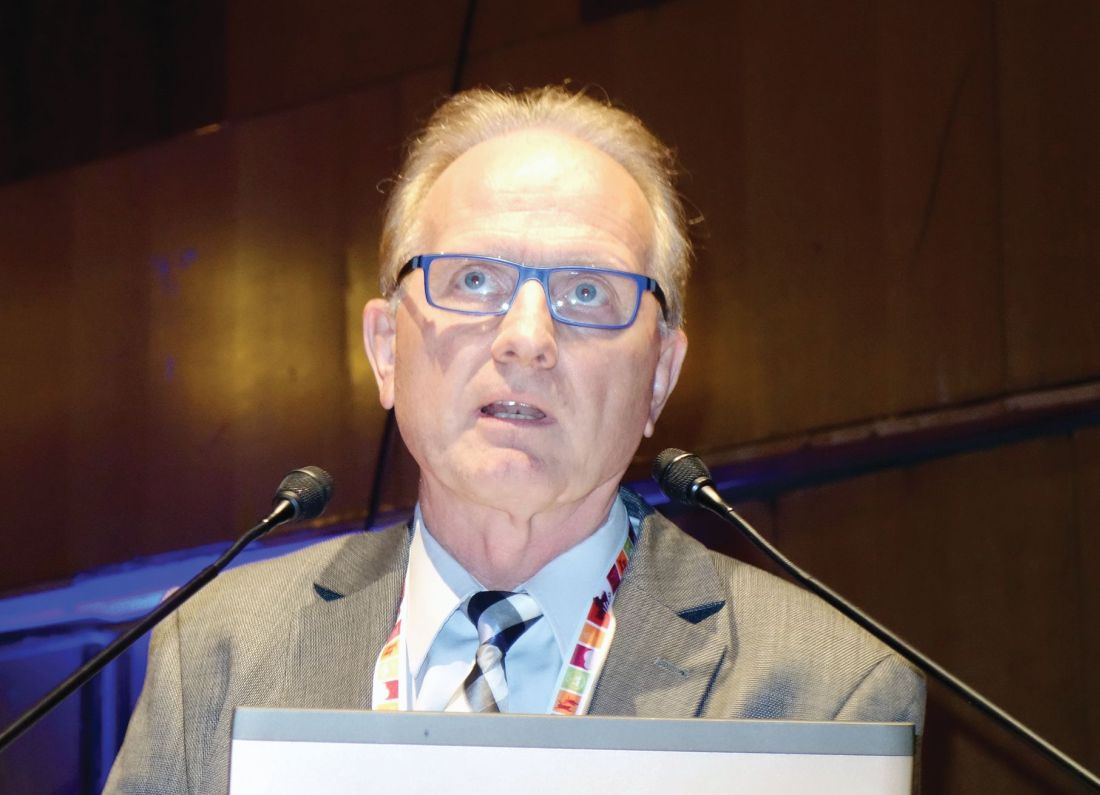
Transient tachypnea of the newborn (TTN) has traditionally been viewed as a benign, self-limited condition involving 1-3 days of respiratory distress. But data from Finland’s comprehensive national health registries indicate that TTN in term babies is associated with significantly increased risk of subsequent bronchiolitis during infancy, Otto Helve, MD, reported at the annual meeting of the European Society for Paediatric Infectious Diseases.
“This association suggests similar pathogenic mechanisms in transient tachypnea of the newborn and bronchiolitis. We suggest that an intrinsic defect in sodium ion–driven pulmonary fluid transport may predispose to clinically significant bronchiolitis during the first year of life,” said Dr. Helve, a pediatrician at the National Institute for Health and Welfare, Helsinki, and the University of Helsinki.
Of more than 1 million term babies born in Finland during 1996-2015, 17,569 were diagnosed with TTN. During the same period, 40,338 infants were hospitalized with a diagnosis of bronchiolitis attributable to respiratory syncytial virus infection.
In a multivariate analysis adjusted for birth year, gender, delivery method, gestational age, and parity, TTN was independently associated with a 1.2-fold increased risk of bronchiolitis during the first year of life.
Dr. Helve reported having no financial conflicts of interest regarding his study.
AT ESPID 2017
Key clinical point:
Major finding: Transient tachypnea of the newborn was independently associated with a 1.2-fold increased risk of bronchiolitis during the first year of life of Finnish term babies.
Data source: This population-based case-control study included 17,569 Finnish term babies diagnosed with transient tachypnea of the newborn and 40,338 diagnosed with bronchiolitis.
Disclosures: Dr. Helve reported having no financial conflicts of interest.
Tipoffs that an infant hospitalized with pertussis may require ICU
MADRID – Infants hospitalized for pertussis are more likely to develop severe disease requiring pediatric ICU admission if they are experiencing apnea, are unvaccinated against pertussis, or are less than 2 months old, Maria Arranz, MD, reported at the annual meeting of the European Society for Paediatric Infectious Diseases.
“The presence of these parameters on admission should warn us of possible severe disease,” said Dr. Arranz of Gregorio Maranon Hospital in Madrid.
Dr. Arranz presented a retrospective observational study of 101 children under 1 year of age who were hospitalized for pertussis at the Madrid tertiary center prior to the hospital’s 2016 shift to a strategy of maternal immunization during pregnancy as a means of preventing pertussis in infancy. Thirteen percent of the children required admission to the pediatric ICU and thus by definition had severe disease.
Half of infants in the study were not vaccinated against pertussis. That proved to be a powerful risk factor for severe disease requiring an ICU stay. Only 8% of children with severe pertussis were vaccinated, compared with a 58% vaccination rate among those who avoided the ICU.
Apneic pauses were noted in 67% of the severe disease group, compared with 28% of the infants who didn’t need the ICU.
The pertussis patients admitted to the pediatric ICU averaged 1 month of age, compared with age 2 months in the nonsevere group.
The maximum leukocyte, lymphocyte, and neutrophil counts during the hospital stay of the severe disease group averaged 23,600 cells/mm3, 18,000/mm3, and 5,000/mm3, respectively, significantly greater than the 15,300, 10,700, and 3,900 cells/mm3 in infants who did not require the ICU. Levels of all three cells also were higher at admission in the severe pertussis group than in those who didn’t make it to the ICU, albeit not statistically significantly so.
Dr. Arranz reported having no financial conflicts of interest regarding her study.
AT ESPID 2017
Key clinical point:
Major finding: Among infants hospitalized for pertussis, apneic pauses were present in two-thirds of those with severe disease, ultimately resulting in admission to the pediatric ICU, compared with 28% of those who didn’t require an ICU stay.
Data source: This retrospective observational study sought to identify factors associated with severe pertussis requiring an ICU stay in a group of 101 Spanish infants hospitalized with the disease.
Disclosures: The presenter reported having no financial conflicts of interest regarding her study.
HCV/HIV-coinfected teens rapidly progress to advanced liver disease
MADRID – Roughly one in four children with vertically transmitted hepatitis C virus (HCV)/HIV coinfection will progress to advanced hepatic fibrosis by age 20 years despite treatment with pegylated interferon plus ribavirin, Carolina Fernández McPhee, MD, reported at the annual meeting of the European Society for Paediatric Infectious Diseases.
This rate was nearly five times higher than in matched children with vertically acquired HCV monoinfection in a multicenter retrospective study, according to Dr. McPhee of Hospital General Universitario Gregorio Marañón in Madrid.
She presented a multicenter retrospective study of liver disease progression in 71 HCV/HIV coinfected children and 71 age- and sex-matched HCV-monoinfected children. The coinfected children are being followed in CORISPES (the Spanish Cohort of HIV-infected Children), where they receive state-of-the-art care.
All was quiet through age 9 years, with no progression to liver fibrosis in either patient group. Among patients followed to age 20 years, however, 9 (24%) of 38 HCV/HIV-coinfected patients showed progression to advanced fibrosis, compared with just 3 (6%) of 54 patients with HCV only.
Of HCV/HIV coinfected patients, 73% were infected with the hard to treat viral genotypes 1 or 4, compared with 93% of patients with HCV-only.
In the study group, 22 patients with HCV/HIV and 52 patients with HCV-only underwent treatment with pegylated interferon and ribavirin. At the time of treatment, three coinfected patients already had cirrhosis, another eight had moderate to advanced fibrosis, and half had no or mild fibrosis. In contrast, only one patient with HCV monoinfection had cirrhosis, four had moderate to advanced fibrosis, and the rest – nearly 90% of the total group – had no or mild fibrosis.
At treatment initiation, 96% of the HCV/HIV group were on antiretroviral therapy, 86% showed suppression of HIV RNA, 44% had AIDS, and 32% had a CD4 count below 500 cells/mm3.
The sustained viral response rate was similar in the two groups of patients – 41% in the HCV/HIV group and 42% in HCV-only patients – despite the fact that the HCV monoinfected patients had a higher prevalence of the tough to treat genotypes.
Roughly 12 million people worldwide are coinfected with HCV and HIV.
Dr. McPhee reported having no relevant financial disclosures.
AT ESPID 2017
Key clinical point:
Major finding: By age 20 years, 24% of a group of patients with vertically transmitted HCV/HIV coinfection had progressed to advanced hepatic fibrosis, compared with 6% of patients with vertically transmitted HCV monoinfection.
Data source: This retrospective, multicenter, observational Spanish study included 71 children with vertically transmitted HCV/HIV coinfection and 71 with vertically transmitted HCV monoinfection.
Disclosures: Dr. McPhee reported having no relevant financial disclosures.
Hypertonic saline for bronchiolitis found ineffective in large RCT study
MADRID – Giving nebulized hypertonic saline (NHS) to infants who present to pediatric emergency departments with a first episode of moderate to severe acute bronchiolitis did not reduce their hospitalization rate in the randomized, multicenter, double-blind GUERANDE trial.
Moreover, mild adverse events – mainly worsening cough – were significantly more frequent in the NHS recipients than in controls given nebulized normal saline, Christele Gras-le Guen, MD, reported at the annual meeting of the European Society for Paediatric Infectious Diseases.
Some previous studies have suggested a modest benefit, but they were underpowered to draw meaningful conclusions. GUERANDE, a 777-patient randomized trial conducted in 24 French pediatric emergency departments, was the first quality study large enough to determine whether the therapy results in fewer hospital admissions, she said.
In GUERANDE, infants with a first episode of acute bronchiolitis were given two 20-minute nebulizations of 3% hypertonic saline or 0.9% normal saline 20 minutes apart.
The primary outcome was the rate of hospital admission within 24 hours after enrollment. The rate was 48% in the NHS group and 52% in controls, a nonsignificant difference which shrunk even more after controlling for age, oxygen saturation, and respiratory syncytial virus infection status.
No serious adverse events occurred in GUERANDE. However, the rate of mild adverse events was 9% in the NHS group, significantly higher than the 4% rate in controls. Notably, cough without respiratory distress occurred 30 times in 26 infants in the NHS group, compared with 4 times in 3 control subjects.
Dr. Gras-le Guin reported having no financial conflicts regarding the GUERANDE study, funded by the French Health Ministry.
AT ESPID 2017
Key clinical point:
Major finding: The rate of hospitalization following administration of nebulized hypertonic saline to infants with a first episode of acute bronchiolitis was 48%, not significantly different from the 52% rate in controls.
Data source: This was a randomized, multicenter, double-blind, controlled clinical trial including 777 infants with acute bronchiolitis.
Disclosures: The GUERANDE study was funded by the French Health Ministry. The presenter reported having no financial conflicts.
Measles immunization is a major challenge in HIV-infected children
MADRID – Impaired response to measles immunization is common in HIV-infected children, and revaccination – while an important strategy – helps only some of them, Ruth del Valle, MD, said at the annual meeting of the European Society for Paediatric Infectious Diseases.
The key predictor of an impaired response to measles immunization appears to be a CD4/CD8 cell ratio of less than 1.0, added Dr. del Valle of Infant Sofia University Hospital in Madrid.
Of note, 48% of the children had a CD4/CD8 ratio of less than 1.0 at baseline. This ratio is gaining credence as a predictor of HIV-positive patients’ immunologic response to a variety of vaccines: most recently, in Dr. del Valle’s study, to measles vaccine and, in two earlier studies published in 2016, to yellow fever vaccine (PLoS Negl Trop Dis. 2016 Dec 12;10[12]:e0005219) and hepatitis B vaccine (Vaccine. 2016 Apr 7;34[16]:1889-95).
In Dr. del Valle’s study, only 52% of HIV-positive children developed protective antibody levels following completion of the standard two-dose MMR schedule. In accord with current Paediatric European Network for Treatment of AIDS (PENTA) guidelines, 41 unresponsive patients then received a booster MMR dose (HIV Med. 2012 Jul;13[6]:333-6). Of the 41, 10 (24%) did not respond to the booster dose, either. Thus, the final measles protection rate in this study was only 77%.
These study findings highlight the importance of routinely testing measles vaccine response in HIV-infected children in clinical practice, she noted.
The CD4/CD8 ratio was 1.3 in good responders to measles immunization and 0.9 in nonresponders. In a multivariate analysis, a CD4/CD8 ratio of less than 1.0 conferred a 3.2 times increased risk of impaired response to the measles vaccine.
Dr. del Valle and her coinvestigators in CORISPES (the Spanish Cohort of HIV-infected Children) plan further studies to determine the duration of protection against measles in those patients who responded to the booster dose.
Session cochair Nigel Klein, MD, commented that Dr. del Valle’s study is “one of a number of reports that HIV-infected children, as they get older, are predisposed to getting infections that we should be able to prevent.”
“I think we don’t know best how to manage this population of patients. You can give more vaccinations, but, as Dr. del Valle has shown, not all of them will respond. And I think this study is further evidence that the better we can maintain our children’s immune systems by treating early, the better our chances of getting good responses when we vaccinate,” said Dr. Klein, professor of pediatric infectious diseases and immunology at Great Ormond Street Children’s Hospital and University College London.
Dr. del Valle reported having no relevant financial disclosures.
AT ESPID 2017
Key clinical point:
Major finding: After two doses of MMR and, if needed, a third booster dose, 23% of a group of HIV-infected children remained unprotected against measles.
Data source: This multicenter study included 120 HIV-infected Spanish children who received two doses of the MMR vaccine and, if still unprotected against measles, a booster dose.
Disclosures: Dr. del Valle reported having no relevant financial disclosures.
All isn’t well with HIV-exposed uninfected infants
MADRID – Children who were HIV-exposed antenatally but not infected are at double the risk of hospitalization for infectious diseases during their first year of life, compared with HIV-unexposed controls, according to what’s believed to be the first prospective study examining the issue in a Western industrialized country.
That’s one key take-away message from the study conducted in Brussels. Another key finding was that the sharply increased risk of hospitalization for infection during infancy was erased if HIV-infected mothers started antiretroviral therapy prior to, rather than during, pregnancy, Catherine Adler, MD, reported at the annual meeting of the European Society for Paediatric Infectious Diseases.
She presented a prospective study of 125 HIV-positive and 119 HIV-negative pregnant Belgian women of comparable ethnic and sociodemographic backgrounds. All of the HIV-positive mothers were on antiretroviral therapy, which they started either prior to or during pregnancy.
The two groups of women gave birth to 132 HEU and 123 HIV-unexposed babies, all born after 35 weeks’ gestation. The babies didn’t differ in terms of gender, prematurity rate, mode of delivery, or the use of antibiotics at delivery. However, 17% of the HEU babies had a birth weight below 2,500 g, compared with just 3% of the HIV-unexposed controls. Also, as a matter of policy, none of the HEU babies were breastfed, while 95% of the controls were, Dr. Adler explained.
The primary outcome in the study was the rate of hospitalization for infection during the first 12 months of life. The rate was 21% in the HEU babies, significantly greater than the 11% rate in HIV-unexposed babies. In a multivariate analysis adjusted for preterm birth, low birth weight, literacy, and maternal age, HEU status was associated with twofold increased risk of hospitalization for infection in infancy.
“The increased susceptibility of HEU infants to infectious disease is not restricted to children born in developing countries,” she declared.
The disparity in hospitalization rates was driven by hospitalization for viral infections, which occurred at a rate of 20% in the HEU group, versus 9% in controls. Particularly notable were the 10 hospitalizations for respiratory syncytial virus infection in the HEU patients, compared with just 1 in the controls.
Dr. Adler and her coinvestigators will continue following the children out to about 3 years of age. After age 12 months, the two groups no longer differed significantly in their risk of hospitalization for infection.
“The first year is a vulnerable period. Our data highlight the importance of a close follow-up of these infants,” she said.
The biggest risk factor for hospitalization for infectious illness in the HEU group was initiation of antiretroviral therapy during pregnancy. The hospitalization rate in HEU infants whose mothers began therapy prior to pregnancy was the same as in HIV-unexposed infants. The inference is that it’s not in utero exposure to antiretroviral drugs that is responsible for the increased risk of hospitalization during infancy.
“This observation supports the notion that it’s the activity of the maternal HIV infection – the exposure to a strongly proinflammatory state in the mother – that contributes to the risk of severe infection in HEU infants, probably by causing changes in innate immunity cells,” according to Dr. Adler.
Even though the increased risk of hospitalization for infectious illnesses in HEU children falls off after age 12 months, she continued, her group is following them out to about age 3 years because “we have the impression that they are at risk for neurodevelopmental problems, including language delay.”
Other researchers in the audience confirmed this risk, reporting that, as they follow HEU children through adolescence, they see an increased rate of attention deficits and associated comorbidities.
Dr. Adler called the administration of antiretroviral therapy to pregnant HIV-infected women in order to prevent maternal-to-child transmission of the disease “one of the major successes of the 21st century.”
“The number of new HIV infections among children has collapsed, leading to an increasing number of HIV-exposed but uninfected children. One million of them are born each year,” she said.
Dr. Adler reported having no financial conflicts of interest regarding her study.
*The article was updated 6/15/17.
AT ESPID 2017
Key clinical point:
Major finding: The rate of hospitalization for a serious infectious illness during the first 12 months of life was 21% in HIV-exposed uninfected children, significantly greater than the 11% rate in HIV-unexposed babies.
Data source: This prospective observational study included 125 HIV-positive and 119 HIV-negative pregnant Belgian women of comparable ethnic and sociodemographic backgrounds and their offspring, followed to date through the infants’ first birthday.
Disclosures: Dr. Adler reported having no financial conflicts of interest.
Whole blood PCR improves diagnosis of pediatric bacterial sepsis
MADRID – Whole blood multiplex polymerase chain reaction (PCR) holds considerable promise as a rapid noninvasive test to improve diagnosis of life-threatening bacterial infections in children with suspected sepsis yet negative blood cultures, Clare Thakker, MD, reported at the annual meeting of the European Society for Paediatric Infectious Diseases.
There are several disadvantages with blood cultures – the current diagnostic standard. The turnaround time is 48 hours or longer. Moreover, false-negative culture results are common because of prior antibiotic therapy. And blood cultures require a considerable amount of blood, which becomes an issue in younger children with small blood volumes. Whole blood PCR is unfettered by these limitations, and it could reduce the need for invasive sampling, such as pleural or joint aspiration or CSF sampling, in blood culture–negative children, observed Dr. Thakker of Imperial College London.
The challenge posed by febrile children is that even though the vast majority will turn out to have a self-limited viral illness, a small proportion will have a life-threatening bacterial infection. And distinguishing between the two groups in timely fashion often remains difficult today, the pediatrician said.
Dr. Thakker reported on 504 EUCLIDS participants with suspected sepsis who had blood cultures and whole blood PCR testing done on the same day. The multiplex PCR, developed by Micropathology of Coventry, England, was set up to test simultaneously for five clinically important bacterial pathogens: Neisseria meningitidis, Streptococcus pneumoniae, S. pyogenes, Staphylococcus aureus, and Haemophilus influenzae. Prior to testing, the blood samples underwent lysozyme/lysostaphin digestion and silica bead disruption in order to break down cell walls and facilitate nucleic acid extraction.
Of the 504 children with suspected sepsis, 438 (87%) had negative blood cultures, among whom were 326 patients (74%) with clinically suspected or definite bacterial infection. The PCR test identified one of the five target pathogens in 25 of the 326 patients (8%) where physicians suspected bacterial sepsis despite negative cultures. Ten blood culture-negative patients (40%) were whole blood PCR positive for N. meningitidis, 6 (24%) for H. influenzae, 5 (20%) for S. pneumoniae, 3 (12%) for S. aureus, and 1 (4%) for S. pyogenes. Three of the 25 positive tests showed poor clinical correlation consistent with environmental contamination. The other 22 positive PCR results were consistent with the patient’s clinical syndrome – for example, N. meningitidis being found in the blood of a child with meningeal encephalitis – or concordant with the results of a culture obtained invasively at a sterile body site.
Ninety-two patients with negative blood cultures were culture positive for a causative bacterial pathogen obtained by invasive sampling of a sterile body site such as the CSF. In 68 of these 92 cases (74%), the pathogen was among those on the multiplex PCR panel.
“Our study results bring into question whether blood culture should be the gold standard; clearly, blood culture is not capturing everything. Our findings also highlight the need for developing additional diagnostic markers, maybe based upon the host inflammatory response, to delineate which of these detections are pathogens and which are passengers,” Dr. Thakker said.
She reported having no relevant financial conflicts regarding the European Union–sponsored study.
AT ESPID 2017
Key clinical point:
Major finding: Twenty-five of 326 children (8%) clinically suspected of having bacterial sepsis despite negative blood cultures proved positive for one of five important bacterial pathogens upon whole blood multiplex PCR testing .
Data source: EUCLIDS, a large, ongoing, 5-year international genomic study aimed at learning why some children with a bacterial infection develop serious illness while others do not.
Disclosures: The European Union sponsored the study. Dr. Thakker reported having no relevant financial conflicts.
Predicting functional outcome after pediatric osteomyelitis
MADRID – Ninety percent of children with acute hematogenous osteomyelitis will do fine after their initial course of antibiotics and don’t require long-term follow-up; and the other 10% can be identified within the first few days of hospitalization, Lawson A. Copley, MD, said at the annual meeting of the European Society for Paediatric Infectious Diseases.
The tool that enables physicians to distinguish the 10% of children at high risk for severe orthopedic sequelae is a validated severity of illness score that can be determined within the first several days of hospitalization. The 0-10 score, developed by Dr. Copley and his coinvestigators (J Pediatr Orthop. 2016 Oct 12. doi: 10.1097/BPO.0000000000000879), awards points for the patient’s initial C-reactive protein level, the C-reactive protein levels on hospital days 2-3 and 4-5, the number of febrile days on antibiotic therapy, the band percentage of WBC, ICU admission, and disseminated disease such as endocarditis, septic pulmonary embolism, and deep venous thrombosis.
There is a dearth of long-term follow-up studies of pediatric osteomyelitis. To address this unmet need, he and his coinvestigators have enrolled 198 children with acute hematogenous osteomyelitis in an ongoing prospective study. All were treated with antibiotics until clinical and laboratory resolution of the infection and achievement of a normal erythrocyte sedimentation rate. All patients are being followed in a specialized multidisciplinary clinic at Texas Scottish Rite Hospital for Children directed by Dr. Copley. To date, 118 patients have been seen for their 2-year follow-up visit, which includes radiographs of the previous infection site, an orthopedic exam, and completion of the Pediatric Quality of Life Inventory and the Pediatric Outcomes Data Collection Instrument.
At follow-up, the children fell into three broad categories. Ten percent had severe radiographic and/or clinical sequelae such as limb length discrepancy, visible deformity, limited range of motion, osteonecrosis, physeal arrest, or joint destruction. Roughly 40% had complete resolution with normal function and no growth disturbance or other sequelae. And 50% had clinical resolution with a completely normal physical exam and excellent outcome measures, but minimal radiographic sequelae, mainly consisting of central physeal tenting.
“We think that they’re probably a low-risk group,” he said of that last group.
Children with severe sequelae had greater severity of illness at presentation and a more complicated course of initial therapy than those with complete resolution at 2 years of follow-up. Their mean severity of illness score was 4.9, compared with 1.8 in the 40% of children with complete resolution and 3.4 in those with mild radiographic sequelae.
In a univariate logistic regression analysis, each point increase in initial disease severity score was associated with a 20% bump in the risk of developing severe sequelae, with a predictive area under the curve of 0.67. A multivariate logistic regression analysis identified other independent predictors of severe sequelae: age below 6 years, being culture positive for methicillin-resistant Streptococcus aureus, and osteomyelitis contiguous with septic arthritis or abscess, which ultimately led to osteonecrosis and destruction. Incorporating these additional risk factors along with the initial severity of illness score improved the predictive area under the curve to 0.85.
About one-half of patients seen in the pediatric osteomyelitis clinic were bacteremic on admission, and of those, roughly half continued to be bacteremic despite antibiotic therapy. However, there was no difference in the prevalence of bacteremia between the groups with mild versus severe illness.
Asked how introduction of the severity-of-illness score has affected his surgical approach, Dr. Copley said he has become selectively more surgically aggressive.
“A lot of our children have abscesses that are pretty substantial,” he noted. “We’ve learned the hard way. I’ve been doing this for about 14 years now, and initially I used to do a lot of simple debridement of the infection. Now we’re much more extensive in our approach, so we do fewer surgeries, but those surgeries are more extensive.”
Dr. Copley reported having no financial conflicts regarding his study.
AT ESPID 2017
Key clinical point:
Major finding: Ninety percent of children with acute hematogenous osteomyelitis require no long-term follow-up after their initial antibiotic therapy.
Data source: An ongoing prospective study of 118 children followed for 2 years after initial treatment of acute hematogenous osteomyelitis.
Disclosures: The study presenter reported having no financial conflicts.
Rapid lab test predicts pediatric pneumococcal pneumonia severity
MADRID – Thomsen-Friedenreich antigen activation is useful as a novel early predictor of empyema in pediatric community-acquired pneumonia, Chi-Jung Chang, MD, reported at the annual meeting of the European Society for Paediatric Infectious Diseases.
In her retrospective study of 142 Taiwanese children and adolescents hospitalized for community-acquired pneumonia (CAP), Thomsen-Friedenreich antigen (TA) activation had 100% specificity, 100% positive predictive value, and 31% sensitivity for Streptococcus pneumoniae as the causative microorganism.
Moreover, the higher the TA activation titer, the more severe the pneumonia complications that followed, according to Dr. Chang of MacKay Children’s Hospital in Taipei, Taiwan.
The value of this lab test lies in its speed and accuracy for detection of S. pneumoniae CAP. Conventional culture methods are relatively slow and have poor sensitivity, because a child often already has been on empiric antimicrobial therapy and the culture specimen is unwittingly obtained from a sterile site, she explained.
Twenty-two of the 142 children and adolescents hospitalized for lobar CAP were TA activation positive at admission. They were considerably sicker than were the 120 patients who were TA activation negative. Their initial C-reactive protein level was 31.9 mg/dL, twice that of the negative group. Their peak CRP during the hospital stay was significantly higher as well, as was their peak WBC.
Hospital lengths of stay were longer in the TA activation–positive group. Eighteen of 22 TA activation–positive patients (82%) were admitted to the ICU for an average of 8 days, compared with 9% of the negative group.
All TA activation–positive patients had complicated pneumonia with parapneumonic effusions, empyema, necrotizing pneumonia, and/or lung abscesses, as did 36% of the negative group.
S. pneumoniae was the most common pathogen in this study of CAP. It was the responsible microbe in all 22 of the TA activation–positive patients and in 29% of the TA activation–negative ones. The most common serotype in the TA activation group was 19A, which accounted for 12 of the 22 cases. This also was the predominant serotype found in CAP across all Taiwan during the first half of this decade, when the study took place.
In a multivariate logistic regression analysis, TA activation was far and away the strongest independent predictor of empyema, with an associated 15.8-fold increased risk. The other two independent predictors – longer fever duration prior to hospitalization and a higher initial CRP level – were far less robust, according to Dr. Chang.
How TA activation works as a predictor
TA is present on the surface of erythrocytes, platelets, and glomeruli, but ordinarily it is covered by a layer of N-acetylneuraminic acid. Streptococcus pneumoniae produces circulating neuraminidases, which cleave the N-acetylneuraminic acid and expose the underlying TA. The TA then quickly becomes activated through interaction with the anti-TA antibodies, which are normally present in plasma. Once activated, the TA stays so for weeks to months.
Other neuraminidase-producing microorganisms include Clostridium perfringens, Escherichia coli, and Bacteroides.
Dr. Chang and her colleagues used the peanut lectin agglutination method in their TA activation testing.
She reported having no financial conflicts regarding her study.
AT ESPID 2017
Key clinical point:
Major finding: A positive Thomsen-Friedenreich antigen activation test in pediatric community-acquired pneumonia had 100% specificity, 100% positive predictive value, and 31% sensitivity for S. pneumoniae as the causative microorganism.
Data source: A retrospective study of 142 Taiwanese children and teens hospitalized for lobular community-acquired pneumonia.
Disclosures: The study presenter reported having no financial conflicts of interest.
Adolescent HCV may be on the cusp of a revolution
MADRID – One hundred percent of adolescents with chronic hepatitis C virus genotypes 1 or 4 had no detectable viral RNA present 12 weeks after completing a course of direct-acting antiviral therapy in the phase III ZIRCON study, Stefan Wirth, MD, reported at the annual meeting of the European Society for Paediatric Infectious Diseases.
Direct-acting antiviral therapy has revolutionized the treatment of chronic hepatitis C virus (HCV) in adults over the past several years as a result of its unprecedented efficacy, safety, brevity, and ease of use. The Study to Evaluate Treatment of Hepatitis C Virus Infection in Pediatric Subjects, also known as ZIRCON, is the first clinical trial to report treatment results in adolescents, noted Dr. Wirth, a pediatric hepatologist at the University of Witten/Herdecke in Germany.
The trial is an ongoing international open-label study of 12 or 24 weeks of ombitasvir/paritaprevir/ritonavir (Viekira) with or without dasabuvir (Exviera) and with or without ribavirin in 3- to 17-year-olds infected with HCV genotypes 1 or 4. Dr. Wirth presented short-term efficacy, safety, and pharmacokinetic results in 38 adolescents, one-third of whom previously had undergone unsuccessful pegylated interferon-based treatment.
The primary efficacy outcome was a sustained virologic response – that is, no detectable virus – 12 weeks after completing the course of treatment. This was achieved in all patients.
The multidrug regimens were well tolerated. There were no serious adverse events, no grade 3 or 4 laboratory abnormalities, and no study discontinuations tied to adverse events.
Dosing was the same as in adults: ombitasvir/paritaprevir/ritonavir at 25 mg/150 mg/100 mg once daily, dasabuvir at 250 mg twice daily, and weight-based ribavirin. The pharmacokinetic endpoints – maximum plasma concentration, trough concentration, and the area under the plasma concentration-time curve – were comparable to those seen in adults.
“That’s an important thing, because it means there doesn’t need to be dosage adjustment in treating adolescents,” Dr. Wirth said.
The seven patients with HCV genotype 4 infection with or without cirrhosis received 12 weeks of ombitasvir/paritaprevir/ritonavir and ribavirin. The 10 teens with genotype 1b infection with or without cirrhosis got 12 weeks of ombitasvir/paritaprevir/ritonavir with dasabuvir, as did 12 with genotype 1a or 1b disease without cirrhosis. Eight patients with genotype 1a disease without cirrhosis received ombitasvir/paritaprevir/ritonavir with dasabuvir and ribavirin.
Asked whether he would recommend that physicians begin treating HCV-infected teens with direct-acting antivirals, Dr. Wirth urged his colleagues to refrain.
“I don’t recommend any treatment now. We have to wait for approval of this new therapy in the adolescents. We should no longer use pegylated interferon with ribavirin, because it’s obsolete. There’s no harm in waiting for the next 1 or 2 years for the adolescent patients to be able to have an indication for the new direct-acting antiviral therapies,” he said.
ZIRCON outcomes in HCV-infected children aged 3-8 and 9-11 years will be reported later. All study participants will be followed for about 3.5 years after completion of treatment.
Dr. Wirth reported receiving research grants from AbbVie, which is funding the ZIRCON study.
AT ESPID 2017
Key clinical point:
Major finding: Twelve weeks after adolescents with hepatitis C genotype 1 or 4 virus infection completed a course of direct-acting antiviral therapy, none had any detectable virus.
Data source: Interim report from an ongoing international open-label phase III study of 38 adolescents with chronic hepatitis C infection treated with direct-acting antiviral agents.
Disclosures: The study presenter reported receiving research grants from AbbVie, which is funding the ongoing ZIRCON study.


