User login
Minimally invasive screening for Barrett’s esophagus offers cost-effective alternative
The high costs of endoscopy make screening patients with gastroesophageal reflux disease (GERD) for Barrett’s esophagus a costly endeavor. But using a minimally invasive test followed by endoscopy only if results are positive could cut costs by up to 41%, according to investigators.
The report is in the September issue of Clinical Gastroenterology and Hepatology (doi: 10.1016/j.cgh.2017.02.017).
The findings mirror those from a prior study (Gastroenterology. 2013 Jan;144[1]:62-73.e60) of the new cytosponge device, which tests surface esophageal tissue for trefoil factor 3, a biomarker for Barrett’s esophagus, said Curtis R. Heberle, of Massachusetts General Hospital in Boston, and his associates. In addition, two separate models found the cytosponge strategy cost effective compared with no screening (incremental cost-effectiveness ratios [ICERs], about $26,000-$33,000). However, using the cytosponge instead of screening all GERD patients with endoscopy would reduce quality-adjusted life-years (QALYs) by about 1.8-5.5 years for every 1,000 patients.
Rates of esophageal adenocarcinoma have climbed more than sixfold in the United States in 4 decades, and 5-year survival rates remain below 20%. Nonetheless, the high cost of endoscopy and 10%-20% prevalence of GERD makes screening all patients for Barrett’s esophagus infeasible. To evaluate the cytosponge strategy, the researchers fit data from the multicenter BEST2 study (PLoS Med. 2015 Jan; 12[1]: e1001780) into two validated models calibrated to high-quality Surveillance, Epidemiology and End Results (SEER) data on esophageal cancer. Both models compared no screening with a one-time screen by either endoscopy alone or cytosponge with follow-up endoscopy in the event of a positive test. The models assumed patients were male, were 60 years old, and had GERD but not esophageal adenocarcinoma.
Without screening, there were about 14-16 cancer cases and about 15,077 quality-adjusted life years (QALYs) for every 1,000 patients. The cytosponge strategy was associated with about 8-13 cancer cases and about 15,105 QALYs. Endoscopic screening produced the most benefit overall – only about 7-12 cancer cases, with more than 15,100 QALYs. “However, greater benefits were accompanied by higher total costs,” the researchers said. For every 1,000 patients, no screening cost about $704,000 to $762,000, the cytosponge strategy cost about $1.5 to $1.6 million, and population-wide endoscopy cost about $2.1 to $2.2 million. Thus, the cytosponge method would lower the cost of screening by 37%-41% compared with endoscopically screening all men with GERD. The cytosponge was also cost effective in a model of 60-year-old women with GERD.
Using only endoscopic screening was not cost effective in either model, exceeding a $100,000 threshold of willingness to pay by anywhere from $107,000 to $330,000. The cytosponge is not yet available commercially, but the investigators assumed it cost $182 based on information from the manufacturer (Medtronic) and Medicare payments for similar devices. Although the findings withstood variations in indirect costs and age at initial screening, they were “somewhat sensitive” to variations in costs of the cytosponge and its presumed sensitivity and specificity in clinical settings. However, endoscopic screening only became cost effective when the cytosponge test cost at least $225.
The models assumed perfect adherence to screening, which probably exaggerated the effectiveness of the cytosponge and endoscopic screening, the investigators said. They noted that cytosponge screening can be performed without sedation during a short outpatient visit.
The National Institutes of Health provided funding. The investigators had no relevant disclosures.
FROM CLINICAL GASTROENTEROLOGY AND HEPATOLOGY
Key clinical point: Using a minimally invasive screen for Barrett’s esophagus and following up with endoscopy if results are positive is a cost-effective alternative to endoscopy alone in patients with gastroesophageal reflux disease.
Major finding: The two-step screening strategy cut screening costs by 37%-41% but was associated with 1.8-5.5 fewer quality-adjusted life years for every 1,000 patients with GERD.
Data source: Two validated models based on Surveillance, Epidemiology, and End Results data, and data from the multicenter BEST2 trial.
Disclosures: The National Institutes of Health provided funding. The investigators had no relevant disclosures.
VIDEO: Large distal nongranular colorectal polyps were most likely to contain occult invasive cancers
Large sessile or flat colorectal polyps or laterally spreading lesions were most likely to contain covert malignancies when their location was rectosigmoid, their Paris classification was 0-Is or 0-IIa+Is, and they were nongranular, according to the results of a multicenter prospective cohort study of 2,106 consecutive patients reported in the September issue of Gastroenterology (doi: 10.1053/j.gastro.2017.05.047).
“Distal nongranular lesions have a high risk of occult SMIC [submucosal invasive cancer], whereas proximal, granular 0-IIa lesions, after a careful assessment for features associated with SMIC, have a very low risk,” wrote Nicholas G. Burgess, MD, of Westmead Hospital, Sydney, with his associates. “These findings can be used to inform decisions [about] which patients should undergo endoscopic submucosal dissection, endoscopic mucosal resection, or surgery.”
Source: American Gastroenterological Association
Many studies of colonic lesions have examined predictors of SMIC. Nonetheless, clinicians need more information on factors that improve clinical decision making, especially as colonic endoscopic submucosal dissection becomes more accessible, the researchers said. Large colonic lesions can contain submucosal invasive SMICs that are not visible on endoscopy, and characterizing predictors of this occurrence could help patients and clinicians decide between endoscopic submucosal dissection and endoscopic mucosal resection. To do so, the researchers analyzed histologic specimens from 2,277 colonic lesions above 20 mm (average size, 37 mm) that lacked overt endoscopic high-risk features. The study ran from 2008 through 2016, study participants averaged 68 years of age, and 53% were male. A total of 171 lesions (8%) had evidence of SMIC on pathologic review, and 138 lesions had covert SMIC. Predictors of overt and occult SMIC included Kudo pit pattern V, a depressed component (0-IIc), rectosigmoid location, 0-Is or 0-IIa+Is Paris classification, nongranular surface morphology, and larger size. After excluding lesions with obvious SMIC features – including serrated lesions and those with depressed components (Kudo pit pattern of V and Paris 0-IIc) – the strongest predictors of occult SMIC included Paris classification, surface morphology, size, and location.
“Proximal 0-IIa G or 0-Is granular lesions had the lowest risk of SMIC (0.7% and 2.3%), whereas distal 0-Is nongranular lesions had the highest risk (21.4%),” the investigators added. Lesion location, size, and combined Paris classification and surface topography showed the best fit in a multivariable model. Notably, rectosigmoid lesions had nearly twice the odds of containing covert SMIC, compared with proximal lesions (odds ratio, 1.9; 95% confidence interval, 1.2-3.0; P = .01). Other significant predictors of covert SMIC in the multivariable model included combined Paris classification, surface morphology (OR, 4.0; 95% CI, 1.2-12.7; P = .02), and increasing size (OR, 1.2 per 10-mm increase; 95% CI, 1.04-1.3; P = .01). Increased size showed an even greater effect in lesions exceeding 50 mm.
Clinicians can use these factors to help evaluate risk of invasive cancer in lesions without overt SMIC, the researchers said. “One lesion type that differs from the pattern is 0-IIa nongranular lesions,” they noted. “Once lesions with overt evidence of SMIC are excluded, these lesions have a low risk (4.2%) of harboring underlying cancer.” Although 42% of lesions with covert SMIC were SM1 (potentially curable by endoscopic resection), no predictor of covert SMIC also predicted SMI status.
Funders included Cancer Institute of New South Wales and Gallipoli Medical Research Foundation. The investigators had no conflicts of interest.
In recent years, substantial efforts have been made to improve both colonoscopy preparation and endoscopic image quality to achieve improved polyp detection. In addition, while large, complex colon polyps (typically greater than 20 mm in size) previously were often referred for surgical resection, improved polyp resection techniques and equipment have led to the ability to remove many such lesions in a piecemeal fashion or en bloc via endoscopic mucosal resection (EMR) and endoscopic submucosal dissection (ESD).

The authors are to be congratulated for their meticulous and sustained efforts in acquiring and analyzing this data. These results provide endoscopists with some important, practical, and entirely visual criteria to assess upon identification of large colon polyps that can aid in determining which type of endoscopy therapy, if any, to embark upon. Avoiding EMR when there is a reasonably high probability of invasive disease will allow for choosing a more appropriate technique such as ESD (which is becoming increasingly available in the West) or surgery. In addition, patients can avoid the unnecessary EMR-related risks of bleeding and perforation when this technique is likely to result in an inadequate resection. Future work should assess whether this information can be widely adopted and utilized to achieve similar predictive accuracy in nonexpert settings.
V. Raman Muthusamy, MD, is director, interventional and general endoscopy, clinical professor of medicine, digestive diseases/gastroenterology, University of California, Los Angeles School of Medicine. He is a consultant for Medtronic and Boston Scientific.
In recent years, substantial efforts have been made to improve both colonoscopy preparation and endoscopic image quality to achieve improved polyp detection. In addition, while large, complex colon polyps (typically greater than 20 mm in size) previously were often referred for surgical resection, improved polyp resection techniques and equipment have led to the ability to remove many such lesions in a piecemeal fashion or en bloc via endoscopic mucosal resection (EMR) and endoscopic submucosal dissection (ESD).
The authors are to be congratulated for their meticulous and sustained efforts in acquiring and analyzing this data. These results provide endoscopists with some important, practical, and entirely visual criteria to assess upon identification of large colon polyps that can aid in determining which type of endoscopy therapy, if any, to embark upon. Avoiding EMR when there is a reasonably high probability of invasive disease will allow for choosing a more appropriate technique such as ESD (which is becoming increasingly available in the West) or surgery. In addition, patients can avoid the unnecessary EMR-related risks of bleeding and perforation when this technique is likely to result in an inadequate resection. Future work should assess whether this information can be widely adopted and utilized to achieve similar predictive accuracy in nonexpert settings.
V. Raman Muthusamy, MD, is director, interventional and general endoscopy, clinical professor of medicine, digestive diseases/gastroenterology, University of California, Los Angeles School of Medicine. He is a consultant for Medtronic and Boston Scientific.
Large sessile or flat colorectal polyps or laterally spreading lesions were most likely to contain covert malignancies when their location was rectosigmoid, their Paris classification was 0-Is or 0-IIa+Is, and they were nongranular, according to the results of a multicenter prospective cohort study of 2,106 consecutive patients reported in the September issue of Gastroenterology (doi: 10.1053/j.gastro.2017.05.047).
“Distal nongranular lesions have a high risk of occult SMIC [submucosal invasive cancer], whereas proximal, granular 0-IIa lesions, after a careful assessment for features associated with SMIC, have a very low risk,” wrote Nicholas G. Burgess, MD, of Westmead Hospital, Sydney, with his associates. “These findings can be used to inform decisions [about] which patients should undergo endoscopic submucosal dissection, endoscopic mucosal resection, or surgery.”
Source: American Gastroenterological Association
Many studies of colonic lesions have examined predictors of SMIC. Nonetheless, clinicians need more information on factors that improve clinical decision making, especially as colonic endoscopic submucosal dissection becomes more accessible, the researchers said. Large colonic lesions can contain submucosal invasive SMICs that are not visible on endoscopy, and characterizing predictors of this occurrence could help patients and clinicians decide between endoscopic submucosal dissection and endoscopic mucosal resection. To do so, the researchers analyzed histologic specimens from 2,277 colonic lesions above 20 mm (average size, 37 mm) that lacked overt endoscopic high-risk features. The study ran from 2008 through 2016, study participants averaged 68 years of age, and 53% were male. A total of 171 lesions (8%) had evidence of SMIC on pathologic review, and 138 lesions had covert SMIC. Predictors of overt and occult SMIC included Kudo pit pattern V, a depressed component (0-IIc), rectosigmoid location, 0-Is or 0-IIa+Is Paris classification, nongranular surface morphology, and larger size. After excluding lesions with obvious SMIC features – including serrated lesions and those with depressed components (Kudo pit pattern of V and Paris 0-IIc) – the strongest predictors of occult SMIC included Paris classification, surface morphology, size, and location.
“Proximal 0-IIa G or 0-Is granular lesions had the lowest risk of SMIC (0.7% and 2.3%), whereas distal 0-Is nongranular lesions had the highest risk (21.4%),” the investigators added. Lesion location, size, and combined Paris classification and surface topography showed the best fit in a multivariable model. Notably, rectosigmoid lesions had nearly twice the odds of containing covert SMIC, compared with proximal lesions (odds ratio, 1.9; 95% confidence interval, 1.2-3.0; P = .01). Other significant predictors of covert SMIC in the multivariable model included combined Paris classification, surface morphology (OR, 4.0; 95% CI, 1.2-12.7; P = .02), and increasing size (OR, 1.2 per 10-mm increase; 95% CI, 1.04-1.3; P = .01). Increased size showed an even greater effect in lesions exceeding 50 mm.
Clinicians can use these factors to help evaluate risk of invasive cancer in lesions without overt SMIC, the researchers said. “One lesion type that differs from the pattern is 0-IIa nongranular lesions,” they noted. “Once lesions with overt evidence of SMIC are excluded, these lesions have a low risk (4.2%) of harboring underlying cancer.” Although 42% of lesions with covert SMIC were SM1 (potentially curable by endoscopic resection), no predictor of covert SMIC also predicted SMI status.
Funders included Cancer Institute of New South Wales and Gallipoli Medical Research Foundation. The investigators had no conflicts of interest.
FROM GASTROENTEROLOGY
Key clinical point: Large sessile or flat colorectal polyps or laterally spreading lesions had the highest risk of occult malignancy when they were distal 0-Is or 0–IIa+Is nongranular lesions. Proximally located 0-Is or 0-IIa granular lesions had the lowest risk.
Major finding: Only 0.7% of proximal 0-IIa granular lesions and 2.3% of 0-Is granular lesions contained occult submucosal invasive malignancies, compared with 21% of distal 0-Is nongranular lesions.
Data source: A multicenter prospective cohort study of 2,277 large colonic lesions from 2,106 consecutive patients.
Disclosures: Funders included Cancer Institute of New South Wales and Gallipoli Medical Research Foundation. The investigators had no conflicts of interest.
VIDEO: High myristic acid intake linked to relapse in ulcerative colitis
High intake of myristic acid approximately tripled the odds of relapse in patients with ulcerative colitis (UC), compared with low intake, according to the results of a 12-month multicenter, prospective, observational study reported in the September 2017 issue of Clinical Gastroenterology and Hepatology (doi: 10.1016/j.cgh.2016.12.036).
Relapsers consumed an average of 2.2 g of this saturated fatty acid daily from sources such as palm and coconut oils, as well as dairy fats, reported Edward L. Barnes, MD, MPH, and his associates at Brigham and Women’s Hospital, Boston, on behalf of the DREAM (Diet’s Role in Exacerbations of Mesalamine Maintenance) investigators. Nonrelapsers averaged 1.4 g/day. “Our broader goal is to determine how alterations in diet can improve the care of people with IBD [inflammatory bowel disease],” the researchers wrote. “These findings can help design interventional dietary studies to determine if supplementation or avoidance of certain compounds might reduce the risk of a flare for patients with ulcerative colitis in remission.”
Dietary factors are thought to underlie relapse in ulcerative colitis, but specific culprits are poorly defined, the investigators said. Therefore, the DREAM study prospectively tracked dietary intake and flares among a homogeneous group of 412 patients with UC from 25 academic and community gastroenterology practices in the United States. Between 2007 and 2014, patients were interviewed by telephone every 3 months for 1 year or until they reported a flare, defined as a Simple Clinical Colitis Activity Index score of at least 5 or a change in disease activity that entailed a change in medication.
Source: American Gastroenterological Association
A total of 34 patients were lost to follow-up, and 45 (11% of those remaining) flared within a year of study enrollment. “When analyzed in tertiles, increasing intake of multiple fatty acids was associated with increasing odds of relapse,” the researchers wrote. Predictors of flare in the univariate analysis included high intake of myristic acid, oleic acid, eicosenoic acid, palmitelaidic acid, total translinoleic acid, saturated fat, monounsaturated fat, and omega-3 fatty acids. These predictors also included moderate or high intake of alpha-linolenic acid. Only high intake of myristic acid maintained a significant dose-response relationship in the multivariable analysis (odds ratio, 3.0; 95% confidence interval, 1.2-7.7; P = .02 for high vs. low intake). Moderate intake of alpha-linolenic acid predicted flare (OR, 5.5; CI, 95%, 1.6-19.3; P = .001) in the multivariable analysis, but high intake did not (OR, 1.3; CI, 95%, 0.3-7.0; P = .4). “Other foods previously implicated in flares of UC, such as processed meat, alcohol, and foods high in sulfur, were not associated with an increased risk of flare,” the researchers wrote.
Study participants were generally in their mid- to late 40s, white, and not current smokers. More than half were male. Most had proctitis or left-sided colitis, not pancolitis. Relapsers averaged 2.4 flares in the 18 months before enrollment (standard deviation, 1.9), compared with 1.8 flares for nonrelapsers (SD, 2.4; P = .003).
This observational study not only was subject to unmeasured confounding, but also excluded many types of patients. Among those excluded were anyone with a history of allergy to salicylates, aminosalicylates, or mesalamine tablets. Also excluded were those who had recent exposure to NSAIDs, oral or parenteral antibiotics, antidiarrheals, antispasmodics, immunosuppressives, biologics, or corticosteroids (except budesonide). Requiring monotherapy with an aminosalicylate might limit the generalizability of the findings, the investigators noted. Patients also were on variable doses of aminosalicylates, and higher doses might have helped inhibit flares.
Actavis and the National Institutes of Health provided funding. The investigators reported having no relevant financial conflicts.
Patients with inflammatory bowel disease commonly ask their physicians if dietary modifications can be made to control their disease. Despite the interest from patients, we have limited data to provide informed recommendations.

These results provide additional information to better guide our discussions with patients regarding diet and disease activity. However, the overall body of information remains sparse, and we should reinforce that dietary manipulation is an adjunct measure, at best, to our current medical therapies.
Rajesh Rasik Shah, MD, is an assistant professor of internal medicine and gastroenterology at Baylor College of Medicine, Houston. He has no conflicts of interest.
Patients with inflammatory bowel disease commonly ask their physicians if dietary modifications can be made to control their disease. Despite the interest from patients, we have limited data to provide informed recommendations.
These results provide additional information to better guide our discussions with patients regarding diet and disease activity. However, the overall body of information remains sparse, and we should reinforce that dietary manipulation is an adjunct measure, at best, to our current medical therapies.
Rajesh Rasik Shah, MD, is an assistant professor of internal medicine and gastroenterology at Baylor College of Medicine, Houston. He has no conflicts of interest.
High intake of myristic acid approximately tripled the odds of relapse in patients with ulcerative colitis (UC), compared with low intake, according to the results of a 12-month multicenter, prospective, observational study reported in the September 2017 issue of Clinical Gastroenterology and Hepatology (doi: 10.1016/j.cgh.2016.12.036).
Relapsers consumed an average of 2.2 g of this saturated fatty acid daily from sources such as palm and coconut oils, as well as dairy fats, reported Edward L. Barnes, MD, MPH, and his associates at Brigham and Women’s Hospital, Boston, on behalf of the DREAM (Diet’s Role in Exacerbations of Mesalamine Maintenance) investigators. Nonrelapsers averaged 1.4 g/day. “Our broader goal is to determine how alterations in diet can improve the care of people with IBD [inflammatory bowel disease],” the researchers wrote. “These findings can help design interventional dietary studies to determine if supplementation or avoidance of certain compounds might reduce the risk of a flare for patients with ulcerative colitis in remission.”
Dietary factors are thought to underlie relapse in ulcerative colitis, but specific culprits are poorly defined, the investigators said. Therefore, the DREAM study prospectively tracked dietary intake and flares among a homogeneous group of 412 patients with UC from 25 academic and community gastroenterology practices in the United States. Between 2007 and 2014, patients were interviewed by telephone every 3 months for 1 year or until they reported a flare, defined as a Simple Clinical Colitis Activity Index score of at least 5 or a change in disease activity that entailed a change in medication.
Source: American Gastroenterological Association
A total of 34 patients were lost to follow-up, and 45 (11% of those remaining) flared within a year of study enrollment. “When analyzed in tertiles, increasing intake of multiple fatty acids was associated with increasing odds of relapse,” the researchers wrote. Predictors of flare in the univariate analysis included high intake of myristic acid, oleic acid, eicosenoic acid, palmitelaidic acid, total translinoleic acid, saturated fat, monounsaturated fat, and omega-3 fatty acids. These predictors also included moderate or high intake of alpha-linolenic acid. Only high intake of myristic acid maintained a significant dose-response relationship in the multivariable analysis (odds ratio, 3.0; 95% confidence interval, 1.2-7.7; P = .02 for high vs. low intake). Moderate intake of alpha-linolenic acid predicted flare (OR, 5.5; CI, 95%, 1.6-19.3; P = .001) in the multivariable analysis, but high intake did not (OR, 1.3; CI, 95%, 0.3-7.0; P = .4). “Other foods previously implicated in flares of UC, such as processed meat, alcohol, and foods high in sulfur, were not associated with an increased risk of flare,” the researchers wrote.
Study participants were generally in their mid- to late 40s, white, and not current smokers. More than half were male. Most had proctitis or left-sided colitis, not pancolitis. Relapsers averaged 2.4 flares in the 18 months before enrollment (standard deviation, 1.9), compared with 1.8 flares for nonrelapsers (SD, 2.4; P = .003).
This observational study not only was subject to unmeasured confounding, but also excluded many types of patients. Among those excluded were anyone with a history of allergy to salicylates, aminosalicylates, or mesalamine tablets. Also excluded were those who had recent exposure to NSAIDs, oral or parenteral antibiotics, antidiarrheals, antispasmodics, immunosuppressives, biologics, or corticosteroids (except budesonide). Requiring monotherapy with an aminosalicylate might limit the generalizability of the findings, the investigators noted. Patients also were on variable doses of aminosalicylates, and higher doses might have helped inhibit flares.
Actavis and the National Institutes of Health provided funding. The investigators reported having no relevant financial conflicts.
FROM CLINICAL GASTROENTEROLOGY AND HEPATOLOGY
Key clinical point: High intake of myristic acid tripled the odds of relapse in patients with ulcerative colitis.
Major finding: Protein, processed meat, alcohol, and sulfur intake were not linked to UC relapse.
Data source: A multicenter prospective study of 412 patients with UC.
Disclosures: Actavis and the National Institutes of Health provided funding. The investigators reported having no relevant financial conflicts.

Liver cancer risk lower after sustained response to DAAs
Individuals with hepatitis C infection who achieved a sustained virologic response (SVR) to treatment with direct-acting antivirals had a significantly lower risk of hepatocellular carcinoma (HCC), a new study suggests.
A retrospective cohort study of 22,500 U.S. veterans with hepatitis C who had been treated with direct-acting antivirals (DAAs) found those with an SVR had a 72% lower risk of HCC, compared with those who did not achieve that response (hazard ratio, 0.28; 95% confidence interval, 0.22-0.36; P less than .0001), even after adjusting for demographics as well as clinical and health utilization factors.
“These data show that successful eradication of HCV [hepatitis C virus] confers a benefit in DAA-treated patients,” wrote Fasiha Kanwal, MD, from the Michael E. DeBakey Veterans Affairs Medical Center in Houston and her coauthors. “Although a few recent studies have raised concerns that DAA might accelerate the risk of HCC in some patients early in the course of treatment, we did not find any factors that differentiated patients with HCC that developed during DAA treatment.”
The results highlighted the importance of early treatment with antivirals, beginning well before the patients showed signs of progressing to advanced fibrosis or cirrhosis, the investigators noted.
“Delaying treatment until patients progress to cirrhosis might be associated with substantial downstream costs incurred as part of lifelong HCC surveillance and/or management of HCC,” they wrote.
Sustained virologic response to DAAs also was associated with a longer time to diagnosis, and patients who didn’t achieve it showed higher rates of cancer much earlier. The most common antivirals used were sofosbuvir (75.2%; 51.1% in combination with ledipasvir), the combination of paritaprevir/ritonavir (23.3%), daclatasvir-based treatments (0.8%), and simeprevir (0.7%).
While the patients achieved SVR that showed similarly beneficial effects on HCC risk in patients with or without cirrhosis, the authors also noted that patients with cirrhosis had a nearly fivefold greater risk of developing cancer than did those without (HR, 4.73; 95% CI, 3.34-6.68). Similarly, patients with a fibrosis score (FIB-4) greater than 3.25 had a sixfold higher risk of HCC, compared with those with a value of 1.45 or lower.
Researchers commented that, at this level of risk, surveillance for HCC in these patients may be cost effective.
“Based on these data, HCC surveillance or risk modification may be needed for all patients who have progressed to cirrhosis or advanced fibrosis (as indicated by high FIB-4) at the time of SVR,” they wrote.
Alcohol use was also associated with a significantly higher annual incidence of HCC (HR, 1.56; 95% CI, 1.11-2.18).
Among the study cohort, 39% had cirrhosis, 29.7% had advanced fibrosis, and nearly one-quarter had previously been treated for hepatitis C infection. More than 40% also had diabetes, 61.4% reported alcohol use, and 54.2% had a history of drug use.
“DAAs offer a chance of cure for all patients with HCV, including patients with advanced cirrhosis, older patients, and those with alcohol use – all characteristics independently associated with risk of HCC in HCV,” the authors explained. “These data show the treated population has changed significantly in the DAA era to include many patients with other HCC risk factors; these differences likely explain why the newer cohorts of DAA-treated patients face higher absolute HCC risk than expected, based on historic data.”
The study was partly supported by the Department of Veteran Affairs’ Center for Innovations in Quality, Effectiveness, and Safety at the Michael E. DeBakey VA Medical Center. No conflicts of interest were declared.
The availability of direct-acting antivirals (DAAs) has revolutionized treatment of hepatitis C. Sustained virologic response (SVR) can be routinely achieved in more than 95% of patients – except in those with decompensated cirrhosis – with a 12-week course of these oral drugs, which have minimal adverse effects. Thus, guidelines recommend that all patients with hepatitis C should be treated with DAAs.1 It was a shock to the medical community when the recent Cochrane review concluded there was insufficient evidence to confirm or reject an effect of DAA therapy on HCV-related morbidity or all-cause mortality.2 The authors cautioned that the lack of valid evidence for DAAs’ effectiveness and the possibility of potential harm should be considered before treating people with hepatitis C with DAAs. Their conclusion was in part based on their rejection of SVR as a valid surrogate for clinical outcome. Previous studies of interferon-based therapies showed that SVR was associated with improvement in liver histology, decreased risk of hepatocellular carcinoma (HCC), and mortality.

Treatment of hepatitis C with DAAs represents one out of a handful of cases in which we can claim that a cure for a chronic disease is possible; however, treatment must be initiated early before advanced fibrosis or cirrhosis to prevent a persistent, though greatly reduced, risk of HCC. Physicians managing patients with hepatitis C should make treatment decisions based on evidence from the entire literature – which supports claims of the DAA treatment’s benefits and refutes allegations of its harmfulness – and should not be swayed by the misguided conclusions of the Cochrane review.
References
1. AASLD-IDSA. Recommendations for testing, managing, and treating hepatitis C. www.hcvguidelines.org. Accessed on July 2, 2017.
2. Jakobsen J.C., Nielsen E.E., Feinberg J., et al. Direct-acting antivirals for chronic hepatitis C. Cochrane Database Syst Rev. 2017 Jun 6;6:CD012143.
3. Curry M.P., O’Leary J.G., Bzowej N., et al. Sofosbuvir and velpatasvir for HCV in patients with decompensated cirrhosis. N Engl J Med. 2015;373(27):2618-28.
4. Kanwal F., Kramer J., Asch S.M., et al. Risk of hepatocellular cancer in HCV patients treated with direct acting antiviral agents. Gastroenterology. 2017 Jun 19. pii: S0016-5085(17)35797.
Anna S. Lok, MD, AGAF, FAASLD, is the Alice Lohrman Andrews Research Professor in Hepatology in the department of internal medicine at the University of Michigan Health System in Ann Arbor. She has received research grants from Bristol-Myers Squibb and Gilead through the University of Michigan.
The availability of direct-acting antivirals (DAAs) has revolutionized treatment of hepatitis C. Sustained virologic response (SVR) can be routinely achieved in more than 95% of patients – except in those with decompensated cirrhosis – with a 12-week course of these oral drugs, which have minimal adverse effects. Thus, guidelines recommend that all patients with hepatitis C should be treated with DAAs.1 It was a shock to the medical community when the recent Cochrane review concluded there was insufficient evidence to confirm or reject an effect of DAA therapy on HCV-related morbidity or all-cause mortality.2 The authors cautioned that the lack of valid evidence for DAAs’ effectiveness and the possibility of potential harm should be considered before treating people with hepatitis C with DAAs. Their conclusion was in part based on their rejection of SVR as a valid surrogate for clinical outcome. Previous studies of interferon-based therapies showed that SVR was associated with improvement in liver histology, decreased risk of hepatocellular carcinoma (HCC), and mortality.
Treatment of hepatitis C with DAAs represents one out of a handful of cases in which we can claim that a cure for a chronic disease is possible; however, treatment must be initiated early before advanced fibrosis or cirrhosis to prevent a persistent, though greatly reduced, risk of HCC. Physicians managing patients with hepatitis C should make treatment decisions based on evidence from the entire literature – which supports claims of the DAA treatment’s benefits and refutes allegations of its harmfulness – and should not be swayed by the misguided conclusions of the Cochrane review.
References
1. AASLD-IDSA. Recommendations for testing, managing, and treating hepatitis C. www.hcvguidelines.org. Accessed on July 2, 2017.
2. Jakobsen J.C., Nielsen E.E., Feinberg J., et al. Direct-acting antivirals for chronic hepatitis C. Cochrane Database Syst Rev. 2017 Jun 6;6:CD012143.
3. Curry M.P., O’Leary J.G., Bzowej N., et al. Sofosbuvir and velpatasvir for HCV in patients with decompensated cirrhosis. N Engl J Med. 2015;373(27):2618-28.
4. Kanwal F., Kramer J., Asch S.M., et al. Risk of hepatocellular cancer in HCV patients treated with direct acting antiviral agents. Gastroenterology. 2017 Jun 19. pii: S0016-5085(17)35797.
Anna S. Lok, MD, AGAF, FAASLD, is the Alice Lohrman Andrews Research Professor in Hepatology in the department of internal medicine at the University of Michigan Health System in Ann Arbor. She has received research grants from Bristol-Myers Squibb and Gilead through the University of Michigan.
Individuals with hepatitis C infection who achieved a sustained virologic response (SVR) to treatment with direct-acting antivirals had a significantly lower risk of hepatocellular carcinoma (HCC), a new study suggests.
A retrospective cohort study of 22,500 U.S. veterans with hepatitis C who had been treated with direct-acting antivirals (DAAs) found those with an SVR had a 72% lower risk of HCC, compared with those who did not achieve that response (hazard ratio, 0.28; 95% confidence interval, 0.22-0.36; P less than .0001), even after adjusting for demographics as well as clinical and health utilization factors.
“These data show that successful eradication of HCV [hepatitis C virus] confers a benefit in DAA-treated patients,” wrote Fasiha Kanwal, MD, from the Michael E. DeBakey Veterans Affairs Medical Center in Houston and her coauthors. “Although a few recent studies have raised concerns that DAA might accelerate the risk of HCC in some patients early in the course of treatment, we did not find any factors that differentiated patients with HCC that developed during DAA treatment.”
The results highlighted the importance of early treatment with antivirals, beginning well before the patients showed signs of progressing to advanced fibrosis or cirrhosis, the investigators noted.
“Delaying treatment until patients progress to cirrhosis might be associated with substantial downstream costs incurred as part of lifelong HCC surveillance and/or management of HCC,” they wrote.
Sustained virologic response to DAAs also was associated with a longer time to diagnosis, and patients who didn’t achieve it showed higher rates of cancer much earlier. The most common antivirals used were sofosbuvir (75.2%; 51.1% in combination with ledipasvir), the combination of paritaprevir/ritonavir (23.3%), daclatasvir-based treatments (0.8%), and simeprevir (0.7%).
While the patients achieved SVR that showed similarly beneficial effects on HCC risk in patients with or without cirrhosis, the authors also noted that patients with cirrhosis had a nearly fivefold greater risk of developing cancer than did those without (HR, 4.73; 95% CI, 3.34-6.68). Similarly, patients with a fibrosis score (FIB-4) greater than 3.25 had a sixfold higher risk of HCC, compared with those with a value of 1.45 or lower.
Researchers commented that, at this level of risk, surveillance for HCC in these patients may be cost effective.
“Based on these data, HCC surveillance or risk modification may be needed for all patients who have progressed to cirrhosis or advanced fibrosis (as indicated by high FIB-4) at the time of SVR,” they wrote.
Alcohol use was also associated with a significantly higher annual incidence of HCC (HR, 1.56; 95% CI, 1.11-2.18).
Among the study cohort, 39% had cirrhosis, 29.7% had advanced fibrosis, and nearly one-quarter had previously been treated for hepatitis C infection. More than 40% also had diabetes, 61.4% reported alcohol use, and 54.2% had a history of drug use.
“DAAs offer a chance of cure for all patients with HCV, including patients with advanced cirrhosis, older patients, and those with alcohol use – all characteristics independently associated with risk of HCC in HCV,” the authors explained. “These data show the treated population has changed significantly in the DAA era to include many patients with other HCC risk factors; these differences likely explain why the newer cohorts of DAA-treated patients face higher absolute HCC risk than expected, based on historic data.”
The study was partly supported by the Department of Veteran Affairs’ Center for Innovations in Quality, Effectiveness, and Safety at the Michael E. DeBakey VA Medical Center. No conflicts of interest were declared.
FROM GASTROENTEROLOGY
Key clinical point:
Major finding: Individuals who achieved an SVR to antiviral treatment for hepatitis C infection had a 72% lower risk of hepatocellular carcinoma than those who do not show a sustained response.
Data source: Retrospective cohort study in 22,500 U.S. veterans with hepatitis C.
Disclosures: The study was partly supported by the Department of Veterans Affairs’ Center for Innovations in Quality, Effectiveness, and Safety at the Michael E. DeBakey VA Medical Center. No conflicts of interest were declared.
VIDEO: Autoimmune hepatitis with cirrhosis tied to hepatocellular carcinoma
The presence of cirrhosis in patients with autoimmune hepatitis markedly increased their risk of hepatocellular carcinoma, according to a systematic review and meta-analysis of 25 cohort studies and 6,528 patients.
Estimated rates of hepatocellular carcinoma (HCC) were 10.1 (6.9-14.7) cases per 1,000 person-years in these patients versus 1.1 (0.6-2.2) cases per 1,000 person-years in patients without cirrhosis at diagnosis, Aylin Tansel, MD, of Baylor College of Medicine in Houston, and associates reported in Clinical Gastroenterology and Hepatology (doi: 10.1016/j.cgh.2017.02.006). Thus, surveillance for HCC “might be cost effective in this population,” they wrote. “However, patients with AIH [autoimmune hepatitis] without cirrhosis at index diagnosis, particularly those identified from general populations, are at an extremely low risk of HCC.”
Source: American Gastroenterological Association
Autoimmune hepatitis may be asymptomatic at presentation or may cause severe acute hepatitis or even fulminant liver failure. Even with immunosuppressive therapy, patients progress to cirrhosis at reported annual rates of 0.1%-8%. HCC is the fastest-growing cause of cancer mortality, and the American Association for the Study of Liver Diseases (AASLD) recommends enhanced surveillance for this disease in patients whose annual estimated risk is at least 1.5%. Although the European Association for the Study of Liver Diseases recommends screening for HCC in patients with autoimmune hepatitis and cirrhosis, AASLD makes no such recommendation, the reviewers noted. To study the risk of HCC in patients with autoimmune hepatitis, they searched PubMed, Embase, and reference lists for relevant cohort studies published through June 2016. This work yielded 20 papers and five abstracts with a pooled median follow-up period of 8 years.
The overall pooled incidence of HCC was 3.1 (95% confidence interval, 2.2-4.2) cases per 1,000 person-years, or 1.007% per year, the reviewers wrote. However, the 95% confidence interval for the annual incidence rate nearly encompassed the 1.5% cutoff recommended by AASLD, they said. Furthermore, 5 of 16 studies that investigated the risk of HCC in patients with concurrent cirrhosis reported incidence rates above 1.5%. Among 93 patients who developed HCC in the meta-analysis, only 1 did not have cirrhosis by the time autoimmune hepatitis was diagnosed.
The meta-analysis also linked HCC to older age and Asian ethnicity among patients with autoimmune hepatitis, as has been reported before. Male sex only slightly increased the risk of HCC, but the studies included only about 1,130 men, the reviewers noted. Although the severity of autoimmune hepatitis varied among studies, higher rates of relapse predicted HCC in two cohorts. Additionally, one study linked alcohol abuse to a sixfold higher risk of HCC among patients with autoimmune hepatitis. “These data support careful monitoring of patients with autoimmune hepatitis, particularly older men, patients with multiple autoimmune hepatitis relapses, and those with ongoing alcohol abuse,” the investigators wrote.
They found no evidence of publication bias, but each individual study included at most 15 cases of HCC, so pooled incidence rates were probably imprecise, especially for subgroups, they said. Studies also inconsistently reported HCC risk factors, often lacked comparison groups, and usually did not report the effects of surveillance for HCC.
Dr. Tansel reported receiving support from the National Institutes of Health. The reviewers had no conflicts of interest.
Serial imaging surveillance facilitates detection of hepatocellular carcinoma (HCC) at a stage amenable to potentially curative resection or liver transplantation. The AASLD, EASL, and APASL recommend surveillance for cirrhotic patients; however, the AASLD stipulates that the incidence of HCC exceed the threshold of cost-effectiveness of 1.5% per year. Whether HCC surveillance in cirrhotic patients with autoimmune hepatitis (AIH) is cost effective remains controversial. The systematic review and meta-analysis by Tansel et al. of 25 rigorously selected cohort studies of AIH addresses this question by calculating incidence rates of HCC per 1,000 person-years using 95% confidence intervals derived from event rates in relation to the duration of follow-up. 
John M. Vierling, MD, FACP, FAASLD, is professor of medicine and surgery, chief of hepatology, Baylor College of Medicine, Houston. He has received grant support from Taiwan J and Novartis and is on the scientific advisory board for Novartis.
Serial imaging surveillance facilitates detection of hepatocellular carcinoma (HCC) at a stage amenable to potentially curative resection or liver transplantation. The AASLD, EASL, and APASL recommend surveillance for cirrhotic patients; however, the AASLD stipulates that the incidence of HCC exceed the threshold of cost-effectiveness of 1.5% per year. Whether HCC surveillance in cirrhotic patients with autoimmune hepatitis (AIH) is cost effective remains controversial. The systematic review and meta-analysis by Tansel et al. of 25 rigorously selected cohort studies of AIH addresses this question by calculating incidence rates of HCC per 1,000 person-years using 95% confidence intervals derived from event rates in relation to the duration of follow-up.
John M. Vierling, MD, FACP, FAASLD, is professor of medicine and surgery, chief of hepatology, Baylor College of Medicine, Houston. He has received grant support from Taiwan J and Novartis and is on the scientific advisory board for Novartis.
The presence of cirrhosis in patients with autoimmune hepatitis markedly increased their risk of hepatocellular carcinoma, according to a systematic review and meta-analysis of 25 cohort studies and 6,528 patients.
Estimated rates of hepatocellular carcinoma (HCC) were 10.1 (6.9-14.7) cases per 1,000 person-years in these patients versus 1.1 (0.6-2.2) cases per 1,000 person-years in patients without cirrhosis at diagnosis, Aylin Tansel, MD, of Baylor College of Medicine in Houston, and associates reported in Clinical Gastroenterology and Hepatology (doi: 10.1016/j.cgh.2017.02.006). Thus, surveillance for HCC “might be cost effective in this population,” they wrote. “However, patients with AIH [autoimmune hepatitis] without cirrhosis at index diagnosis, particularly those identified from general populations, are at an extremely low risk of HCC.”
Source: American Gastroenterological Association
Autoimmune hepatitis may be asymptomatic at presentation or may cause severe acute hepatitis or even fulminant liver failure. Even with immunosuppressive therapy, patients progress to cirrhosis at reported annual rates of 0.1%-8%. HCC is the fastest-growing cause of cancer mortality, and the American Association for the Study of Liver Diseases (AASLD) recommends enhanced surveillance for this disease in patients whose annual estimated risk is at least 1.5%. Although the European Association for the Study of Liver Diseases recommends screening for HCC in patients with autoimmune hepatitis and cirrhosis, AASLD makes no such recommendation, the reviewers noted. To study the risk of HCC in patients with autoimmune hepatitis, they searched PubMed, Embase, and reference lists for relevant cohort studies published through June 2016. This work yielded 20 papers and five abstracts with a pooled median follow-up period of 8 years.
The overall pooled incidence of HCC was 3.1 (95% confidence interval, 2.2-4.2) cases per 1,000 person-years, or 1.007% per year, the reviewers wrote. However, the 95% confidence interval for the annual incidence rate nearly encompassed the 1.5% cutoff recommended by AASLD, they said. Furthermore, 5 of 16 studies that investigated the risk of HCC in patients with concurrent cirrhosis reported incidence rates above 1.5%. Among 93 patients who developed HCC in the meta-analysis, only 1 did not have cirrhosis by the time autoimmune hepatitis was diagnosed.
The meta-analysis also linked HCC to older age and Asian ethnicity among patients with autoimmune hepatitis, as has been reported before. Male sex only slightly increased the risk of HCC, but the studies included only about 1,130 men, the reviewers noted. Although the severity of autoimmune hepatitis varied among studies, higher rates of relapse predicted HCC in two cohorts. Additionally, one study linked alcohol abuse to a sixfold higher risk of HCC among patients with autoimmune hepatitis. “These data support careful monitoring of patients with autoimmune hepatitis, particularly older men, patients with multiple autoimmune hepatitis relapses, and those with ongoing alcohol abuse,” the investigators wrote.
They found no evidence of publication bias, but each individual study included at most 15 cases of HCC, so pooled incidence rates were probably imprecise, especially for subgroups, they said. Studies also inconsistently reported HCC risk factors, often lacked comparison groups, and usually did not report the effects of surveillance for HCC.
Dr. Tansel reported receiving support from the National Institutes of Health. The reviewers had no conflicts of interest.
FROM CLINICAL GASTROENTEROLOGY AND HEPATOLOGY
Key clinical point: Patients with autoimmune hepatitis and cirrhosis are at increased risk of hepatocellular carcinoma.
Major finding: For every 1,000 person-years, there were 3.1 (95% CI, 2.2-4.2) cases of hepatocellular carcinoma overall, 10.1 (6.9-14.7) cases in patients who also had cirrhosis at diagnosis, and 1.1 (0.6-2.2) cases in patients who did not have cirrhosis at diagnosis.
Data source: A systematic review and meta-analysis of 25 studies of 6,528 patients with autoimmune hepatitis.
Disclosures: Dr. Tansel reported receiving support from the National Institutes of Health. The investigators disclosed no conflicts.

VIDEO: Meta-analysis favors anticoagulation for patients with cirrhosis and portal vein thrombosis
Patients with cirrhosis and portal vein thrombosis (PVT) who received anticoagulation therapy had nearly fivefold greater odds of recanalization compared with untreated patients, and were no more likely to experience major or minor bleeding, in a pooled analysis of eight studies published in the August issue of Gastroenterology (doi: 10.1053/j.gastro.2017.04.042).
Rates of any recanalization were 71% in treated patients and 42% in untreated patients (P less than .0001), wrote Lorenzo Loffredo, MD, of Sapienza University, Rome, and his coinvestigators. Rates of complete recanalization were 53% and 33%, respectively (P = .002), rates of spontaneous variceal bleeding were 2% and 12% (P = .04), and bleeding affected 11% of patients in each group. Together, the findings “show that anticoagulants are efficacious and safe for treatment of portal vein thrombosis in cirrhotic patients,” although larger, interventional clinical trials are needed to pinpoint the clinical role of anticoagulation in cirrhotic patients with PVT, the reviewers reported.
Source: American Gastroenterological Association
Bleeding from portal hypertension is a major complication in cirrhosis, but PVT affects about 20% of patients and predicts poor outcomes, they noted. Anticoagulation in this setting can be difficult because patients often have concurrent coagulopathies that are hard to assess with standard techniques, such as PT-INR (international normalized ratio). Although some studies support anticoagulating these patients, data are limited. Therefore, the reviewers searched PubMed, the ISI Web of Science, SCOPUS, and the Cochrane database through Feb. 14, 2017, for trials comparing anticoagulation with no treatment in patients with cirrhosis and PVT.
This search yielded eight trials of 353 patients who received low-molecular-weight heparin, warfarin, or no treatment for about 6 months, with a typical follow-up period of 2 years. The reviewers found no evidence of publication bias or significant heterogeneity among the trials. Six studies evaluated complete recanalization, another set of six studies tracked progression of PVT, a third set of six studies evaluated major or minor bleeding events, and four studies evaluated spontaneous variceal bleeding. Compared with no treatment, anticoagulation was tied to a significantly greater likelihood of complete recanalization (pooled odds ratio, 3.4; 95% confidence interval, 1.5-7.4; P = .002), a significantly lower chance of PVT progressing (9% vs. 33%; pooled odds ratio, 0.14; 95% CI, 0.06-0.31; P less than .0001), no difference in bleeding rates (11% in each pooled group), and a significantly lower risk of spontaneous variceal bleeding (OR, 0.23; 95% CI, 0.06-0.94; P = .04).
“Metaregression analysis showed that duration of anticoagulation did not influence outcomes,” the reviewers wrote. “Low-molecular-weight heparin, but not warfarin, was significantly associated with a complete PVT resolution as compared to untreated patients, while both low-molecular-weight heparin and warfarin were effective in reducing PVT progression.” That finding merits careful interpretation, however, because most studies on warfarin were retrospective and lacked data on the quality of anticoagulation, they added.
“It is a challenge to treat patients with cirrhosis using anticoagulants because of the perception that the coexistent coagulopathy could promote bleeding,” the researchers wrote. Nonetheless, their analysis suggests that anticoagulation has significant benefits and does not increase bleeding risk, regardless of the severity of liver failure, they concluded.
The reviewers reported having no funding sources or conflicts of interest.
FROM GASTROENTEROLOGY
Key clinical point: Anticoagulation produced favorable outcomes with no increase in bleeding risk in patients with cirrhosis and portal vein thrombosis.
Major finding: Rates of any recanalization were 71% in treated patients and 42% in untreated patients (P less than .0001); rates of complete recanalization were 53% and 33%, respectively (P = .002), rates of spontaneous variceal bleeding were 2% and 12% (P = .04), and bleeding affected 11% of patients in each group.
Data source: A systematic review and meta-analysis of eight studies of 353 patients with cirrhosis and portal vein thrombosis.
Disclosures: The reviewers reported having no funding sources or conflicts of interest.
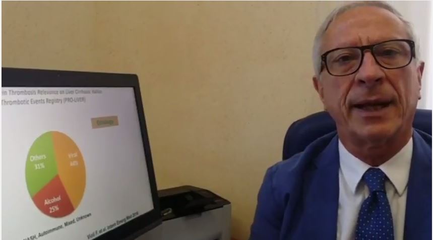
Heritability of colorectal cancer estimated at 40%
Thus, CRC “has a substantial heritable component, and may be more heritable in women than men,” Rebecca E. Graff, ScD, of the Harvard T.H. Chan School of Public Health, Boston, and the University of California, San Francisco, and her colleagues wrote. “Siblings, and particularly monozygotic cotwins, of individuals with colon or rectal cancer should consider personalized screening,” they added.
The model that best fits the data accounted for additive genetic and environmental effects, the researchers said. Based on this model, genetic factors explained about 40% (95% CI, 33%-48%) of variation in the risk of CRC. Thus, the heritability of CRC was about 40%. The estimated heritability of colon cancer was 16% and that of rectal cancer was 15%, but confidence intervals were wide, ranging from 0% to 47% or 50%, respectively. Individual environmental factors accounted for most of the remaining risk of colon and rectal cancers.
“Heritability was greater among women than men and greatest when colorectal cancer combining all subsites together was analyzed,” the researchers noted. Monozygotic twins of CRC patients were at greater risk of both colon and rectal cancers than were same-sex dizygotic twins, indicating that both types of cancer might share inherited genetic risk factors, they added. Variants identified by genomewide association studies “do not come close to accounting for the disease’s heritability,” but, in the meantime, twins of affected cotwins “might benefit particularly from diligent screening, given their excess risk relative to the general population.”
Funders included the Ellison Foundation, the Odense University Hospital AgeCare program, the Academy of Finland, and US BioSHaRE-EU, and the European Union’s Seventh Framework Programme, BioSHaRE-EU, the Swedish Ministry for Higher Education, and the Karolinska Institutet, and the National Cancer Institute. Dr. Graff had no conflicts of interest.
FROM CLINICAL GASTROENTEROLOGY AND HEPATOLOGY
Key clinical point: If a monozygotic twin developed colorectal cancer, his or her twin had about a threefold higher risk of CRC than did the overall cohort (familial risk ratio, 3.1; 95% CI, 2.4-3.8).
Major finding: Genetic factors explained about 40% (95% CI, 33%-48%) of variation in the risk of colorectal cancer.
Data source: An analysis of 39,990 monozygotic and 61,443 same-sex dizygotic twins from the Nordic Twin Study of Cancer.
Disclosures: Funders included the Ellison Foundation, the Odense University Hospital AgeCare program, the Academy of Finland, US BioSHaRE-EU, and the European Union’s Seventh Framework Programme, BioSHaRE-EU, the Swedish Ministry for Higher Education, and the Karolinska Institutet, and the National Cancer Institute. Dr. Graff had no conflicts of interest.
Endoscopists who took 1 minute longer detected significantly more upper GI neoplasms
Endoscopists who took about a minute longer during screening esophagogastroduodenoscopy (EGD) detected significantly more upper gastrointestinal neoplasms than their quicker colleagues, in a retrospective study reported in the August issue of Gastroenterology (doi: 10.1053/j.gastro.2017.05.009).
Detection rates were 0.28% and 0.20%, respectively (P = .005), and this difference remained statistically significant after the investigators controlled for numerous factors that also can affect the chances of detecting gastric neoplasms during upper endoscopy, reported Jae Myung Park, MD, and his colleagues at Seoul (Korea) St. Mary’s Hospital.
Gastric cancer is the second leading cause of cancer mortality worldwide, they noted. Although EGD has been practiced globally for decades, there is no consensus on which performance metrics optimize diagnostic yield. Both Korea and Japan have adopted screening EGD to help diagnose gastric cancer, but interval cancers and false-positive results remain concerns, and the field lacks rigorous analyses comparing EGD time with gastric cancer detection rates. Therefore, the researchers studied 111,962 consecutive patients who underwent EGD at one hospital in Seoul from 2009 through 2015. Fourteen board-certified endoscopists performed these procedures at a single endoscopy unit.A total of 262 patients (0.23%) were diagnosed with gastric neoplasia or dysplasia, duodenal neoplasia, or esophageal carcinoma. This finding closely resembled a prior report from Korea, the investigators noted. Detection rates among individual endoscopists ranged from 0.14% to 0.32%, and longer EGD time was associated with a higher detection rate with an R value of 0.54 (P = .046). During the first year of the study, EGDs without biopsies averaged 2 minutes, 53 seconds. Based on this finding, the researchers set a cutoff time of 3 minutes and classified eight endoscopists as fast (mean EGD duration, 2 minutes and 38 seconds; standard deviation, 21 seconds) and six endoscopists as slow (mean duration, 3 minutes and 25 seconds; SD, 19 seconds). Slower endoscopists were significantly more likely to detect gastric adenomas or carcinomas, even after the investigators controlled for the biopsy rates and experience level of these endoscopists and patients’ age, sex, smoking status, alcohol use, and family history of cancer (odds ratio, 1.52; 95% confidence interval, 1.17-1.97; P = .0018).
Older age, male sex, and smoking also were significant correlates of cancer detection in the multivariable model. Biopsy rates among endoscopists ranged from 7% to 28%, and taking a biopsy was an even stronger predictor of detecting an upper GI neoplasm than was EGD duration, with an R value of 0.76 (P = .002). Although this study was observational and retrospective and lacked data on prior EGD examinations, surveillance intervals, and rates of false-negative results, it “supports the hypothesis that examination duration affects the diagnostic accuracy of EGD,” Dr. Park and his coinvestigators reported. “Prolonging the examination time will allow more careful examination by the endoscopist, which will enable the detection of more subtle lesions. Based on our data, we believe that these indicators can improve the quality of EGD.”
Funders included the Ministry of Education, Science, and Technology; the Ministry of Science, ICT, and Future Planning; and the Ministry of Health & Welfare, Republic of Korea. The investigators reported having no conflicts of interest.
Screening and surveillance practices remain one of the major indications for performing upper endoscopy in patients to detect esophageal (adenocarcinoma in the West; squamous cell in the East) and gastric cancer (in the East). The goal of the initial endoscopy is to detect precancerous lesions (such as Barrett's esophagus and gastric intestinal metaplasia) and, if detected, to grade them properly and evaluate for the presence of dysplasia and cancer in subsequent surveillance examinations.

Prateek Sharma, MD, is a professor of medicine in the division of gastroenterology and hepatology, Veterans Affairs Medical Center, University of Kansas, Kansas City, Mo. He has no conflicts of interest.
Screening and surveillance practices remain one of the major indications for performing upper endoscopy in patients to detect esophageal (adenocarcinoma in the West; squamous cell in the East) and gastric cancer (in the East). The goal of the initial endoscopy is to detect precancerous lesions (such as Barrett's esophagus and gastric intestinal metaplasia) and, if detected, to grade them properly and evaluate for the presence of dysplasia and cancer in subsequent surveillance examinations.
Prateek Sharma, MD, is a professor of medicine in the division of gastroenterology and hepatology, Veterans Affairs Medical Center, University of Kansas, Kansas City, Mo. He has no conflicts of interest.
Endoscopists who took about a minute longer during screening esophagogastroduodenoscopy (EGD) detected significantly more upper gastrointestinal neoplasms than their quicker colleagues, in a retrospective study reported in the August issue of Gastroenterology (doi: 10.1053/j.gastro.2017.05.009).
Detection rates were 0.28% and 0.20%, respectively (P = .005), and this difference remained statistically significant after the investigators controlled for numerous factors that also can affect the chances of detecting gastric neoplasms during upper endoscopy, reported Jae Myung Park, MD, and his colleagues at Seoul (Korea) St. Mary’s Hospital.
Gastric cancer is the second leading cause of cancer mortality worldwide, they noted. Although EGD has been practiced globally for decades, there is no consensus on which performance metrics optimize diagnostic yield. Both Korea and Japan have adopted screening EGD to help diagnose gastric cancer, but interval cancers and false-positive results remain concerns, and the field lacks rigorous analyses comparing EGD time with gastric cancer detection rates. Therefore, the researchers studied 111,962 consecutive patients who underwent EGD at one hospital in Seoul from 2009 through 2015. Fourteen board-certified endoscopists performed these procedures at a single endoscopy unit.A total of 262 patients (0.23%) were diagnosed with gastric neoplasia or dysplasia, duodenal neoplasia, or esophageal carcinoma. This finding closely resembled a prior report from Korea, the investigators noted. Detection rates among individual endoscopists ranged from 0.14% to 0.32%, and longer EGD time was associated with a higher detection rate with an R value of 0.54 (P = .046). During the first year of the study, EGDs without biopsies averaged 2 minutes, 53 seconds. Based on this finding, the researchers set a cutoff time of 3 minutes and classified eight endoscopists as fast (mean EGD duration, 2 minutes and 38 seconds; standard deviation, 21 seconds) and six endoscopists as slow (mean duration, 3 minutes and 25 seconds; SD, 19 seconds). Slower endoscopists were significantly more likely to detect gastric adenomas or carcinomas, even after the investigators controlled for the biopsy rates and experience level of these endoscopists and patients’ age, sex, smoking status, alcohol use, and family history of cancer (odds ratio, 1.52; 95% confidence interval, 1.17-1.97; P = .0018).
Older age, male sex, and smoking also were significant correlates of cancer detection in the multivariable model. Biopsy rates among endoscopists ranged from 7% to 28%, and taking a biopsy was an even stronger predictor of detecting an upper GI neoplasm than was EGD duration, with an R value of 0.76 (P = .002). Although this study was observational and retrospective and lacked data on prior EGD examinations, surveillance intervals, and rates of false-negative results, it “supports the hypothesis that examination duration affects the diagnostic accuracy of EGD,” Dr. Park and his coinvestigators reported. “Prolonging the examination time will allow more careful examination by the endoscopist, which will enable the detection of more subtle lesions. Based on our data, we believe that these indicators can improve the quality of EGD.”
Funders included the Ministry of Education, Science, and Technology; the Ministry of Science, ICT, and Future Planning; and the Ministry of Health & Welfare, Republic of Korea. The investigators reported having no conflicts of interest.
FROM GASTROENTEROLOGY
Key clinical point: Adding about 1 minute to an upper endoscopy might significantly increase the detection of upper gastrointestinal neoplasms.
Major finding: Slow endoscopists (with a mean duration for the procedure of 3 minutes and 25 seconds) had a detection rate of 0.28%, while fast endoscopists (2 minutes and 38 seconds) had a detection rate of 0.20% (P = .005).
Data source: A single-center retrospective study of 111,962 individuals who underwent esophagogastroduodenoscopy in Korea between 2009 and 2015.
Disclosures: Funders included the Ministry of Education, Science, and Technology; the Ministry of Science, ICT, and Future Planning; and the Ministry of Health & Welfare, Republic of Korea. The investigators reported having no conflicts of interest.
Obeticholic acid fails to prevent liver damage in an animal model of short-bowel syndrome
Obeticholic acid failed to prevent the development of short-bowel syndrome–associated liver disease in a preliminary study using piglet models. The findings were published in the July issue of Cellular and Molecular Gastroenterology and Hepatology (doi: 10.1016/j.jcmgh.2017.02.008).
Current treatment options for short-bowel syndrome-associated liver disease are limited, wrote Prue M. Pereira-Fantini, PhD, of Murdoch Childrens Research Institute, Victoria, Australia, and colleagues. However, the farnesoid X receptor, which regulates genes involved in bile acid synthesis, absorption, and transport in the intestine and liver, has shown promise as a pharmaceutical target.
“Recently, we described SBS-ALD-associated alterations in bile acid composition associated with disrupted farnesoid X receptor (FXR) signaling mechanisms,” the researchers said. Obeticholic acid (OCA) has been shown to prevent liver disease in mouse models and human disease, and the researchers explored whether it would be effective in the context of short-bowel syndrome associated liver disease (SBS-ALD).
The researchers randomized piglets into four groups to receive small-bowel resection or sham surgery, and either a daily dose of 2.4 mg/kg per day of OCA or no treatment. The pigs were euthanized 2 weeks after their surgeries, and the researchers collected portal plasma samples, bile samples, and liver samples.
OCA treatment in piglets in the SBS surgery group was associated with decreased stool fat that suggested improved fat absorption, but impacted liver morphology, the researchers noted. “Untreated, sham-operated piglets showed normal liver histology when compared with SBS piglets who showed decreased hepatic lobule area and small clusters of inflammatory cells together with mild-to-moderate vesicular zone 2 lipidosis,” they wrote.
Overall, OCA treatment prevented the depletion of taurine; taurine concentration was approximately 8 ng/mL for piglets with SBS treated with OCA compared with 8 ng/mL in the sham group, 9 ng/mL in the sham plus OCA group, and 3 ng/mL in the SBS-only group. However, bile acid dysmetabolism occurred as shown by HDCA levels, which increased with OCA treatment compared to sham controls but were significantly reduced in SBS piglets treated with OCA vs. untreated SBS piglets.
In addition, the researchers found that small-bowel resection did not impact gene expression levels of FXR targets in the intestine or liver. However, “intestinal FXR gene expression was 11-fold higher in untreated SBS piglets when compared with untreated sham piglets,” they wrote. OCA treatment had no significant impact on FXR gene expression in the OCA-treated group vs. the untreated group and in the OCA-treated SBS group.
Although the findings were limited by use of an animal model, the results suggest that OCA treatment may have clinical benefits for SBS patients by reducing fat malabsorption, which remains a challenge, the researchers wrote. However, OCA “did not prevent the development of SBS-ALD, thereby limiting the potential therapeutic benefit in patients with SBS,” they concluded.
The researchers had no financial conflicts to disclose. The study was supported in part by the National Health and Medical Research Council of Australia and by a research grant from the Science Foundation Ireland.
FROM CMGH
Key clinical point: Treatment with obeticholic acid improved absorption and altered bile acid, but did not prevent liver damage in a piglet model of short-bowel syndrome.
Major finding: Overall, taurine concentration was approximately 8 ng/mL for piglets with SBS treated with OCA compared with 8 ng/mL in the sham surgery group, 9 ng/mL in the sham treated with OCA group, and 3 ng/mL in the SBS-only group.
Data source: The data come from piglets treated with obeticholic acid or untreated, and randomized to a small-bowel resection or a sham surgery.
Disclosures: The researchers had no financial conflicts to disclose,
For chronic abdominal pain, THC resembled placebo
Seven weeks of treatment with delta-9-atetrahydrocannabinol (THC) did not improve chronic abdominal pain in a placebo-controlled trial of 65 adults.
Treatment “was safe and well tolerated,” but did not significantly reduce pain scores or secondary efficacy outcomes, Marjan de Vries, MSc, and her associates wrote in the July issue of Clinical Gastroenterology and Hepatology (doi: 10.1016/j.cgh.2016.09.147). Studies have not clearly shown that THC improves central pain sensitization, a key mechanism in chronic abdominal pain, they noted. Future studies of THC and central sensitization include quantitative sensory testing or electroencephalography, they added.
Source: American Gastroenterological Association
Treatment-refractory chronic abdominal pain is common after abdominal surgery or in chronic pancreatitis, wrote Ms. de Vries of Radboud University Medical Center, Nijmegen, the Netherlands. Affected patients tend to develop central sensitization, or hyper-responsive nociceptive central nervous system pathways. When this happens, pain no longer couples reliably with peripheral stimuli, and therapy targeting central nociceptive pathways is indicated. The main psychoactive compound of Cannabis sativa is THC, which interacts with CB1 receptors in the central nervous system, including in areas of the brain that help regulate emotions, such as the amygdala. Emotion-processing circuits are often overactive in chronic pain, and disrupting them might help modify pain perception, the investigators hypothesized. Therefore, they randomly assigned 65 adults with at least 3 months of abdominal pain related to chronic pancreatitis or abdominal surgery to receive oral placebo or THC tablets three times daily for 50-52 days. The 31 patients in the THC group received step-up dosing (3 mg per dose for 5 days, followed by 5 mg per dose for 5 days) followed by stable dosing at 8 mg. Both groups continued other prescribed analgesics as usual, including oxycontin, fentanyl, morphine, codeine, tramadol, paracetamol, anti-epileptics, and nonsteroidal anti-inflammatories. All but two study participants were white, 25 were male, and 24 were female.
At baseline, all patients reported pain of at least 3 on an 11-point visual analogue scale (VAS). By days 50-52, average VAS scores decreased by 1.6 points (40%) in the THC group and by 1.9 points (37%) in the placebo group (P = .9). Although a strong placebo effect is common in studies of visceral pain, that did not prevent pregabalin from significantly outperforming placebo in another similarly designed randomized clinical trial of patients from this study group with chronic pancreatitis, the investigators noted.
The THC and placebo groups also resembled each other on various secondary outcome measures, including patient global impression of change, pain catastrophizing, pain-related anxiety, measures of depression and generalized anxiety, and subjective impressions of alertness, mood, feeling “high,” drowsiness, and difficulties in controlling thoughts. The only exception was that the THC group showed a trend toward improvement on the Short Form 36, compared with the placebo group (P = .051).
Pharmacokinetic analysis showed good oral absorption of THC. Dizziness, somnolence, and headache were common in both groups, but were more frequent with THC than placebo, as was nausea, dry mouth, and visual impairment. There were no serious treatment-related adverse events, although seven patients stopped THC because they could not tolerate the maximum dose.
Some evidence suggests that the shift from acute to chronic pain entails a transition from nociceptive to cognitive, affective, and autonomic sensitization, the researchers noted. “Therefore, an agent targeting particular brain areas related to the cognitive emotional feature of chronic pain, such as THC, might be efficacious in our chronic pain population, but might be better measured by using affective outcomes of pain,” they concluded.
The trial was supported by a grant from the European Union, the European Fund for Regional Development, and the Province of Gelderland. The THC was provided by Echo Pharmaceuticals, Nijmegen, the Netherlands. The investigators reported having no conflicts of interest.
FROM CLINICAL GASTROENTEROLOGY AND HEPATOLOGY
Key clinical point: Tetrahydrocannabinol did not improve chronic abdominal pain more than did placebo.
Major finding: By days 50-52, average VAS scores decreased by 1.6 points (40%) in the THC group and by 1.9 points (37%) in the placebo group (P = .9).
Data source: A phase II, placebo-controlled study of 65 patients with chronic abdominal pain for at least 3 months who received either placebo or delta-9-atetrahydrocannabinol (THC), 8 mg three times daily.
Disclosures: The trial was supported by a grant from the European Union, the European Fund for Regional Development, and the Province of Gelderland. The THC was provided by Echo Pharmaceuticals, Nijmegen, the Netherlands. The investigators reported having no conflicts of interest.