User login
Potential treatment on the horizon for cold agglutinin disease
In a first-in-human trial, sutimlimab rapidly halted hemolysis, corrected anemia, precluded the need for transfusion, and caused no serious adverse effects in patients with cold agglutinin disease.
Sutimlimab also “induced clinically meaningful increases in hemoglobin levels, even in patients with multiple previous lines of therapy,” according to investigators.
The European Medicines Agency and the U.S. Food and Drug Administration (FDA) awarded sutimlimab orphan drug status based on these results. The FDA also granted sutimlimab breakthrough therapy designation.
Sutimlimab is a humanized anti-C1s IgG4 monoclonal antibody that blocks the classical complement pathway–specific protease C1s and prevents further hemolysis in patients with cold agglutinin disease.
Cold agglutinin disease is a rare, acquired chronic autoimmune hemolytic condition that destroys red blood cells. It leads to chronic anemia, severe fatigue, and potentially fatal thrombotic events. No drug has yet been approved to treat cold agglutinin disease.
The phase 1 trial of sutimlimab (formerly BIVV009 and TNT009) in cold agglutinin disease was conducted at the Medical University of Vienna in Austria and reported in Blood.
The study (NCT02502903) involved 10 patients, ages 56 to 76, who were previously treated with multiple lines of therapy, including two patients who failed treatment with eculizumab.
Of the 10 patients, eight were female, eight were Caucasian, one was Asian, and one was Hispanic. Patients had cold agglutinin disease for a median of 5 years (range, 1 – 20).
At baseline, the median hemoglobin level was 7.8 g/dL, the median number of reticulocytes was 133 x 109/L, the median bilirubin was 2.0 mg/dL, and the median haptoglobin was less than 12 mg/dL.
The patients received an initial dose of 10 mg/kg intravenous sutimlimab as a test dose to allow rapid wash-out of the drug if unforeseen adverse effects occurred with the first infusion.
One to 4 days later, they received the full dose of 60 mg/kg, followed by three additional weekly doses.
Investigators observed the patients for 49 to 53 days.
Results
Within the first week, patients’ median hemoglobin levels increased by 1.6 g/dL (P=0.007), and the median best response was an increase of 3.9 g/dL (P=0.005) after 6 weeks.
Seven patients had increased hemoglobin levels by more than 2 g/dL, and this included those who recently failed to respond or relapsed after rituximab, rituximab plus bendamustine, or eculizumab.
In five patients, hemoglobin increased by 4 g/dL or more. In four patients, it completely normalized to 12 g/dL.
In the first 24 hours after sutimlimab infusion, reticulocyte counts increased by a median of 41% and then gradually declined as hemoglobin levels rose.
In four patients, haptoglobin levels normalized within 1 to 2 weeks. In eight patients who had abnormal bilirubin levels at baseline, sutimlimab decreased the median bilirubin levels by 61%, normalizing levels in most patients within 24 hours of the first infusion (P=0.007).
When sutimlimab was washed out, bilirubin levels increased again, which demonstrated the recurrence of hemolysis.
Approximately 3 to 4 weeks after the last dose of sutimlimab, hemolysis and anemia recurred in all responders.
When patients were re-exposed to sutimlimab, rapid and complete inhibition of hemolysis occurred once again.
None of the patients required packed red blood cell transfusions during treatment.
Safety
The investigators reported that all infusions were well tolerated without premedication and without relevant drug-related adverse effects.
They reported few adverse events during the trial. All were mild or moderate in severity, and most were considered unrelated or unlikely related to sutimlimab.
Two adverse events—one mild purpural rash on both hands and one case of moderate hair loss (each occurring in one patient)—were possibly related to sutimlimab.
While the investigators considered the safety data encouraging, they recommended interpreting the data “cautiously in light of the limited duration of the trial.”
“Provided that safety results remain positive, sutimlimab could become the first approved treatment for cold agglutinin disease,” said corresponding author Bernd Jilma, MD, of the Medical University of Vienna in Austria.
“The drug clearly addresses an unmet medical need, as we have seen rapid, strong responses in patients for whom multiple prior therapies have failed.”
This study was funded by True North Therapeutics, Inc, now part of Bioverativ, a Sanofi company.
Some of the authors disclosed financial relationships, including employment, with True North Therapeutics and Bioverativ.
A phase 3 trial of sutimlimab is underway with top-line results due in 2019.
Team reports long-term effects of blood management
An initiative that reduced red blood cell (RBC) transfusions and increased moderate anemia in hospital did not adversely impact patients long-term, according to an analysis.
Researchers found that an increase in moderate in-hospital anemia did not increase subsequent RBC use, readmission, or mortality over the next 6 months.
However, authors of a related editorial argued that additional factors must be assessed to truly determine the effects of moderate anemia on patient outcomes.
The study and the editorial were published in the Annals of Internal Medicine.
Study: Long-term outcomes
Nareg H. Roubinian, MD, of Kaiser Permanente Northern California in Oakland, and colleagues sought to evaluate the impact of blood management programs—starting in 2010—that included blood-sparing surgical and medical techniques, increased use of hemostatic and cell salvage agents, and treatment of suboptimal iron stores before surgery.
In previous retrospective cohort studies, the researchers had found that blood conservation strategies did not impact in-hospital or 30-day mortality rates, which was consistent with short-term safety data from clinical trials and other observational studies.
Their new report on longer-term outcomes was based on data from Kaiser Permanente Northern California for 445,371 adults who had 801,261 hospitalizations with discharges between 2010 and 2014.
In this cohort, moderate anemia (hemoglobin between 7 g/dL and 10 g/dL) at discharge occurred in 119,489 patients (27%) and 187,440 hospitalizations overall (23%).
Over the 2010-2014 period, RBC transfusions decreased by more than 25% in the inpatient and outpatient settings. In parallel, the prevalence of moderate anemia at hospital discharge increased from 20% to 25%.
However, the risks of subsequent RBC transfusions and rehospitalization after discharge with anemia decreased during the study period, and mortality rates stayed steady or decreased slightly.
Among patients with moderate anemia, the proportion with subsequent RBC transfusions within 6 months decreased from 18.9% in 2010 to 16.8% in 2014 (P<0.001), while the rate of rehospitalization within 6 months decreased from 36.5% to 32.8% over that same time period (P<0.001).
The adjusted 6-month mortality rate likewise decreased from 16.1% to 15.6% (P=0.004) over that time period among patients with moderate anemia.
“These data support the efficacy and safety of practice recommendations to limit red blood cell transfusion in patients with anemia during and after hospitalization,” the researchers wrote.
However, they also said additional studies are needed to guide anemia management, particularly since persistent anemia has impacts on quality of life that are “likely to be substantial” and linked to the severity of that anemia.
This study was supported by a grant from the National Heart, Lung, and Blood Institute. Dr. Roubinian and several coauthors reported grants from the National Institutes of Health.
Editorial: Aim to treat anemia, not tolerate it
Dr. Roubinian and his colleagues’ findings warrant some scrutiny, according to Aryeh Shander, MD, of Englewood Hospital and Medical Center in New Jersey, and Lawrence Tim Goodnough, MD, of Stanford University in California.
“Missing here is a wide spectrum of morbidity outcomes and issues related to diminished quality of life that do not reach the level of severity that would necessitate admission but nonetheless detract from patients’ health and well-being,” Drs. Shander and Goodnough wrote in a related editorial.
They also noted that transfusion rate is not a clinical outcome, adding that readmission and mortality are important outcomes, but they do not accurately or fully reflect patient well-being.
While blood management initiatives may be a safe practice, as the study suggests, proper management of anemia after discharge may actually improve outcomes, given the many consequences of anemia, Drs. Shander and Goodnough wrote.
The pair suggested that, instead of again testing whether restricting transfusions is acceptable because of lack of impact on outcomes, future studies could evaluate a “more sensible” hypothesis that proper anemia management, especially post-discharge, could improve outcomes.
“Let’s increase efforts to prevent and treat anemia properly, rather than requiring patients to tolerate it,” Drs. Shander and Goodnough wrote.
Dr. Shander reported consulting fees from Vifor and AMAG. Dr. Goodnough reported having no relevant financial disclosures.
FDA approves ravulizumab for PNH
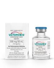
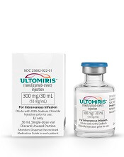
The U.S. Food and Drug Administration (FDA) has approved ravulizumab-cwvz (Ultomiris) to treat adults with paroxysmal nocturnal hemoglobinuria (PNH).
Ravulizumab is a long-acting C5 complement inhibitor, administered every 8 weeks, that has been shown to prevent hemolysis.
The prescribing information for ravulizumab includes a boxed warning stating that meningococcal infections/sepsis have occurred in patients treated with the drug, and these adverse effects can become life-threatening or fatal if not recognized and treated early.
Ravulizumab is available only through a restricted program under a Risk Evaluation and Mitigation Strategy.
The FDA previously granted the application for ravulizumab priority review, and the product received orphan drug designation from the FDA.
The FDA granted the approval of ravulizumab to Alexion Pharmaceuticals.
The FDA’s approval of ravulizumab is based on results from two phase 3 studies, one in patients who had previously received treatment with a complement inhibitor and one in patients who were complement-inhibitor-naïve. Both studies were recently published in Blood.
Efficacy in inhibitor-experienced patients
In one study (NCT03056040), researchers compared ravulizumab administered every 8 weeks to eculizumab administered every 2 weeks in complement-inhibitor-experienced patients.
The trial included 195 PNH patients who were taking eculizumab for more than 6 months. They were randomized to switch to ravulizumab (n=97) or continue on eculizumab (n=98).
Ravulizumab proved noninferior to eculizumab for all endpoints studied (P<0.0006), including:
- Percentage change in lactate dehydrogenase (LDH): difference, 9.21% (95% CI: -0.42 to 18.84; P=0.058 for superiority)
- Breakthrough hemolysis: difference, 5.1 (95% CI: -8.89 to 18.99)
- Change in FACIT-Fatigue score: difference, 1.47 (95% CI: -0.21 to 3.15)
- Transfusion avoidance: difference, 5.5 (95% CI: -4.27 to 15.68)
- Stabilized hemoglobin: difference, 1.4 (95% CI: -10.41 to 13.31).
Efficacy in inhibitor-naïve patients
In another study (NCT02946463), researchers compared ravulizumab and eculizumab in 246 PNH patients who had not previously received a complement inhibitor.
Ravulizumab was noninferior to eculizumab for all endpoints (P<0.0001), including:
- Transfusion avoidance: 73.6% vs 66.1%; difference of 6.8% (95% CI: -4.66 to 18.14)
- LDH normalization: 53.6% vs 49.4%; odds ratio=1.19 (95% CI: 0.80 to 1.77)
- Percent reduction in LDH: -76.8% vs -76.0%; difference of -0.83% (95% CI: -5.21 to 3.56)
- Change in FACIT-Fatigue score: 7.07 vs 6.40; difference of 0.67 (95% CI: -1.21 to 2.55)
- Breakthrough hemolysis: 4.0% vs 10.7%; difference of -6.7% (95% CI: -14.21 to 0.18)
- Stabilized hemoglobin: 68.0% vs 64.5%; difference of 2.9 (95% CI: -8.80 to 14.64).
Safety in both trials
The safety data from both trials included 441 adults who received ravulizumab (n=222) or eculizumab (n=219) for a median of 6 months.
The most frequent adverse events in both arms (ravulizumab and eculizumab, respectively) were upper respiratory tract infection (39% and 39%) and headache (32% and 26%).
Serious adverse events occurred in 15 (6.8%) patients treated with ravulizumab. These events included hyperthermia and pyrexia.
There was one fatal case of sepsis in a patient treated with ravulizumab.
FDA approves ravulizumab for treatment of paroxysmal nocturnal hemoglobinuria
The Food and Drug Administration has approved ravulizumab (Ultomiris) injection for the treatment of adult patients with paroxysmal nocturnal hemoglobinuria (PNH).
“The approval of Ultomiris will change the way that patients with PNH are treated. Prior to this approval, the only approved therapy for PNH required treatment every 2 weeks, which can be burdensome for patients and their families. Ultomiris uses a novel formulation so patients only need treatment every 8 weeks, without compromising efficacy,” Richard Pazdur, MD, director of the FDA’s Oncology Center of Excellence, said in a press release from the agency.
Patients with PNH, a rare disorder, lack a protein which protects red blood cells from being destroyed in the immune system. Episodes can be triggered by stresses on the body such as infection or physical exertion, and symptoms include severe anemia, profound fatigue, shortness of breath, intermittent episodes of dark-colored urine, kidney disease, or recurrent pain.
FDA approval for ravulizumab is based on results from a pair of clinical trials. In the first, 246 treatment-naive PNH patients received either ravulizumab or eculizumab, the current standard of care; ravulizumab was noninferior, with no patients undergoing a transfusion and all patients having similar incidence of hemolysis. In the second trial, 195 patients who had clinically stable PNH after receiving eculizumab for 6 months were randomized to receive ravulizumab or continue eculizumab; again, ravulizumab was noninferior.
The most common adverse events associated with ravulizumab were headache and respiratory tract infection. Caution is recommended when prescribing ravulizumab to patients with any type of infection.
Find the full press release on the FDA website.
CHMP backs lusutrombopag for severe thrombocytopenia
The European Medicines Agency’s Committee for Medicinal Products for Human Use (CHMP) has recommended approval for lusutrombopag to treat severe thrombocytopenia in adults with chronic liver disease who are undergoing invasive procedures.
Lusutrombopag is a thrombopoietin (TPO) receptor agonist that acts on the transmembrane domain of TPO receptors to induce proliferation and differentiation of megakaryocyte progenitor cells, thus leading to thrombocytopoiesis.
Lusutrombopag is intended to reduce the need for platelet transfusions before an invasive procedure and for rescue therapy for bleeding in the 7 days after the procedure.
The CHMP’s recommendation for lusutrombopag will be reviewed by the European Commission, which has the authority to approve medicines for use in the European Union, Norway, Iceland, and Liechtenstein.
The European Commission usually makes a decision within 67 days of a CHMP recommendation.
Lusutrombopag trials
The efficacy of lusutrombopag was evaluated in two phase 3 trials—L-PLUS1 (1304M0631) and L-PLUS2 (1423M0634, NCT02389621).
The trials included 312 patients with chronic liver disease, severe thrombocytopenia (platelet counts below 50,000/μL), and a scheduled invasive procedure. The patients received lusutrombopag or placebo once daily for up to 7 days.
In L-PLUS1, 78% (38/49) of patients receiving lusutrombopag did not require platelet transfusions prior to the primary invasive procedure. The same was true for 13% (6/48) of patients who received placebo (P<0.0001).
In L-PLUS2 , 65% (70/108) of patients who received lusutrombopag did not require platelet transfusions prior to the primary invasive procedure or rescue therapy for bleeding in the 7 days after the procedure. The same was true for 29% (31/107) of patients receiving placebo (P<0.0001).
The safety of lusutrombopag was evaluated in three trials—L‐PLUS 1, L‐PLUS 2, and M0626 (1208M062).
The most common adverse event (AE) in these trials (n=341) was headache, which occurred in 5% of patients on lusutrombopag and 4% of patients on placebo.
Serious AEs occurred in 5% of patients on lusutrombopag and 7% of patients on placebo. The most common serious AE was portal vein thrombosis, which occurred in 1% of patients in both treatment groups.
None of the patients discontinued lusutrombopag due to AEs.
The trials were sponsored by Shionogi & Co., Ltd.
FDA aims to boost safety of platelets for transfusion
The Food and Drug Administration is asking for comments on its
The draft document, “Bacterial Risk Control Strategies for Blood Collection Establishments and Transfusion Services to Enhance the Safety and Availability of Platelets for Transfusion,” will be open for public comment through Feb. 4, 2019.
It is the first update to the policy document since 2016.
In the draft guidance, the FDA recommended three strategies for platelets stored for 5 days from collection. For apheresis platelets and prestorage pools, the FDA suggested an initial primary culture followed by a secondary culture on day 3 or day 4 or an initial primary culture followed by secondary testing with a rapid test. The third strategy – for apheresis platelets – is pathogen reduction alone.
The FDA also outlined three strategies for testing platelets stored for 7 days, all of which apply to apheresis platelets. The methods include an initial primary culture followed by a secondary culture no earlier than day 4, using a device labeled as a safety measure; an initial primary culture followed by a secondary rapid test, labeled as a safety measure; or large volume delayed sampling.
The supply of blood and blood components in the United States is among the safest in the world, FDA Commissioner Scott Gottlieb, MD, said in a statement. The FDA’s continuously updated protocols are intended to keep it that way.
“Blood and blood components are some of the most critical medical products American patients depend upon,” Dr. Gottlieb wrote. “But there remains risk, albeit uncommon, of contamination with infectious diseases, particularly with blood products that are stored at room temperature. While we’ve made great strides in reducing the risk of blood contamination through donor screening and laboratory testing, we continue to support innovations and blood product alternatives that can better keep pace with emerging pathogens and reduce some of the logistical challenges and costs associated with ensuring the safety of blood products.”
Since the 2016 guidance document was issued, new strategies for bacterial detection have become available that could potentially reduce the risk of contamination of platelets and permit extension of platelet dating up to 7 days, including bacterial testing strategies using culture-based devices, rapid bacterial detection devices, and the implementation of pathogen reduction technology.
The recommendations in the draft guidance incorporate ideas put forth during a July 2018 meeting of the agency’s Blood Products Advisory Committee. Committee members were asked to discuss the advantages and disadvantages of various strategies to control the risk of bacterial contamination in platelets, including the scientific evidence and the operational considerations involved. Their comments have been incorporated into the new draft guidance document.
In late November 2018, the FDA held a public workshop to encourage a scientific discussion on a range of pathogen reduction topics, including the development of novel technologies. “The ideal pathogen reduction technology would: be relatively inexpensive, be simple to implement on whole blood, allow treated blood to subsequently be separated into components or alternatively could be performed on apheresis products, inactivate a broad range of pathogens, and would have no adverse effect on product safety or product yield,” the FDA noted in a statement.
Drug may be new option for transfusion-dependent β-thalassemia
SAN DIEGO—Luspatercept can produce “clinically meaningful” results in transfusion-dependent adults with β-thalassemia, according to a speaker at the 2018 ASH Annual Meeting.
In the phase 3 BELIEVE trial, β-thalassemia patients were significantly more likely to experience a reduction in transfusion burden if they were treated with luspatercept rather than placebo.
“Luspatercept showed a statistically significant and clinically meaningful . . . reduction in transfusion burden compared with placebo at any 12- or 24-week [period] along this study,” said Maria Domenica Cappellini, MD, of the University of Milan in Italy.
“At this point, we believe [luspatercept] is a potential new treatment for adult patients with β-thalassemia who are requiring regular blood transfusions.”
Dr. Cappellini presented these results at ASH as abstract 163.
The BELIEVE trial (NCT02604433) enrolled 336 patients from 65 sites in 15 countries. All patients had β-thalassemia or hemoglobin E/β‑thalassemia. They required regular transfusions of six to 20 red blood cell (RBC) units in the 24 weeks prior to randomization, and none had a transfusion-free period lasting 35 days or more.
The patients were randomized 2:1 to receive luspatercept—at a starting dose of 1.0 mg/kg with titration up to 1.25 mg/kg—(n=224) or placebo (n=112) subcutaneously every 3 weeks for at least 48 weeks.
All patients continued to receive RBC transfusions and iron chelation therapy as necessary (so they maintained the same baseline hemoglobin level).
The median age was 30 in both treatment arms (range, 18-66). More than half of patients were female—58.9% in the luspatercept arm and 56.3% in the placebo arm.
A similar percentage of patients in both arms had the β0, β0 genotype—30.4% in the luspatercept arm and 31.3% in the placebo arm.
The median hemoglobin level at baseline was 9.31 g/dL in the luspatercept arm and 9.15 g/dL in the placebo arm. The median RBC transfusion burden was 6.12 units/12 weeks and 6.27 units/12 weeks, respectively.
Other baseline characteristics were similar as well.
Efficacy
“[L]uspatercept showed a statistically significant improvement in the primary endpoint,” Dr. Cappellini noted.
The primary endpoint was at least a 33% reduction in transfusion burden—of at least two RBC units—from week 13 to week 24, as compared to the 12-week baseline period.
This endpoint was achieved by 21.4% (n=48) of patients in the luspatercept arm and 4.5% (n=5) in the placebo arm (odds ratio=5.79; P<0.0001).
“Statistical significance was also demonstrated with luspatercept versus placebo for all the key secondary endpoints,” Dr. Cappellini said.
There were more patients in the luspatercept arm than the placebo arm who achieved at least a 33% reduction in transfusion burden from week 37 to 48—19.6% and 3.6%, respectively (P<0.0001).
Similarly, there were more patients in the luspatercept arm than the placebo arm who achieved at least a 50% reduction in transfusion burden from week 13 to 24—7.6% and 1.8%, respectively (P=0.0303)—and from week 37 to 48—10.3% and 0.9%, respectively (P=0.0017).
During any 12-week interval, 70.5% of luspatercept-treated patients and 29.5% of placebo-treated patients achieved at least a 33% reduction in transfusion burden (P<0.0001), and 40.2% and 6.3%, respectively (P<0.0001), achieved at least a 50% reduction in transfusion burden.
During any 24-week interval, 41.1% of luspatercept-treated patients and 2.7% of placebo-treated patients achieved at least a 33% reduction in transfusion burden (P<0.0001), and 16.5% and 0.9%, respectively (P<0.0001), achieved at least a 50% reduction in transfusion burden.
Safety
Ninety-six percent of patients in the luspatercept arm and 92.7% in the placebo arm had at least one treatment-emergent adverse event (TEAE).
Grade 3 or higher TEAEs occurred in 29.1% of patients in the luspatercept arm and 15.6% of those in the placebo arm. Serious TEAEs occurred in 15.2% and 5.5%, respectively.
One patient in the placebo arm had a TEAE-related death (acute cholecystitis), but there were no treatment-related deaths in the luspatercept arm.
TEAEs leading to treatment discontinuation occurred in 5.4% of luspatercept-treated patients and 0.9% of placebo-treated patients.
TEAEs that occurred more frequently in the luspatercept arm than in the placebo arm (respectively) included bone pain (19.7% and 8.3%), arthralgia (19.3% and 11.9%), and dizziness (11.2% and 4.6%).
Grade 3/4 TEAEs (in the luspatercept and placebo arms, respectively) included anemia (3.1% and 0%), increased liver iron concentration (2.7% and 0.9%), hyperuricemia (2.7% and 0%), hypertension (1.8% and 0%), syncope (1.8% and 0%), back pain (1.3% and 0.9%), bone pain (1.3% and 0%), blood uric acid increase (1.3% and 0%), increased aspartate aminotransferase (1.3% and 0%), increased alanine aminotransferase (0.9% and 2.8%), and thromboembolic events (0.9% and 0%).
Dr. Cappellini noted that thromboembolic events occurred in eight luspatercept-treated patients and one placebo-treated patient. In all cases, the patients had multiple risk factors for thrombosis.
This study was sponsored by Celgene Corporation and Acceleron Pharma. Dr. Cappellini reported relationships with Novartis, Celgene, Sanofi-Genzyme, and Vifor.
In-hospital blood saving strategy appears safe with anemia
A blood management initiative that reduced RBC transfusions in the hospital did not adversely impact long-term outcomes after discharge, a retrospective analysis of an extensive patient database suggested.
Tolerating moderate in-hospital anemia did not increase subsequent RBC use, readmission, or mortality over the next 6 months, according to results of the study, which drew on nearly half a million patient records.
In fact, modest mortality decreases were seen over time for patients with moderate anemia, perhaps because of concomitant initiatives that targeted infectious and circulatory conditions, reported Nareg H. Roubinian, MD, of Kaiser Permanente Northern California in Oakland and the University of California, San Francisco, and coinvestigators.
“These data support the efficacy and safety of practice recommendations to limit red blood cell transfusion in patients with anemia during and after hospitalization,” Dr. Roubinian and colleagues wrote in their report, which appears in the Annals of Internal Medicine.
However, additional studies are needed to guide anemia management, they wrote, particularly since persistent anemia has impacts on quality of life that are “likely substantial” and linked to the severity of that anemia.
Dr. Roubinian and colleagues sought to evaluate the impact of blood management programs – initiated starting in 2010 – that included blood-sparing surgical and medical techniques, increased use of hemostatic and cell salvage agents, and treatment of suboptimal iron stores before surgery.
In previous retrospective cohort studies, the researchers had found that the blood conservation strategies did not impact in-hospital or 30-day mortality rates, which was consistent with short-term safety data from clinical trials and other observational studies.
Their latest report on longer-term outcomes was based on data from Kaiser Permanente Northern California for 445,371 adults who had 801,261 hospitalizations with discharges between 2010 and 2014. In this cohort, moderate anemia (hemoglobin between 7 g/dL and 10 g/dL) at discharge occurred in 119,489 patients (27%) and 187,440 hospitalizations overall (23%).
Over the 2010-2014 period, RBC transfusions decreased by more than 25% in the inpatient and outpatient settings; and in parallel, the prevalence of moderate anemia at hospital discharge increased from 20% to 25%.
However, the risks of subsequent RBC transfusions and rehospitalization after discharge with anemia decreased during the study period, and mortality rates stayed steady or decreased slightly.
Among patients with moderate anemia, the proportion with subsequent RBC transfusions within 6 months decreased from 18.9% in 2010 to 16.8% in 2014 (P less than .001), while the rate of rehospitalization within 6 months decreased from 36.5% to 32.8% over that same time period (P less than .001).
The adjusted 6-month mortality rate likewise decreased from 16.1% to 15.6% (P = .004) over that time period among patients with moderate anemia.
The study was supported by a grant from the National Heart, Lung, and Blood Institute. Dr. Roubinian and several coauthors reported grants during the conduct of the study from the National Institutes of Health.
SOURCE: Roubinian NH et al. Ann Intern Med. 2018 Dec 18. doi: 10.7326/M17-3253.
Some scrutiny is warranted of the observation of Roubinian et al. that long-term transfusion, readmission, and mortality outcomes were apparently unaffected by decreased in-hospital RBC transfusions, according to the authors of an accompanying editorial.
“Missing here is a wide spectrum of morbidity outcomes and issues related to diminished quality of life that do not reach the level of severity that would necessitate admission but nonetheless detract from patients’ health and well-being,” wrote Aryeh Shander, MD, and Lawrence Tim Goodnough, MD.
Moreover, transfusion rate is not a clinical outcome, they noted, adding that readmission and mortality are important outcomes but that they do not accurately or fully reflect patient well-being.
While blood management initiatives may be a safe practice, as Roubinian et al. found, proper management of anemia after discharge may actually improve outcomes, given the many consequences of anemia.
Instead of again testing whether restricting transfusions is acceptable because of lack of impact on outcomes, future studies could evaluate a “more sensible” hypothesis that proper anemia management – especially post discharge – could improve outcomes.
“Let’s increase efforts to prevent and treat anemia properly, rather than requiring patients to tolerate it,” they wrote.
Dr. Shander is with Englewood (N.J.) Hospital and Medical Center; Dr. Goodnough is with Stanford (Calif.) University. Dr. Shander reported consulting fees from Vifor and AMAG. Dr. Goodnough reported having no relevant financial disclosures. Their comments are taken from an accompanying editorial (Ann Intern Med. 2018 Dec 18. doi: 10.7326/M18-3145).
A blood management initiative that reduced RBC transfusions in the hospital did not adversely impact long-term outcomes after discharge, a retrospective analysis of an extensive patient database suggested.
Tolerating moderate in-hospital anemia did not increase subsequent RBC use, readmission, or mortality over the next 6 months, according to results of the study, which drew on nearly half a million patient records.
In fact, modest mortality decreases were seen over time for patients with moderate anemia, perhaps because of concomitant initiatives that targeted infectious and circulatory conditions, reported Nareg H. Roubinian, MD, of Kaiser Permanente Northern California in Oakland and the University of California, San Francisco, and coinvestigators.
“These data support the efficacy and safety of practice recommendations to limit red blood cell transfusion in patients with anemia during and after hospitalization,” Dr. Roubinian and colleagues wrote in their report, which appears in the Annals of Internal Medicine.
However, additional studies are needed to guide anemia management, they wrote, particularly since persistent anemia has impacts on quality of life that are “likely substantial” and linked to the severity of that anemia.
Dr. Roubinian and colleagues sought to evaluate the impact of blood management programs – initiated starting in 2010 – that included blood-sparing surgical and medical techniques, increased use of hemostatic and cell salvage agents, and treatment of suboptimal iron stores before surgery.
In previous retrospective cohort studies, the researchers had found that the blood conservation strategies did not impact in-hospital or 30-day mortality rates, which was consistent with short-term safety data from clinical trials and other observational studies.
Their latest report on longer-term outcomes was based on data from Kaiser Permanente Northern California for 445,371 adults who had 801,261 hospitalizations with discharges between 2010 and 2014. In this cohort, moderate anemia (hemoglobin between 7 g/dL and 10 g/dL) at discharge occurred in 119,489 patients (27%) and 187,440 hospitalizations overall (23%).
Over the 2010-2014 period, RBC transfusions decreased by more than 25% in the inpatient and outpatient settings; and in parallel, the prevalence of moderate anemia at hospital discharge increased from 20% to 25%.
However, the risks of subsequent RBC transfusions and rehospitalization after discharge with anemia decreased during the study period, and mortality rates stayed steady or decreased slightly.
Among patients with moderate anemia, the proportion with subsequent RBC transfusions within 6 months decreased from 18.9% in 2010 to 16.8% in 2014 (P less than .001), while the rate of rehospitalization within 6 months decreased from 36.5% to 32.8% over that same time period (P less than .001).
The adjusted 6-month mortality rate likewise decreased from 16.1% to 15.6% (P = .004) over that time period among patients with moderate anemia.
The study was supported by a grant from the National Heart, Lung, and Blood Institute. Dr. Roubinian and several coauthors reported grants during the conduct of the study from the National Institutes of Health.
SOURCE: Roubinian NH et al. Ann Intern Med. 2018 Dec 18. doi: 10.7326/M17-3253.
FROM THE ANNALS OF INTERNAL MEDICINE
Key clinical point:
Major finding: The adjusted 6-month mortality rate decreased from 16.1% to 15.6% (P = .004) in the 4-year period following implementation of blood conservation strategies.
Study details: A retrospective cohort study including 445,371 adults hospitalized and discharged between 2010 and 2014.
Disclosures: The study was supported by a grant from the National Heart, Lung, and Blood Institute. Several authors reported grants during the conduct of the study from the National Institutes of Health.
Source: Roubinian NH et al. Ann Intern Med. 2018 Dec 18. doi: 10.7326/M17-3253.
Romiplostim now approved for children with ITP
The thrombopoietin receptor agonist romiplostim (NPlate®) is now approved by the U.S. Food and Drug Administration (FDA) to treat pediatric patients 1 year and older who have had immune thrombocytopenia (ITP) for at least 6 months and have not responded sufficiently to corticosteroids, immunoglobulins, or splenectomy.
Romiplostim was originally FDA-approved in 2008 to treat adult patients with chronic ITP who had an insufficient response to the same treatments.
Romiplostim is manufactured by Amgen, Inc.
The FDA based its approval on two double-blind, placebo-controlled clinical trials.
NCT01444417
The phase 3 study (NCT01444417) enrolled 62 pediatric patients 1 year and older who had ITP for at least 6 months. They were refractory to or relapsed after at least one prior therapy.
Investigators randomized them 2:1 to receive romiplostim (n=42) or placebo (n=20).
The starting dose was 1 μg/kg weekly for all ages. The dose was titrated up over a 24-week period to a maximum of 10 μg/kg weekly.
Patients were a median age of 9.5 years (range, 3–17), and 57% were female. A little over half (58%) had baseline platelet counts of 20 x 109/L or less, which was similar in both treatment arms.
Eighty-one percent of romiplostim-treated patients had at least two prior ITP therapies, compared with 70% in the placebo group. One patient in each group had undergone splenectomy.
Twenty-two (52%) of the romiplostim-treated patients had durable platelet responses of 50 x 109/L or greater for at least six weekly assessments during weeks 18 through 25 of treatment. Two (10%) patients in the placebo arm achieved durable platelet responses.
Thirty (71%) romiplostim-treated patients achieved an overall platelet response, defined as a durable or transient platelet response. This compared with four (20%) patients in the placebo group.
Romiplostim-treated patients had platelet counts of at least 50 x 109/L for a median of 12 weeks, compared to 1 week for patients in the placebo arm.
All response endpoints were significant at P<0.05.
NCT00515203
The phase 1/2 study (NCT00515203) enrolled 22 patients who had ITP for at least 6 months prior to study enrollment and were relapsed from or refractory to prior treatment.
Investigators randomized the patients 3:1 to romiplostim (n=17) or placebo (n=5).
Patients were a median age of 10 years (range, 1–17), and 27.3% were female.
Approximately 82% of patients had baseline platelet counts of 20 x 109/L or less, which was similar between the treatment arms.
Eighty-eight percent of patients in the romiplostim arm had at least two prior ITP therapies, as did 100% in the placebo group.
Six patients in the romiplostim group and two in the placebo group had undergone splenectomy.
Of the 17 patients treated with romiplostim, 15 (88.2%) achieved a platelet count of 50 x 109/L or great for 2 consecutive weeks.
The same 15 patients also achieved an increase in platelet count of 20 x 109/L or greater above baseline for 2 consecutive weeks during the treatment period.
None of the placebo-treated patients achieved either endpoint.
The adverse events profile in pediatric patients was compiled from the two trials and reflects a median drug exposure of 168 days for 59 patients.
The most common adverse events, occurring in 25% or more of romiplostim-treated patients, were contusion (41%), upper respiratory tract infection (31%), and oropharyngeal pain (25%). These occurred with an incidence at least 5% higher than in the placebo group.
Dosing
The recommended starting dose for pediatric patients is 1 µg/kg based on actual body weight and administered as a weekly subcutaneous injection.
The dose should be adjusted in increments of 1 µg/kg until the patient achieves a platelet count of 50 x 109/L or greater.
The prescribing information recommends reassessing patients’ body weight every 12 weeks.
Test proves accurate for sickle cell screening
SAN DIEGO—An inexpensive, rapid, and easy-to-use blood test accurately detected sickle cell disease in young children in Uganda, according to a speaker at the 2018 ASH Annual Meeting.
The test, called HemoTypeSC, uses monoclonal antibodies to detect hemoglobins A, S, and C in a drop of whole blood.
HemoTypeSC costs less than $2 to the end user and delivers results in about 10 minutes.
“This is ideal for use in resource-constrained regions of high prevalence, such as Africa and central India,” said Erik Serrao, PhD, of Silver Lake Research in Azusa, California.
“Early screening plus treatment plus counseling equals saving millions of lives over the coming decades, and we believe HemoTypeSC can form an integral part of the initial part of this equation.”
Dr. Serrao reported results from a study of HemoTypeSC during the late-breaking abstracts session at ASH (abstract LBA-3).
He and his colleagues compared results with the HemoTypeSC test to results with hemoglobin electrophoresis for detection of the phenotypes HbAA (normal), HbAS (sickle cell trait), and HbSS (sickle cell disease).
The investigators compared these two testing methods in 1,000 children between the ages of 1 month and 5 years who were prospectively recruited from hospital wards and outpatient clinics in Uganda.
Results
The initial analysis suggested HemoTypeSC had an overall accuracy of 99.8%, correctly identifying 998 of 1,000 phenotypes as initially determined by electrophoresis.
HemoTypeSC correctly identified 100% of the 720 HbAA specimens and 100% of 182 HbAS specimens.
HemoTypeSC identified as HbSS 98% (96/98) of specimens that were identified as HbSS by electrophoresis. This left two discordant samples, both of which HemoTypeSC identified as HbAS.
Investigators subsequently discovered that both individuals with the discordant samples had previously been diagnosed with sickle cell disease and had received recent transfusions with HbAA blood.
“Therefore, the true phenotype at the time of testing for these samples was HbAS, as hemoglobin A and S were both in the blood,” Dr. Serrao said.
This brought the accuracy rate of HemoTypeSC up to 100%.
Dr. Serrao noted that this study excluded newborns. However, a different study of HemoTypeSC, recently published in the American Journal of Hematology, demonstrated 100% accuracy across multiple phenotypes in the setting of newborn screening.
Implications
Of all the late-breaking abstracts at ASH this year, the study by Dr. Serrao and his colleagues is the one with the potential to save the most lives, according to Mark Crowther, MD, of McMaster University in Hamilton, Ontario, Canada.
He said using current gold-standard methods for diagnosing sickle cell disease is, at minimum, challenging and “frankly impossible” in many low-resource settings because of the cost and the requirement for sophisticated equipment and reliable electricity.
However, he believes HemoTypeSC could change that.
“The ability to diagnose sickle cell disease early and intervene early will result in potentially thousands of infants, who would otherwise die in infancy or early childhood, surviving into adulthood,” Dr. Crowther said.
Dr. Serrao noted that sickle cell disease screening programs have been projected to be cost-effective in Africa. Such programs could even save money for governments over time as budgets are reallocated toward screening, with less money needed for treatment of patients presenting with severe complications in hospitals.
Dr. Serrao reported that he is an employee of Silver Lake Research, which funded this study, approved the study design, and donated HemoTypeSC tests.
