User login
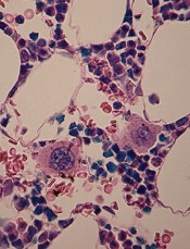
Drivers of cardiac complications in sickle cell anemia

with sickle cell anemia
Image courtesy of the
University of Michigan
Preclinical research has revealed malfunctioning molecular pathways associated with cardiac anomalies in sickle cell anemia (SCA) that lead to sudden death.
Researchers used a mouse model of SCA and identified a unique “restrictive cardiomyopathy” that is superimposed on the anemia-associated heart enlargement.
They also found upregulated gene expression that fuels myocardial fibrosis and electrophysiological changes.
The researchers believe these findings, published in PNAS, will aid the development of new targeted therapies to treat cardiac dysfunction in patients with SCA.
“Sickle cell anemia is associated with significant morbidity and mortality, including a high incidence of unexplained, sudden death in young adults,” said study author Punam Malik, MD, of Cincinnati Children’s Hospital Medical Center in Ohio.
“Our findings may provide a unifying cardiac pathophysiology that explains reported cardiac abnormalities and sudden death seen in humans with SCA.”
To find pathologies specific to SCA, Dr Malik and his colleagues compared mice bred to have SCA and wild-type mice with experimentally induced chronic anemia.
The mice underwent serial comprehensive cardiac analysis, including detailed cardiac imaging (MRI), electrocardiography, and microscopic cross-section analysis of heart tissues (histopathology and electron microscopy).
The researchers said that, in the SCA mice, they observed a distinctive sickle cardiomyopathy in which restrictive physiology was superimposed on chronic anemia and predisposed the mice to sudden death.
The SCA mice had progressive left atrial enlargement and diastolic dysfunction but preserved systolic function. This restrictive physiology appeared to be caused by ischemic-scattered cardiomyocyte loss, myocardial fibrosis, and cardiac remodeling.
The researchers also conducted transcriptome analysis. This revealed that SCA mice had upregulation of genes that cause increased oxidation, hypoxia, and fibrosis in heart tissues. It also showed a downregulation of genes associated with electrophysiological function.
Finally, the team observed progressive corrected QT prolongation, arrhythmias, and ischemic changes in the SCA mice shortly before a significant number of the animals experienced sudden death.
The researchers are continuing to study the molecular mechanisms and pathways that trigger myocardial fibrosis in SCA, using a variety of knockout mice.
In addition, the successful use of noninvasive cardiac imaging techniques in this study inspired the researchers to launch a clinical trial to test these early diagnostic techniques on people with SCA. Enrollment in that study is now complete.
“It is incredibly exciting to see that this work has inspired clinical trials and other research studies,” said study author Nihal Bakeer, MD, of the Indiana Hemophilia and Thrombosis Center in Indianapolis.
“Our goal has always been [to] find the underling pathobiology of cardiac complications in sickle cell anemia and help find new diagnostics and therapeutics to decrease the morbidity and rate of sudden cardiac death in young adults with SCA.” ![]()
How hydroxyurea fights sickle cell disease

Photo by Zak Hubbard
Researchers say they have uncovered hydroxyurea’s main mechanism of action in sickle cell disease (SCD).
The drug’s mechanism has been a topic of debate, with some researchers claiming hydroxyurea works by reactivating fetal hemoglobin and others saying it increases the volume of red blood cells (RBCs), thereby reducing the concentration of sickle hemoglobin.
Now, research published in PNAS suggests the latter mechanism is the dominant one.
“Our findings shine a light on the mechanism behind hydroxyurea action, which has long been debated in the scientific community,” said study author Ming Dao, PhD, of the Massachusetts Institute of Technology in Cambridge.
“It’s exciting to see that, using the latest optical imaging tools, we can now confirm which one is the dominating mechanism. Understanding the key mechanism of action will allow us to explore novel and improved therapeutic approaches for sickle cell disease.”
For this study, the researchers analyzed blood samples from patients with SCD.
The team used common-path interferometric microscopy to assess the biophysical properties (shape, surface area, and volume) and biomechanical properties (flexibility and stickiness) of RBCs.
The researchers separated RBCs into 4 groups based on their density. Normal, disc-shaped cells were the least dense, while severely sickled cells were the densest.
The team then compares samples from patients who were taking hydroxyurea and those who were not.
The RBCs of patients receiving treatment showed an improvement in all of the biophysical and biomechanical properties tested across all density levels.
Improvement in the physical properties of RBCs from patients treated with hydroxyurea correlated more with an increase in RBC volume than with levels of fetal hemoglobin.
The researchers hope these biophysical markers can be combined with biochemical and molecular-level markers to assess the severity of a patient’s disease, determine whether or not a patient will respond to hydroxyurea, and monitor the effectiveness of that treatment.
“There is a critical need for patient-specific biomarkers that can be used to assess the effectiveness of treatments for sickle cell disease,” said study author Subra Suresh, ScD, of Carnegie Mellon University in Pittsburgh, Pennsylvania.
“This study shows how techniques commonly used in engineering and physics can help us to better understand how the red blood cells in people with sickle cell disease react to treatment, which could lead to improved diagnostics and therapies.” ![]()
Teva launches generic imatinib tablets in US

Photo by Steven Harbour
Teva Pharmaceutical Industries Ltd. has announced the US launch of imatinib mesylate, the generic equivalent of Novartis’s Gleevec®, in 100 mg and 400 mg tablets.
In the US, imatinib is approved to treat newly diagnosed Philadelphia-chromosome-positive (Ph+) chronic myeloid leukemia in chronic phase, blast crisis, and accelerated phase, as well as Ph+
chronic myeloid leukemia in chronic phase after failure of interferon-alpha therapy.
Imatinib is also approved to treat adults with relapsed or refractory Ph+ acute lymphoblastic leukemia, adults with myelodysplastic syndromes or myeloproliferative neoplasms associated with platelet-derived growth factor receptor gene re-arrangements, and adults with aggressive systemic mastocytosis without the D816V c-Kit mutation or with unknown c-Kit mutational status.
In addition, imatinib is approved to treat adults with hypereosinophilic syndrome and/or chronic eosinophilic leukemia (regardless of whether they have the FIP1L1-PDGFRα fusion kinase) and adults with unresectable, recurrent, and/or metastatic dermatofibrosarcoma protuberans.
Finally, the drug is approved as an adjuvant treatment following complete gross resection of Kit (CD117)-positive gastrointestinal stromal tumors in adults.
For more details on imatinib, see the full prescribing information. ![]()
Products granted orphan designation for use in HSCT
The European Commission has granted orphan drug designation for the T-cell therapy product candidate BPX-501 and the small molecule rimiducid.
BPX-501 consists of genetically modified donor T cells incorporating the CaspaCIDe safety switch, which is designed to eliminate the T cells in the event of toxicity.
Rimiducid is used to activate the CaspaCIDe safety switch, which consists of the CID-binding domain coupled to the signaling domain of caspase-9, an enzyme that is part of the apoptotic pathway.
The goal of this therapy is to allow physicians to more safely perform haploidentical hematopoietic stem cell transplant (haplo-HSCT).
Haplo-HSCT recipients receive BPX-501 to speed immune reconstitution and provide control over viral infections. And rimiducid is used to eliminate BPX-501 alloreactive T cells if severe graft-vs-host disease (GVHD) occurs.
If a patient develops severe GVHD, rimiducid is used to trigger activation of the domain of caspase-9, which leads to selective apoptosis of the CaspaCIDe-containing cells.
About orphan designation
Orphan drug designation from the European Commission provides regulatory and financial incentives for companies to develop and market therapies that treat serious or life-threatening conditions that affect no more than 5 in 10,000 people in the European Union (EU), and where no treatment is currently approved.
In addition to a 10-year period of marketing exclusivity in the EU upon product approval, orphan drug designation provides fee waivers, protocol assistance, and marketing authorization under the centralized procedure granting approval in all EU countries.
BPX-501/rimiducid development
BPX-501 and rimiducid are being developed by Bellicum Pharmaceuticals.
The company has met with regulatory authorities in Europe to discuss the potential approval pathway for BPX-501 and rimiducid for the treatment of immunodeficiency and GVHD following haplo-HSCT in pediatric patients with leukemias, lymphomas, and rare inherited blood diseases who do not have a matched donor.
These discussions have resulted in an initial agreement regarding the company’s development plans, subject to further refinement in a formal protocol assistance process that is available for orphan drug products.
Based on regulatory discussions, Bellicum believes that data from the European arm of its BP-004 trial, with a 6-month follow-up time and expanded to enroll additional patients, could form the basis of marketing authorization applications for BPX-501 and rimiducid.
The European Medicines Agency’s Committee for Medicinal Products for Human Use has agreed that review and approval under “exceptional circumstances” may be suitable, recognizing that a randomized trial may not be feasible in the pediatric setting. In place of a randomized trial, Bellicum intends to collect data from a concurrent observational study of allogeneic HSCT outcomes in the pediatric setting.
The European Medicines Agency can grant early market authorization to orphan drug products under exceptional circumstances. Exceptional circumstances can be granted for medicines that treat very rare diseases or where controlled studies are impractical or not consistent with accepted principles of medical ethics.
BP-004 trial
BP-004 is a phase 1/2 dose-escalation trial of BPX-501 and rimiducid in pediatric patients with malignant and nonmalignant diseases. Interim results from this trial were reported in 2 presentations at the 42nd Annual Meeting of the European Society for Blood and Marrow Transplantation in April 2016.
One presentation involved patients with acute leukemia who received BPX-501 after haplo-HSCT. At a median follow-up of 7 months, 16 of the 17 patients were alive and disease-free. There were several cases of GVHD, but nearly all were resolved.
The other presentation covered patients with nonmalignant disorders who received BPX-501 after haplo-HSCT. At a median follow-up of 7 months, all 24 patients studied were still alive and disease-free. The incidence of GVHD was considered “very low.” ![]()
Sickle cell trait doesn’t increase risk of death, study suggests
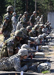
Results of a large study contradict the view that having sickle cell trait increases a person’s risk of death.
Health records of nearly 50,000 active-duty US Army soldiers showed no significant difference in the risk of death between soldiers who had sickle cell trait and those who did not.
The risk of exertional rhabdomyolysis (ER) was 54% higher among soldiers with sickle cell trait than those without it.
But the study suggested that tobacco use, obesity, and taking certain drugs also incur a heightened risk of ER.
Lianne Kurina, PhD, of Stanford University School of Medicine in California, and her colleagues reported these findings in NEJM.
Previous studies have suggested the health consequences of sickle cell trait might be dire, including higher mortality from ER. ER is characterized by the severe breakdown of skeletal-muscle tissue due to extreme physical exertion. The condition has been known to affect athletes and soldiers.
To assess the risk of ER and death among people with sickle cell trait, Dr Kurina and her colleagues reviewed the health records of 47,944 African-American soldiers who served on active duty between 2011 and 2014 and for whom sickle cell status was known.
The team found no significant difference in the risk of death among soldiers with sickle cell trait and those without. The hazard ratio (HR) was 0.99 (95% confidence interval [CI], 0.46 to 2.13; P=0.97).
Sickle cell trait was associated with a significantly higher adjusted risk of ER, with an HR of 1.54 (95% CI, 1.12 to 2.12; P=0.008).
However, the risk of ER was also higher for the following groups:
- Soldiers who used tobacco (HR=1.54, 95% CI, 1.23 to 1.94; P<0.001)
- Those with a body mass index of 30 or higher, as compared to 25 or lower (HR=1.39, 95% CI, 1.04 to 1.86; P=0.03)
- Those who recently used a statin (HR=2.89, 95% CI, 1.51 to 5.55; P=0.001)
- Those who recently used an antipsychotic agent (HR=3.02, 95% CI, 1.34 to 6.82; P=0.008).
Dr Kurina said the reason the results of this study differ from those of previous studies may be better safety for active-duty soldiers.
As of 2003, soldiers who are engaged in strenuous exercise are required to drink plenty of fluids, build up to strenuous exercise gradually, and take regular rests when it’s hot. All of these measures are known to reduce exercise-related fatality rates, regardless of whether individuals have sickle cell trait, the researchers said.
“Another critical difference between our study and the earlier, population-based studies is that, in our study, we knew the sickle cell status of everyone in the population,” Dr Kurina said.
She and her team looked only at soldiers whose sickle cell status was confirmed by blood tests taken during their years of service, instead of from self-reported sickle cell status or past medical history, as had been done in the other studies.
“The most important thing to come out of this study is the really reassuring news that, under conditions of universal precautions against dehydration and overheating, we don’t see an elevation in the risk of mortality in people with sickle cell trait,” Dr Kurina said.
She added that the study’s results call into question the need to screen service members with sickle cell trait, especially with better safety precautions during intense exertion. ![]()
Method provides more accurate diagnosis of MDS, team says

Next-generation sequencing (NGS) of cell-free DNA should be the method of choice to confirm the diagnosis of myelodysplastic syndromes (MDS), according to researchers.
The team found that using NGS to analyze samples from MDS patients yielded more accurate results than Sanger sequencing.
And sequencing cell-free DNA rather than peripheral blood cell DNA increased the likelihood of detecting mutations associated with MDS.
The team reported these findings in Genetic Testing and Molecular Biomarkers. This research was funded by NeoGenomics Laboratories.
For this study, the researchers performed NGS on a panel of 14 target genes using total nucleic acid extracted from the plasma of 16 patients with early MDS (blasts <5%). The team also performed Sanger sequencing and NGS on peripheral blood cell DNA from the same patients.
The researchers found that NGS of cell-free DNA confirmed the diagnosis of MDS in all 16 patients.
In addition, NGS of cell-free DNA revealed abnormalities in 5 patients (31%) that were not detected by Sanger sequencing of peripheral blood cell DNA.
NGS of peripheral blood cell DNA produced the same results as NGS of cell-free DNA for 4 of the 5 patients. However, NGS of peripheral blood cell DNA did not detect a mutation in the RUNX1 gene that was evident in cell-free DNA from 1 patient.
Overall, the researchers found that mutant allele frequency was significantly higher in cell-free DNA than cellular DNA (P=0.008).
The team therefore concluded that cell-free DNA is more reliable than peripheral blood cell DNA for detecting molecular abnormalities in patients with MDS, and NGS is more accurate than Sanger sequencing. ![]()
Light-based therapy may treat thrombocytopenia
in the bone marrow
A low-intensity type of laser therapy could provide a non-invasive, drug-free treatment option for thrombocytopenia, according to research published in Science Translational Medicine.
Researchers found that low-level laser (LLL) therapy increased the generation of platelets from megakaryocytes in vitro and had the same effect in mouse models of thrombocytopenia.
The team also identified the probable mechanism underlying this effect.
“Our study reveals, for the first time, that low-level laser therapy enhances platelet production in animals with thrombocytopenia but not in normal controls,” said study author Mei X. Wu, PhD, of Massachusetts General Hospital in Boston.
“This result suggests that a safe, drug-free method that does not depend on donated blood products can be developed for treating or preventing thrombocytopenia.”
LLLs emit low-powered laser light that does not heat its target tissue. LLLs are known to protect the function of mitochondria, and several conditions associated with impaired platelet production are characterized by abnormalities in the mitochondria of cells that give rise to platelets.
Dr Wu and her colleagues conducted a number of experiments to investigate whether LLLs’ ability to protect mitochondrial function could mitigate several forms of thrombocytopenia.
The team found that LLL treatment of megakaryocytes increased their size, accelerated the formation of proplatelets, and doubled the production of platelets.
Infusion of LLL-treated megakaryocytes into mice led to greater platelet production than did infusion of megakaryocytes treated with normal light.
One of the keys to determining the number of platelets generated from megakaryocytes was mitochondrial production of the energy molecule ATP.
LLL treatment greatly increased mitochondrial generation in polyploid megakaryocytes, but the increase was only slight in less mature megakaryocytes with only 2 copies of each chromosome.
Whole-body LLL treatment of mice with radiation-induced thrombocytopenia spurred the rapid maturation of megakaryocytes and restored platelet levels in a light-dose-dependent fashion.
Platelets from LLL-treated mice had normal structure and function. LLL treatment of normal mice did not raise levels of either megakaryocytes or platelets.
LLL treatment also restored platelet levels in mice with the autoimmune form of thrombocytopenia or with thrombocytopenia caused by chemotherapy.
In cultured human megakaryocytes, LLL treatment at dosage levels similar to those used in mice increased ATP production and platelet generation.
Dr Wu noted that LLL’s lack of an effect in animals without thrombocytopenia indicates it would probably avoid the potential complications of current drug treatments for thrombocytopenia, which act by increasing the production of megakaryocytes from their progenitors in the bone marrow.
“Directly stimulating the differentiation of [megakaryocytes] the way all current drugs do risks clotting if platelet levels rise too high,” Dr Wu said. “LLL appears to enhance [megakaryocytes’] inherent ability to produce platelets most effectively in response to low platelet levels in the circulation, a response that stops when platelet levels are normalized.”
“The fact that treatment only has an effect in polyploid cells, which are very rare, implies that it would not increase production of mitochondria in cancer cells or other cells. In fact, while LLL has been employed in research and in clinical treatment for decades, this is the first study reporting that it can promote mitochondrial biogenesis.”
Dr Wu added that the current primary obstacle to testing LLL in humans is the lack of a device large enough to treat either the entire body or enough bones to stimulate sufficient platelet production by megakaryocytes within the bone marrow, something her team plans to address.
She also noted that, while LLL will probably be beneficial for treatment of many forms of acquired thrombocytopenia, it may not be effective when the condition is caused by inborn genetic defects. ![]()
in the bone marrow
A low-intensity type of laser therapy could provide a non-invasive, drug-free treatment option for thrombocytopenia, according to research published in Science Translational Medicine.
Researchers found that low-level laser (LLL) therapy increased the generation of platelets from megakaryocytes in vitro and had the same effect in mouse models of thrombocytopenia.
The team also identified the probable mechanism underlying this effect.
“Our study reveals, for the first time, that low-level laser therapy enhances platelet production in animals with thrombocytopenia but not in normal controls,” said study author Mei X. Wu, PhD, of Massachusetts General Hospital in Boston.
“This result suggests that a safe, drug-free method that does not depend on donated blood products can be developed for treating or preventing thrombocytopenia.”
LLLs emit low-powered laser light that does not heat its target tissue. LLLs are known to protect the function of mitochondria, and several conditions associated with impaired platelet production are characterized by abnormalities in the mitochondria of cells that give rise to platelets.
Dr Wu and her colleagues conducted a number of experiments to investigate whether LLLs’ ability to protect mitochondrial function could mitigate several forms of thrombocytopenia.
The team found that LLL treatment of megakaryocytes increased their size, accelerated the formation of proplatelets, and doubled the production of platelets.
Infusion of LLL-treated megakaryocytes into mice led to greater platelet production than did infusion of megakaryocytes treated with normal light.
One of the keys to determining the number of platelets generated from megakaryocytes was mitochondrial production of the energy molecule ATP.
LLL treatment greatly increased mitochondrial generation in polyploid megakaryocytes, but the increase was only slight in less mature megakaryocytes with only 2 copies of each chromosome.
Whole-body LLL treatment of mice with radiation-induced thrombocytopenia spurred the rapid maturation of megakaryocytes and restored platelet levels in a light-dose-dependent fashion.
Platelets from LLL-treated mice had normal structure and function. LLL treatment of normal mice did not raise levels of either megakaryocytes or platelets.
LLL treatment also restored platelet levels in mice with the autoimmune form of thrombocytopenia or with thrombocytopenia caused by chemotherapy.
In cultured human megakaryocytes, LLL treatment at dosage levels similar to those used in mice increased ATP production and platelet generation.
Dr Wu noted that LLL’s lack of an effect in animals without thrombocytopenia indicates it would probably avoid the potential complications of current drug treatments for thrombocytopenia, which act by increasing the production of megakaryocytes from their progenitors in the bone marrow.
“Directly stimulating the differentiation of [megakaryocytes] the way all current drugs do risks clotting if platelet levels rise too high,” Dr Wu said. “LLL appears to enhance [megakaryocytes’] inherent ability to produce platelets most effectively in response to low platelet levels in the circulation, a response that stops when platelet levels are normalized.”
“The fact that treatment only has an effect in polyploid cells, which are very rare, implies that it would not increase production of mitochondria in cancer cells or other cells. In fact, while LLL has been employed in research and in clinical treatment for decades, this is the first study reporting that it can promote mitochondrial biogenesis.”
Dr Wu added that the current primary obstacle to testing LLL in humans is the lack of a device large enough to treat either the entire body or enough bones to stimulate sufficient platelet production by megakaryocytes within the bone marrow, something her team plans to address.
She also noted that, while LLL will probably be beneficial for treatment of many forms of acquired thrombocytopenia, it may not be effective when the condition is caused by inborn genetic defects.
Blood disorders prove costly for European economy

chemotherapy
Photo by Rhoda Baer
Malignant and non-malignant blood disorders cost 31 European countries a total of €23 billion in 2012, according to a pair of papers published in The Lancet Haematology.
Healthcare costs accounted for €16 billion of the total costs, with €7 billion for hospital inpatient care and €4 billion for medications.
Informal care (from friends and relatives) cost €1.6 billion, productivity losses due to mortality cost €2.5 billion, and morbidity cost €3 billion.
Researchers determined these figures by analyzing data from international health organizations (WHO and EUROSTAT), as well as national ministries of health and statistical institutes.
The team estimated the economic burden of malignant and non-malignant blood disorders in 2012 for all 28 countries in the European Union (EU), as well as Iceland, Norway, and Switzerland.
The costs considered were healthcare costs (primary care, accident and emergency care, hospital inpatient and outpatient care, and drugs), informal care costs (from friends and relatives), and productivity losses (due to premature death and people being unable to work due to illness).
Malignant blood disorders
In one paper, the researchers noted that the total economic cost of blood cancers to the 31 countries studied was €12 billion in 2012. Healthcare costs measured €7.3 billion (62% of total costs), productivity losses cost €3.6 billion (30%), and informal care cost €1 billion (8%).
In the 28 EU countries, blood cancers represented 8% of the total cancer costs (€143 billion), meaning that blood cancers are the fourth most expensive type of cancer after lung (15%), breast (12%), and colorectal (10%) cancers.
When considering healthcare costs alone, blood cancers were second only to breast cancers (12% vs 13% of healthcare costs for all cancers).
In 2012, blood cancers cost, on average, €14,674 per patient in the EU (€15,126 in all 31 countries), which is almost 2 times higher than the average cost per patient across all cancers (€7929 in the EU).
The researchers said this difference may be due to the longer length of hospital stay observed for patients with blood cancers (14 days, on average, compared to 8 days across all cancers).
Another potential reason is that blood cancers are increasingly treated with complex, long-term treatments (including stem cell transplants, multi-agent chemotherapy, and radiotherapy) and diagnosed via extensive procedures.
The costs of blood cancers varied widely between the countries studied, but the reasons for this were unclear. For instance, the average healthcare costs in Finland were nearly twice as high as in Belgium (€18,014 vs €9596), despite both countries having similar national income per capita.
Non-malignant blood disorders
In the other paper, the researchers said the total economic cost of non-malignant blood disorders to the 31 countries studied was €11 billion in 2012. Healthcare costs accounted for €8 billion (75% of total costs), productivity losses for €2 billion (19%), and informal care for €618 million (6%).
Averaged across the population studied, non-malignant blood disorders represented an annual healthcare cost of €159 per 10 citizens.
“Non-malignant blood disorders cost the European economy nearly as much as all blood cancers combined,” said Jose Leal, DPhil, of the University of Oxford in the UK.
“We found wide differences in the cost of treating blood disorders in different countries, likely linked to the significant differences in the access and delivery of care for patients with blood disorders. Our findings suggest there is a need to harmonize care of blood disorders across Europe in a cost-effective way.”
Radiologists no longer have higher risk of cancer-related death

Photo by Rhoda Baer
Radiologists who graduated from medical school after 1940 do not have an increased risk of dying from radiation-related causes such as cancers, according to a study published in Radiology.
However, the study suggested that male radiologists who graduated before 1940 had a higher risk of death from certain cancers, including acute myeloid leukemia and non-Hodgkin lymphoma.
Researchers said these findings point to the success of efforts to reduce occupational radiation doses over the past several decades.
The team noted that female radiologists did not have an increased risk of all-cause mortality or cancer-related mortality, regardless of when they graduated from medical school.
However, the small number of women in this study prevented the researchers from studying the subjects’ mortality rates in detail. And very few female radiologists worked during the early period of the study, when radiation exposures were likely highest.
To conduct this study, the researchers analyzed records from the American Medical Association Physician Masterfile, a database established in 1906 that has grown to include current and historical data for more than 1.4 million physicians, residents, and medical students in the US.
The team compared cancer incidence and mortality rates between 43,763 radiologists and 64,990 psychiatrists who graduated from medical school between 1916 and 2006. Psychiatrists were chosen as a comparison group because they are unlikely to have had occupational radiation exposure.
“Our most important finding is that radiologists have lower death rates from all causes of death combined, compared to psychiatrists, and had similar risks of cancer deaths overall,” said study author Martha Linet, MD, of the National Cancer Institute in Bethesda, Maryland.
Results in males
The researchers found that, among male subjects who graduated after 1940, the risk of all-cause mortality was lower for the radiologists than the psychiatrists (relative risk [RR]=0.94; 95% CI: 0.90, 0.97), and the risk of death from cancer was similar (RR=1.00; 95% CI: 0.93, 1.07).
In contrast, male radiologists who graduated before 1940 had higher mortality rates from certain cancers.
They had a higher risk of skin cancer mortality (RR=6.38; 95% CI: 1.75, 23.20) that was driven by an excess of melanoma (RR=8.75; 95% CI: 1.89, 40.53).
They had an increased risk of death from all myeloid leukemias (RR=1.43; 95% CI: 1.00, 2.05) that was driven by acute myeloid leukemia and/or myelodysplastic syndromes (RR=4.68; 95% CI: 0.91, 24.18).
And they had an increased risk of death from lymphomas (RR=2.24; 95% CI: 1.31, 3.86) that was driven by non-Hodgkin lymphoma (RR=2.69; 95% CI: 1.33, 5.45).
The researchers also found an increased risk of cerebrovascular deaths in the male radiologists who graduated before 1940 (RR=1.49; 95% CI: 1.11, 2.01).
The team said the reduced health risks for more recent radiology graduates are likely due to developments and improvements in radiation protection and monitoring, along with improvements in equipment safety.
“Most of the findings of increased risk were in the earlier radiologists,” Dr Linet noted. “We do feel there is evidence that decreases in dose in the United States and other countries seem to have paid off, reducing risks in recent graduates.”
Results in females
The researchers said there were no clear increases in mortality in the female radiologists compared with the female psychiatrists.
The risk of all-cause mortality was lower in the radiologists, as was the risk of death from circulatory diseases, but the risk of cancer-related mortality was similar between the radiologists and the psychiatrists.
However, the researchers said the relatively small number of female deaths in this study prevented detailed investigation. Only 2% of female radiologists (208/8851) and 3% of female psychiatrists (524/17,493) died, compared to 12% of male radiologists (4260/43,763) and 16% of male psychiatrists (7815/47,443).
Study challenges previous notions about RBC disposal

A new study contradicts previous beliefs about how the body disposes of red blood cells (RBCs) and recycles their iron.
The work suggests the accumulation and removal of aged or damaged RBCs largely takes place in the liver rather than the spleen, and the same is true for iron
recycling.
Researchers believe this discovery, published in Nature Medicine, could lead to improved treatment or prevention of anemia or iron toxicity.
“Textbooks tell us that red blood cells are eliminated in the spleen by specialized macrophages that live in that organ, but our study shows that the liver—not the spleen—is the major on-demand site of red blood cell elimination and iron recycling,” said study author Filip Swirski, PhD, of Massachusetts General Hospital in Boston.
“In addition to identifying the liver as the primary site of these processes, we also identified a transient population of bone-marrow-derived immune cells as the recycling cells.”
Dr Swirski and his colleagues used several different models of RBC damage to investigate the mechanisms involved in RBC clearance and the recycling of their iron.
Experiments in mice revealed that the presence of damaged RBCs in the bloodstream led to a rapid increase in a specific population of monocytes. These cells took up the damaged RBCs and traveled to both the liver and the spleen.
Several hours later, almost all of those RBCs were located within a population of specialized macrophages that were observed only in the liver. Those macrophages eventually disappeared once they were no longer needed.
The researchers also showed that expression of chemokines draws RBC-ingesting monocytes to the liver, resulting in the accumulation of the iron-recycling macrophages.
Blocking that process led to several indicators of impaired RBC clearance, including toxic levels of free iron and hemoglobin and signs of liver and kidney damage.
“The fact that the liver is the main organ of RBC removal and iron recycling is surprising, as is the fact that the liver relies on a buffer system consisting of bone marrow-derived monocytes that consume damaged red blood cells in the blood and settle in the liver, where they become the transient macrophages capable of iron recycling,” Dr Swirski said.
“The mechanism we identified could be either helpful or damaging, depending on the conditions. If overactive, it could remove too many RBCs, but if it’s sluggish or otherwise impaired, it could lead to iron toxicity. Further study could provide us with details of how this mechanism occurs in the first place and help us understand how to harness or suppress it in various conditions.”