User login
Avatrombopag cut procedure-related transfusions in patients with thrombocytopenia, chronic liver disease
Once-daily treatment with the oral second-generation thrombopoietin agonist avatrombopag (Doptelet) significantly reduced the need for platelet transfusion and rescue therapy for up to 7 days after patients with chronic liver disease and thrombocytopenia underwent scheduled procedures, according to the results of two international, randomized, double-blind, phase III, placebo-controlled trials reported in the September issue of Gastroenterology.
SOURCE: AMERICAN GASTROENTEROLOGICAL ASSOCIATION
In the ADAPT-1 trial, 66% of patients in the 60-mg arm met this primary endpoint, as did 88% of patients who received 40 mg for less severe thrombocytopenia, versus 23% and 38% of the placebo arms, respectively (P less than .001 for each comparison). In the ADAPT-2 trial, 69% of the 60-mg group met the primary endpoint, as did 88% of the 40-mg group, versus 35% and 33% of the respective placebo groups (P less than .001 for each comparison).
These results led the Food and Drug Administration to approve avatrombopag in May 2018 under its priority review process. The novel therapy “may be a safe and effective alternative to platelet transfusions” that could simplify the clinical management of patients with chronic liver disease and thrombocytopenia, Norah Terrault, MD, MPH, and her associates wrote in Gastroenterology.
The ADAPT-1 study included 231 patients, while ADAPT-2 included 204 patients. In each trial, patients were randomized on a 2:1 basis to receive oral avatrombopag or placebo once daily for 5 consecutive days. Patients in the intervention arms received 60 mg avatrombopag if their baseline platelet count was less than 40 x 109 per liter, and 40 mg if their baseline platelet count was 40-50 x 109 per liter. Procedures were scheduled for 10-13 days after treatment initiation.
“Platelet counts increased by [treatment] day 4, peaked at days 10-13, and then returned to baseline levels by day 35,” the researchers reported. Among ADAPT-1 patients with low baseline counts, 69% of avatrombopag recipients reached a prespecified target of at least 50 x 109 platelets per liter on their procedure day, versus 4% of placebo recipients (P less than .0001). Corresponding proportions in ADAPT-2 were 67% and 7%, respectively (P less than .0001). Among patients with higher baseline counts, 88% and 20% achieved the target, respectively, in ADAPT-1 (P less than .0001), as did 93% versus 39%, respectively, in ADAPT-2 (P less than .0001).
Avatrombopag and placebo produced similar rates of treatment-emergent adverse events. These most often consisted of abdominal pain, dyspepsia, nausea, pyrexia, dizziness, and headache. Only three avatrombopag patients developed platelet counts above 200 x 109 per liter, and they all remained asymptomatic, the investigators said.
Dova Pharmaceuticals makes avatrombopag and funded medical writing support. Dr. Terrault and three coinvestigators disclosed ties to AbbVie, Allergan, Bristol-Myers Squibb, Eisai, Gilead, Merck, and other pharmaceutical companies. One coinvestigator is chief medical officer of Dova, helped analyze the data and write the manuscript, and gave final approval of the submitted version.
SOURCE: Terrault N et al. Gastroenterology. 2018 May 17. doi: 10.1053/j.gastro.2018.05.025.
Thrombocytopenia in cirrhosis is frequent and multifactorial and includes sequestration in the spleen, reduced liver-derived thrombopoietin, bone marrow toxicity, and autoimmunity towards platelets. Severe thrombocytopenia (less than 50/nL) is rare in cirrhotic patients, but when it occurs may prevent required procedures from being performed or require platelet transfusions, which are associated with significant risks.
Previous attempts to increase platelets in cirrhotic patients with thrombopoietin agonists were halted because of increased frequency of portal vein thrombosis and hepatic decompensation.
Now avatrombopag has been specifically licensed with a 5-day regimen to increase platelets prior to elective interventions in severely thrombocytopenic (less than 50/nL) patients with chronic liver disease with a seemingly better safety profile than earlier treatments and good efficacy. The patient groups studied in the licensing trial had slightly milder but not significantly different liver disease, compared with those in the eltrombopag studies. The key difference was a pretreatment requirement of a portal vein flow of more than 10 cm/sec prior to enrollment, which likely reduced the risk of portal vein thrombosis. It is important that providers ready to use avatrombopag are aware of this.
Importantly, no data are currently available for patients with a Model for End-Stage Liver Disease score greater than 24, and very limited data are available for patients with Child B and Child C cirrhosis.
Given this limitation, careful judgment will be needed; a pretreatment portal vein flow may be advisable, though not a label requirement.
An observational study, NCT03554759, in patients with chronic liver disease and thrombocytopenia is ongoing and will further confirm the likely safety of avatrombopag.
Hans L. Tillmann, MD, is a clinical associate professor, East Carolina University, Greenville, and staff physician, Greenville (N.C.) VA Health Care Center. He has no relevant conflicts of interest.
Once-daily treatment with the oral second-generation thrombopoietin agonist avatrombopag (Doptelet) significantly reduced the need for platelet transfusion and rescue therapy for up to 7 days after patients with chronic liver disease and thrombocytopenia underwent scheduled procedures, according to the results of two international, randomized, double-blind, phase III, placebo-controlled trials reported in the September issue of Gastroenterology.
SOURCE: AMERICAN GASTROENTEROLOGICAL ASSOCIATION
In the ADAPT-1 trial, 66% of patients in the 60-mg arm met this primary endpoint, as did 88% of patients who received 40 mg for less severe thrombocytopenia, versus 23% and 38% of the placebo arms, respectively (P less than .001 for each comparison). In the ADAPT-2 trial, 69% of the 60-mg group met the primary endpoint, as did 88% of the 40-mg group, versus 35% and 33% of the respective placebo groups (P less than .001 for each comparison).
These results led the Food and Drug Administration to approve avatrombopag in May 2018 under its priority review process. The novel therapy “may be a safe and effective alternative to platelet transfusions” that could simplify the clinical management of patients with chronic liver disease and thrombocytopenia, Norah Terrault, MD, MPH, and her associates wrote in Gastroenterology.
The ADAPT-1 study included 231 patients, while ADAPT-2 included 204 patients. In each trial, patients were randomized on a 2:1 basis to receive oral avatrombopag or placebo once daily for 5 consecutive days. Patients in the intervention arms received 60 mg avatrombopag if their baseline platelet count was less than 40 x 109 per liter, and 40 mg if their baseline platelet count was 40-50 x 109 per liter. Procedures were scheduled for 10-13 days after treatment initiation.
“Platelet counts increased by [treatment] day 4, peaked at days 10-13, and then returned to baseline levels by day 35,” the researchers reported. Among ADAPT-1 patients with low baseline counts, 69% of avatrombopag recipients reached a prespecified target of at least 50 x 109 platelets per liter on their procedure day, versus 4% of placebo recipients (P less than .0001). Corresponding proportions in ADAPT-2 were 67% and 7%, respectively (P less than .0001). Among patients with higher baseline counts, 88% and 20% achieved the target, respectively, in ADAPT-1 (P less than .0001), as did 93% versus 39%, respectively, in ADAPT-2 (P less than .0001).
Avatrombopag and placebo produced similar rates of treatment-emergent adverse events. These most often consisted of abdominal pain, dyspepsia, nausea, pyrexia, dizziness, and headache. Only three avatrombopag patients developed platelet counts above 200 x 109 per liter, and they all remained asymptomatic, the investigators said.
Dova Pharmaceuticals makes avatrombopag and funded medical writing support. Dr. Terrault and three coinvestigators disclosed ties to AbbVie, Allergan, Bristol-Myers Squibb, Eisai, Gilead, Merck, and other pharmaceutical companies. One coinvestigator is chief medical officer of Dova, helped analyze the data and write the manuscript, and gave final approval of the submitted version.
SOURCE: Terrault N et al. Gastroenterology. 2018 May 17. doi: 10.1053/j.gastro.2018.05.025.
FROM GASTROENTEROLOGY
Key clinical point: Once-daily treatment with oral avatrombopag significantly reduced the need for platelet transfusion and rescue therapy for up to 7 days after patients with chronic liver disease and thrombocytopenia underwent scheduled procedures.
Major finding: In the ADAPT-1 trial, 66% of patients in the 60-mg arm met this primary endpoint, as did 88% of patients who received 40 mg for less severe thrombocytopenia versus 23% and 38% of the placebo arms, respectively (P less than .001 for each comparison). In the ADAPT-2 trial, 69% of the 60-mg group met the primary endpoint, as did 88% of the 40-mg group versus 35% and 33% of the respective placebo groups (P less than .001 for each comparison).
Study details: ADAPT-1 and ADAPT-2, international, randomized, double-blind, placebo-controlled, phase III trials.
Disclosures: Dova Pharmaceuticals makes avatrombopag and funded medical writing support. Dr. Terrault and three coinvestigators disclosed ties to AbbVie, Allergan, BMS, Eisai, Gilead, Merck, and other pharmaceutical companies. One coinvestigator is chief medical officer of Dova, helped analyze the data and write the manuscript, and gave final approval of the submitted version.
Source: Terrault N et al. Gastroenterology. 2018 May 17. doi: 10.1053/j.gastro.2018.05.025.
FDA approves new factor VIII product for hemophilia A
Jivi (formerly BAY94-9027), a DNA-derived, factor VIII concentrate, is now approved for use in previously treated adults and adolescents – aged 12 years and older – with hemophilia A.
The product is also approved for on-demand treatment and control of bleeding episodes, for perioperative management of bleeding, and as routine prophylaxis to reduce the frequency of bleeding episodes.
The initial recommended prophylactic regimen is dosing twice weekly (30-40 IU/kg) with the ability to dose every 5 days (45-60 IU/kg) and to further individually adjust to less or more frequent dosing based on bleeding episodes.
The FDA’s approval of Jivi is based on results from the phase 2/3 PROTECT VIII trial. Some results from this trial were published in the Journal of Thrombosis and Haemostasis (2017 Mar;15[3]:411-419). Additional results are available in the prescribing information for Jivi. The product is marketed by Bayer.
PROTECT VIII enrolled previously treated adults and adolescents with severe hemophilia A. In part A, researchers evaluated different dosing regimens for Jivi used as prophylaxis and on-demand treatment. An optional extension study was available to patients who completed part A. In part B, researchers evaluated Jivi for perioperative management.
In part A, there were 132 patients in the intent‐to‐treat population – 112 in the prophylaxis group and 20 in the on-demand group.
Patients received Jivi for 36 weeks. For the first 10 weeks, patients in the prophylaxis group received twice-weekly dosing at 25 IU/kg.
Patients with more than one bleed during this time went on to receive 30-40 IU/kg twice weekly. Patients with one or fewer bleeds were eligible for randomization to dosing every 5 days (45-60 IU/kg) or every 7 days (60 IU/kg).
The median annualized bleeding rate (ABR) for the patients who were treated twice weekly and were not eligible for randomization (n = 13) improved after their dose increase. Their median ABR decreased from 17.4 to 4.1.
The median ABR for patients who were randomized to treatment every 5 days (n = 43) was 1.9. The median ABR was also 1.9 for patients who were eligible for randomization but continued on twice-weekly treatment (n = 11). The median ABR for patients who completed prophylaxis with dosing every 7 days (32/43) was 0.96. The median ABR for patients treated on demand was 24.1.
There were 388 treated bleeds in the on-demand group and 317 treated bleeds in the prophylaxis group.
The safety data in the prescribing information includes 221 patients from three studies. The 17 patients who received Jivi for perioperative management were excluded from the pooled safety analysis but included in the analysis for inhibitor development.
In the 148 patients aged 12 years and older who were exposed to Jivi, adverse events included abdominal pain, nausea, vomiting, injection site reactions, pyrexia, hypersensitivity, dizziness, headache, insomnia, cough, erythema, pruritus, rash, and flushing.
A factor VIII inhibitor was reported in one adult patient, but repeat testing did not confirm the report.
One adult patient with asthma had a clinical hypersensitivity reaction and a transient increase of IgM anti-PEG antibody titer, which was negative upon retesting.
Emicizumab beats factor VIII prophylaxis by a wide margin
In patients with hemophilia A who do not have factor VIII inhibitors, emicizumab therapy outperformed factor VIII prophylaxis, according to a results of a randomized, open-label, phase 3 trial.
Patients treated with emicizumab had a significantly lower bleeding rate than without prophylaxis, and more than half of the treated patients had no treated bleeding events during the trial, reported lead author Johnny Mahlangu, MD, of the University of the Witwatersrand in Johannesburg, South Africa, and his colleagues.
For patients with severe hemophilia A, factor VIII infusions are standard prophylaxis for bleeding events; however, because of the short half-life of factor VIII, multiple infusions are needed each week, and bleeding events may still occur.
“Treatments with a high efficacy and reduced burden are needed,” the authors wrote in the New England Journal of Medicine.
Emicizumab may serve as such a treatment. It is a monoclonal antibody that joins activated factor IX with factor X. This combination restores hemostasis by replacing missing factor VIII. In 2017, the FDA approved emicizumab for patients with hemophilia A and anti-factor VIII alloantibodies (inhibitors).
The HAVEN 3 trial involved 152 patients with hemophilia A who did not have inhibitors. Patients were divided into four groups. The first three groups (A, B, and C) consisted of patients who had previously been receiving episodic factor VIII therapy. During the trial, group A received emicizumab 1.5 mg/kg per week, group B received emicizumab 3.0 mg/kg every 2 weeks, and group C received no prophylaxis. The fourth group (D) consisted of patients who had previously received factor VIII prophylaxis. This group received emicizumab 1.5 mg/kg per week.
Patients who received no prophylaxis had an annualized bleeding rate of 38.2 events. For patients treated with emicizumab, the annualized bleeding rates were 1.5 events for group A (96% lower than no prophylaxis; P less than .001) and 1.3 events for group B (97% lower than no prophylaxis; P less than .001).
More than half of the patients treated with emicizumab had no treated bleeding events during the trial; in comparison, all of the patients who did not receive prophylaxis had bleeding events. An intraindividual comparison of 48 patients (group D) showed that individuals treated with emicizumab had a 68% lower annualized bleeding rate than when they were receiving factor VIII prophylaxis (decrease from 4.8 events to 1.5 events; P less than .001).
The most common adverse event associated with emicizumab was injection-site reaction in 38 patients (25%). No thrombotic events, thrombotic microangiopathies, or deaths occurred.
“Although it is difficult to predict how the treatment approach will evolve, emicizumab therapy represents one option that holds promise to improve the care of patients with hemophilia A,” the researchers wrote.
The HAVEN 3 trial was funded by F. Hoffmann-La Roche and Chugai Pharmaceutical. The researchers reported support from Bayer, Baxalta, CSL Behring, and others.
SOURCE: Mahlangu et al. N Engl J Med. 2018;379:811-22.
The HAVEN 3 trial by Mahlangu et al. showed that emicizumab can significantly decrease the incidence of bleeding events in patients with hemophilia A, but questions remain about impacts on factor VIII tolerance and the role of emicizumab in long-term management, according to Margaret V. Ragni, MD.
The HAVEN 3 results showed that emicizumab was highly effective for hemophilia A patients who did not have inhibitors. In addition, thrombosis or thrombotic microangiopathy did not occur in the trial, as breakthrough bleeding was treated with factor VIII concentrate instead of prothrombin concentrate, which was used in previous trials.
“The absence of thrombosis with factor VIII concentrate is probably related to the fact that factor VIII has an affinity for binding factors IXa and X that is 10 times as high as that of emicizumab, so it displaces emicizumab, thereby avoiding potential additive toxicity,” Dr. Ragni wrote.
A survey conducted in the trial showed that patients preferred emicizumab over factor VIII, but Dr. Ragni noted that “there was no direct comparison, nor was the basis specified for the preference, such as a simpler route of administration or a reduction in the incidence of joint bleeding events or pain severity.”
Questions surrounding joint disease, efficacy for acute bleeding, and cost should be addressed before emicizumab replaces factor VIII therapy as standard of care. Additionally, the relationship between emicizumab and inhibitor immune tolerance remains unclear. One trial participant exhibited recurrence of anti-factor VIII antibody, “a finding that suggests incomplete inhibitor suppression by emicizumab prophylaxis.”
“Until more is known, it will be important for discussions between providers and patients to focus on risk, benefit, cost, and participation in observational studies and clinical trials,” Dr. Ragni wrote.
Margaret V. Ragni, MD , is with the division of hematology/oncology at the University of Pittsburgh. Dr. Ragni reported funding from Bayer, Biomarin, NovoNordisk, and others. These comments are adapted from an accompanying editorial ( N Engl J Med. 2018;379:880-2 ).
In patients with hemophilia A who do not have factor VIII inhibitors, emicizumab therapy outperformed factor VIII prophylaxis, according to a results of a randomized, open-label, phase 3 trial.
Patients treated with emicizumab had a significantly lower bleeding rate than without prophylaxis, and more than half of the treated patients had no treated bleeding events during the trial, reported lead author Johnny Mahlangu, MD, of the University of the Witwatersrand in Johannesburg, South Africa, and his colleagues.
For patients with severe hemophilia A, factor VIII infusions are standard prophylaxis for bleeding events; however, because of the short half-life of factor VIII, multiple infusions are needed each week, and bleeding events may still occur.
“Treatments with a high efficacy and reduced burden are needed,” the authors wrote in the New England Journal of Medicine.
Emicizumab may serve as such a treatment. It is a monoclonal antibody that joins activated factor IX with factor X. This combination restores hemostasis by replacing missing factor VIII. In 2017, the FDA approved emicizumab for patients with hemophilia A and anti-factor VIII alloantibodies (inhibitors).
The HAVEN 3 trial involved 152 patients with hemophilia A who did not have inhibitors. Patients were divided into four groups. The first three groups (A, B, and C) consisted of patients who had previously been receiving episodic factor VIII therapy. During the trial, group A received emicizumab 1.5 mg/kg per week, group B received emicizumab 3.0 mg/kg every 2 weeks, and group C received no prophylaxis. The fourth group (D) consisted of patients who had previously received factor VIII prophylaxis. This group received emicizumab 1.5 mg/kg per week.
Patients who received no prophylaxis had an annualized bleeding rate of 38.2 events. For patients treated with emicizumab, the annualized bleeding rates were 1.5 events for group A (96% lower than no prophylaxis; P less than .001) and 1.3 events for group B (97% lower than no prophylaxis; P less than .001).
More than half of the patients treated with emicizumab had no treated bleeding events during the trial; in comparison, all of the patients who did not receive prophylaxis had bleeding events. An intraindividual comparison of 48 patients (group D) showed that individuals treated with emicizumab had a 68% lower annualized bleeding rate than when they were receiving factor VIII prophylaxis (decrease from 4.8 events to 1.5 events; P less than .001).
The most common adverse event associated with emicizumab was injection-site reaction in 38 patients (25%). No thrombotic events, thrombotic microangiopathies, or deaths occurred.
“Although it is difficult to predict how the treatment approach will evolve, emicizumab therapy represents one option that holds promise to improve the care of patients with hemophilia A,” the researchers wrote.
The HAVEN 3 trial was funded by F. Hoffmann-La Roche and Chugai Pharmaceutical. The researchers reported support from Bayer, Baxalta, CSL Behring, and others.
SOURCE: Mahlangu et al. N Engl J Med. 2018;379:811-22.
FROM THE NEW ENGLAND JOURNAL OF MEDICINE
Key clinical point:
Major finding: Treatment with emicizumab resulted in 68% fewer bleeding events, compared with factor VIII prophylaxis (P less than .001).
Study details: HAVEN 3 was a randomized, open-label phase 3 trial involving 152 patients who had hemophilia A without inhibitors.
Disclosures: The study was funded by F. Hoffmann-La Roche and Chugai Pharmaceutical. The authors reported support from Bayer, Baxalta, CSL Behring, and others.
Source: Mahlangu et al. N Engl J Med. 2018;379:811-22.
Marzeptacog alfa reduces bleeding episodes in hemophilia with inhibitors
The activated factor VIIa variant according to researchers.
To date, the trial has enrolled five patients with hemophilia A or B and inhibitors. Catalyst would not disclose how many patients have hemophilia A and how many have hemophilia B. Three patients have completed dosing with marzeptacog alfa in a phase 2/3 study. None of these patients experienced bleeding during treatment, and none have developed antidrug antibodies or reported injection site reactions. As for the other two patients enrolled in this study, one withdrew consent, and one died of an adverse event unrelated to marzeptacog alfa.
Howard Levy, chief medical officer of Catalyst Biosciences, which has been developing this drug and sponsored the trial, presented these data at the 2018 Hemophilia Drug Development Summit in Boston.
The goal of this ongoing trial is to determine whether daily subcutaneous injections of marzeptacog alfa can eliminate or minimize spontaneous bleeding episodes. The primary endpoint is a reduction in annualized bleed rate (ABR), compared with each individual’s recorded historical ABR.
One patient with a historic ABR of 26.7 experienced a bleed on day 46 when receiving marzeptacog alfa at 30 mcg/kg but then had no bleeds after 50 days of treatment with marzeptacog alfa at 60 mcg/kg. This patient did experience a bleed 16 days after the end of dosing at 60 mcg/kg.
A second patient with a historic ABR of 16.6 had no bleeds when receiving marzeptacog alfa at 30 mcg/kg for 50 days.
And a third patient with a historic ABR of 15.9 had no bleeds when receiving marzeptacog alfa at 30 mcg/kg for 44 days.
“The data from these 3 individuals support the efficacy of [marzeptacog alfa] to reduce annualized bleed rates after daily subcutaneous injections,” said Nassim Usman, PhD, chief executive officer of Catalyst Biosciences. “Importantly, to date, we have not observed any injection site reactions nor any antidrug antibodies after more than 200 subcutaneous doses of [marzeptacog alfa].”
A fourth patient with a historic ABR of 18.3 had a fatal hemorrhagic stroke on day 11 that was considered unrelated to marzeptacog alfa. The patient had previously treated hypertension that was going untreated at the time of death.
A fifth patient with a historic ABR of 12.2 withdrew consent.
Blood disorders researcher is finalist for Trailblazer Prize
Daniel Bauer, MD, PhD, a pediatric hematologist and blood disorders researcher in Boston, is one of three finalists for the inaugural Trailblazer Prize for Clinician-Scientists, which is awarded by the Foundation for the National Institutes of Health.
Dr. Bauer, of Dana-Farber/Boston Children’s Cancer and Blood Disorders Center and Harvard Medical School, was selected based on his research using genome editing to tease out the causes of blood disorders, such as sickle cell disease and beta-thalassemia.
All three finalists for the Trailblazer Prize are early career clinician-scientists whose work has the potential to or has led to innovations in patient care, according to the Foundation for the National Institutes of Health.
The other two finalists are Jaehyuk Choi, MD, PhD, of Northwestern University in Chicago and Michael Fox, MD, PhD, of Beth Israel Deaconess Medical Center in Boston.
Dr. Choi was selected for using genomics to identify mutations in skin cells that can lead to autoinflammatory diseases and cancer. Dr. Fox was selected for the development of innovative techniques to map human brain connectivity that can be used in novel treatments for Parkinson’s disease and depression.
The winner will be announced during a ceremony in Washington on Oct. 24, 2018, and will receive a $10,000 honorarium.
FDA grants priority review to drug for PNH
The US Food and Drug Administration (FDA) has accepted for priority review the biologics license application (BLA) for ALXN1210, a long-acting C5 complement inhibitor.
With this BLA, Alexion Pharmaceuticals, Inc., is seeking approval for ALXN1210 for the treatment of patients with paroxysmal nocturnal hemoglobinuria (PNH).
The FDA grants priority review to applications for products that may provide significant improvements in the treatment, diagnosis, or prevention of serious conditions.
The agency intends to take action on a priority review application within 6 months of receiving it rather than the standard 10 months.
The FDA expects to make a decision on the BLA for ALXN1210 by February 18, 2019.
The application is supported by data from a pair of phase 3 trials—PNH-301 and the Switch trial. Alexion released topline results from the PNH-301 trial in March and the Switch trial in April.
PNH-301 trial
This study enrolled 246 adults (age 18+) with PNH who were naïve to treatment with a complement inhibitor. Patients received ALXN1210 (n=125) or eculizumab (n=121).
Patients in the ALXN1210 arm received a single loading dose of ALXN1210, followed by regular maintenance weight-based dosing every 8 weeks. Patients in the eculizumab arm received 4 weekly induction doses, followed by regular maintenance dosing every 2 weeks.
Both arms were treated for 26 weeks. All patients enrolled in an extension study of up to 2 years, during which they will receive ALXN1210 every 8 weeks.
The study’s co-primary endpoints are:
- Transfusion avoidance, which was defined as the proportion of patients who remain transfusion-free and do not require a transfusion per protocol-specified guidelines through day 183
- Normalization of lactate dehydrogenase (LDH) levels as directly measured every 2 weeks by LDH levels ≤ 1 times the upper limit of normal from day 29 through day 183.
ALXN1210 proved non-inferior to eculizumab for both primary endpoints. Specifically, 73.6% of patients in the ALXN1210 arm and 66.1% in the eculizumab arm were able to avoid transfusion. LDH normalization occurred in 53.6% and 49.4%, respectively.
ALXN1210 also demonstrated non-inferiority to eculizumab on all 4 key secondary endpoints:
- Percentage change from baseline in LDH levels (-76.8% and -76.0%, respectively)
- Change from baseline in quality of life as assessed by the Functional Assessment of Chronic Illness Therapy (FACIT)-Fatigue scale (7.1 and 6.4, respectively)
- Proportion of patients with breakthrough hemolysis (4.0% and 10.7%, respectively)
- Proportion of patients with stabilized hemoglobin levels (68.0% and 64.5%, respectively).
Alexion said there were no notable differences in the safety profiles for ALXN1210 and eculizumab. The most frequently observed adverse event (AE) was headache, and the most frequently observed serious AE was pyrexia.
One anti-drug antibody (ADA) was observed in the ALXN1210 arm and 1 in the eculizumab arm. There were no neutralizing antibodies detected and no cases of meningococcal infection.
Switch study
This study is a comparison of ALXN1210 and eculizumab in 195 adults (18+). At baseline, patients had a confirmed diagnosis of PNH, had LDH levels ≤ 1.5 times the upper limit of normal, and had been treated with eculizumab for at least the past 6 months.
ALXN1210 was administered every 8 weeks, and eculizumab was administered every 2 weeks. The 26-week treatment period is followed by an extension period, in which all patients will receive ALXN1210 every 8 weeks for up to 2 years.
Alexion did not provide any efficacy data in its announcement of results. However, the company said ALXN1210 proved non-inferior to eculizumab based on the primary endpoint of change in LDH levels.
Alexion also said ALXN1210 demonstrated non-inferiority on all 4 key secondary endpoints:
- The proportion of patients with breakthrough hemolysis
- The change from baseline in quality of life as assessed via the FACIT-Fatigue Scale
- The proportion of patients avoiding transfusion
- The proportion of patients with stabilized hemoglobin levels.
ALXN1210 had a safety profile that is consistent with that seen for eculizumab, according to Alexion. The most frequently observed AEs were headache and upper respiratory infection. The most frequently observed serious AEs were pyrexia and hemolysis.
There were no treatment-emergent ADAs in the ALXN1210 arm, but one patient in the eculizumab arm did have ADAs. There were no neutralizing antibodies and no cases of meningococcal infection in either arm.
Integrated pain program reduced LOS for sickle cell patients
WASHINGTON – Pediatric patients who received interdisciplinary outpatient care for sickle cell disease–related chronic pain experienced a reduction in average length of stay for pain-related hospitalizations, according to an exploratory analysis of patient outcomes at a single center.
Experiences at Children’s Mercy Hospital in Kansas City, Mo., have added to the body of evidence supporting integrative care for sickle cell disease (SCD) pain, Derrick L. Goubeaux, DO, said during an interview at the annual symposium of the Foundation for Sickle Cell Disease Research.
With time, chronic pain can become an overlay on pain from vasoocclusive crises as patients with SCD age, shifting the way that patients and providers think about pain, said Dr. Goubeaux, a pediatric hematology/oncology fellow at Children’s Mercy Hospital in Kansas City.
Using a collaborative approach that pulls in psychologists, social workers, and pain management specialists, the hospital’s multidisciplinary Sickle Cell Integrated Pain Program (SCIPP) seeks to optimize pain control by adding nonpharmacologic measures to medications, he said.
Dr. Goubeaux and his colleagues conducted a retrospective chart review that looked at individuals who received care from, or were referred to, the institution’s SCD program. Included in the study were patients who received care for SCD for at least 2 years before their referral to the SCIPP clinic, so that investigators could compare care for those patients before and after SCIPP clinic integration. The study also included patients who had not yet been integrated into SCIPP clinic care, for comparison.
Though the seven patients who were integrated into the SCIPP clinic did not have fewer hospitalizations than the five who were not referred, average length of stay (LOS) for the SCIPP patients dropped from 11 days to 8 days. Mean LOS also decreased for the non-SCIPP patients, from 7.4 to 5.8 days. The number of admissions per month for both groups increased over the study period, from a mean of 0.41 to 0.84 admissions per month for SCIPP patients, and from 0.27 to 0.43 for non-SCIPP patients.
The patients, who ranged in age from 138 to 253 months, mostly had HbSS SCD, but HbSbeta0, HbSD, and HbSC patients were also included. Four patients in the SCIPP group and two of the non-SCIPP patients were taking hydroxyurea.
Noting that data collection is still in the early stages, Dr. Goubeaux and his collaborators observed that “the LOS has shortened by 3 days in the integrated group, compared to 1.6 days in the [non-SCIPP] group.” They are currently also investigating whether costs per admission and admission-associated opioid use differs for patients integrated into the SCIPP clinic.
Aside from the small number of patients studied, Dr. Goubeaux and his colleagues acknowledged that even non-SCIPP patients are likely to have had pain management and psychology consultations during their inpatient stays – and these consults are conducted by SCIPP-associated providers.
Dr. Goubeaux reported no relevant disclosures or outside sources of funding.
REPORTING FROM FSCDR 2018
Key clinical point:
Major finding: Mean length of stay dropped from 11 days to 8 days after patients were referred to a multidisciplinary care clinic.
Study details: A retrospective chart review of 12 pediatric patients with chronic sickle cell disease-related pain.
Disclosures: The authors reported no conflicts of interest or outside sources of funding.
Marzeptacog alfa may prevent bleeds in hemophilia A/B with inhibitors
BOSTON—The activated factor VIIa variant marzeptacog alfa has demonstrated efficacy as prophylaxis for patients with hemophilia A or B who also have inhibitors, according to researchers.
Three patients have completed dosing with marzeptacog alfa in a phase 2/3 study.
None of these patients experienced bleeding during treatment, and none have developed antidrug antibodies or reported injection site reactions.
As for the other 2 patients enrolled in this study, 1 withdrew consent, and 1 died of an adverse event unrelated to marzeptacog alfa.
Howard Levy, chief medical officer of Catalyst Biosciences, Inc., presented these data at the 2018 Hemophilia Drug Development Summit.
The trial is sponsored by Catalyst Biosciences, the company developing marzeptacog alfa.
Results
The goal of this ongoing trial is to determine whether daily subcutaneous injections of marzeptacog alfa can eliminate or minimize spontaneous bleeding episodes. The primary endpoint is a reduction in annualized bleed rate (ABR) compared to each individual’s recorded historical ABR.
Thus far, the trial has enrolled 5 patients with hemophilia A or B and inhibitors. (Catalyst would not disclose how many patients have hemophilia A and how many have hemophilia B).
One patient with a historic ABR of 26.7 completed the trial with no bleeds after 50 days of treatment with marzeptacog alfa at 60 µg/kg.
This patient had previously experienced a bleed on day 46 when receiving marzeptacog alfa at 30 µg/kg, and the patient experienced another bleed 16 days after the end of dosing at 60 µg/kg.
A second patient with a historic ABR of 16.6 had no bleeds when receiving marzeptacog alfa at 30 µg/kg for 50 days.
And a third patient with a historic ABR of 15.9 had no bleeds when receiving marzeptacog alfa at 30 µg/kg for 44 days.
“The data from these 3 individuals support the efficacy of [marzeptacog alfa] to reduce annualized bleed rates after daily subcutaneous injections,” said Nassim Usman, PhD, chief executive officer of Catalyst Biosciences.
“Importantly, to date, we have not observed any injection site reactions nor any anti-drug antibodies after more than 200 subcutaneous doses of [marzeptacog alfa].”
A fourth patient with a historic ABR of 18.3 had a fatal hemorrhagic stroke on day 11 that was considered unrelated to marzeptacog alfa. The patient had previously treated hypertension that was going untreated at the time of death.
A fifth patient with a historic ABR of 12.2 withdrew consent.
Feds aim to streamline gene therapy oversight
“As the NIH, the FDA, and research entities have moved to strengthen their individual oversight efforts, some overlaps have occurred,” Dr. Collins and Dr. Gottlieb wrote Aug. 15 in the New England Journal of Medicine. “Substantial duplication has arisen in the submission of initial protocols, annual reports, amendments, and reports of serious adverse events. Originally, these overlaps – which affect no other field in biomedical research – were viewed as harmonized reporting that enabled FDA to conduct regulatory oversight while maintaining confidentiality with sponsors and allowed NIH to provide transparency with regard to the research.”
With three approved treatments – two CAR T therapies and a treatment for a genetic retinal dystrophy – and more than 700 active investigational new drug applications in front of the FDA, “it seems reasonable to envision a day when gene therapy will be a mainstay of treatment for many diseases,” according to the editorial.
To remedy the situation, NIH officials Aug. 16 posted a proposed update to NIH guidance that seeks “to reduce the duplicative oversight burden by further limiting the role of NIH and RAC [Recombinant DNA Advisory Committee] in assessing gene therapy protocols and reviewing their safety information,” Dr. Collins and Dr. Gottlieb wrote. “Specifically, these proposals will eliminate RAC review and reporting requirements to the NIH for human gene therapy protocols. They will also revise the responsibility of institutional Biosafety Committees, which have local oversight for this research, making their review of human gene therapy protocols consistent with review of other research subject to the NIH Guidelines. Such streamlining will also appropriately place the focus of the NIH Guidelines squarely back on laboratory biosafety.”
RAC was originally established in 1974 to advise the NIH director on research involving manipulation of nucleic acids, but was later expanded to review and discuss protocols for gene therapy in humans.
The proposed structure provides an opportunity “to return the RAC to the spirit in which it was founded. ... The NIH envisions using the RAC as an advisory board on today’s emerging technologies, such as gene editing, synthetic biology, and neurotechnology, while harnessing the attributes that have long ensured its transparency,” they wrote.
This latest proposal follows a suite of FDA draft guidances on gene therapies issued in July that proposed new guidance on manufacturing issues, long-term follow-up, and pathways for clinical development in certain areas.
The guidance is scheduled for publication in the Federal Register on Aug. 17.
SOURCE: Collins FS and Gottlieb S. N Engl J Med. doi: 10.1056/NEJMp1810628.
Key clinical point: NIH and FDA are looking to streamline gene therapy development oversight.
Major finding: The proposal would return the function of the Recombinant DNA Advisory Committee (RAC) to a function more in line with its original mission.
Study details: Full proposal details are to be published in the Federal Register on Aug. 17.
Disclosures: The agency leaders reported no relevant disclosures in the production of the proposal.
Source: Collins FS and Gottlieb S. N Engl J Med. doi: 10.1056/NEJMp1810628.
Health Canada approves emicizumab
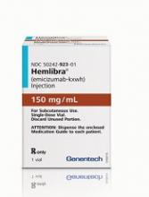
Health Canada has approved emicizumab (Hemlibra®) for use as routine prophylaxis to prevent or reduce bleeding episodes in hemophilia A patients with factor VIII inhibitors.
Emicizumab is a bispecific factor IXa- and factor X-directed antibody. It bridges activated factor IX and factor X to restore the natural function of missing activated factor VIII that is needed for effective blood clotting.
Emicizumab is given as a once-weekly subcutaneous injection.
“Preventing bleeds in patients with hemophilia A can be extremely challenging, usually requiring patients to self-infuse medications multiple times a week, or even daily,” said Jayson Stoffman, MD, of the University of Manitoba in Winnipeg.
“The development of inhibitors adds a significant challenge, with more demanding treatments that are often less effective. Hemlibra offers these patients the chance to effectively reduce the frequency of their bleeds with a once-weekly injection at home. This could significantly improve the quality of life for inhibitor patients, and particularly children and their families.”
Trial results
The Health Canada approval of emicizumab is based on data from a pair of phase 3 trials—HAVEN 1 and HAVEN 2.
Results from HAVEN 1 were published in NEJM and presented at the 26th ISTH Congress in July 2017. Updated results from HAVEN 2 were presented at the 2017 ASH Annual Meeting in December.
HAVEN 1
This study enrolled 109 patients (age 12 and older) with hemophilia A and factor VIII inhibitors who were previously treated with bypassing agents (BPAs) on demand or as prophylaxis.
The patients were randomized to receive emicizumab prophylaxis or no prophylaxis. On-demand treatment of breakthrough bleeds with BPAs was allowed.
There was an 87% reduction in treated bleeds with emicizumab compared to no prophylaxis (P<0.0001). And there was an 80% reduction in all bleeds with emicizumab (P<0.0001).
The proportion of patients with 0 treated bleeds was 62.9% among emicizumab recipients and 5.6% among patients who did not receive prophylaxis.
Adverse events (AEs) occurring in at least 5% of patients treated with emicizumab were local injection site reactions, headache, fatigue, upper respiratory tract infection, and arthralgia.
Two patients experienced thromboembolic events (TEs), and 3 had thrombotic microangiopathy (TMA) while receiving emicizumab prophylaxis and more than 100 µ/kg/day of activated prothrombin complex concentrate, on average, for 24 hours or more before the event. Two of these patients had also received recombinant factor VIIa.
Neither TE required anticoagulation therapy, and 1 patient restarted emicizumab. The cases of TMA observed were transient, and 1 patient restarted emicizumab.
There was 1 death, but it was considered unrelated to emicizumab. The patient had developed TMA but died of rectal hemorrhage.
HAVEN 2
In this single-arm trial, researchers evaluated emicizumab prophylaxis in 60 patients, ages 1 to 17, who had hemophilia A with factor VIII inhibitors.
The efficacy analysis included 57 patients who were younger than 12. The 3 older patients were only included in the safety analysis.
Of the 57 patients, 64.9% had 0 bleeds, 94.7% had 0 treated bleeds, and 98.2% had 0 treated spontaneous bleeds/treated joint bleeds. None of the patients had treated target joint bleeds.
A subset of 23 patients received emicizumab for at least 12 weeks (median treatment duration of 38.1 weeks; range, 12.7 to 41.6 weeks).
Of these 23 patients, 34.8% had 0 bleeds, 87.0% had 0 treated bleeds, and 95.7% had 0 treated spontaneous bleeds/treated joint bleeds.
There were 40 patients who had a total of 201 AEs. The most common of these were viral upper respiratory tract infections (16.7%) and injection site reactions (16.7%).
There were no TEs or TMA events, and none of the patients tested positive for anti-drug antibodies. None of the 7 serious AEs in this trial were considered treatment-related.


