User login
Fludarabine deemed important for CD30.CAR T-cell therapy
SAN DIEGO—Fludarabine is “very important” for lymphodepletion prior to CD30-directed chimeric antigen receptor (CAR) T-cell therapy, according to a presentation at the 2018 ASH Annual Meeting.
A phase 1/2 study showed that bendamustine alone was not sufficient as lymphodepletion.
However, adding fludarabine to bendamustine could enhance responses to CD30.CAR T-cell therapy and improve progression-free survival (PFS) in patients with Hodgkin or non-Hodgkin lymphoma.
Natalie S. Grover, MD, of the University of North Carolina in Chapel Hill, presented these results as abstract 681.*
This trial (NCT02690545) included patients with relapsed/refractory, CD30+ Hodgkin lymphoma or T-cell non-Hodgkin lymphoma.
Twenty-four adult patients have been treated thus far. Twenty-two had classical Hodgkin lymphoma, one had Sézary syndrome, and one had enteropathy-associated T-cell lymphoma.
The patients’ median age at baseline was 34.5 years (range, 23-69), and they had received a median of 7.5 prior lines of therapy (range, 3-17).
Prior treatments included brentuximab vedotin (n=23), checkpoint inhibitors (n=16), autologous transplant (n=17), and allogeneic transplant (n=7).
In this trial, patients could receive bridging therapy while their T cells were being processed. They then underwent lymphodepletion and received CAR T-cell therapy at one of two doses.
Bendamustine alone
Eight patients received lymphodepletion with 2 days of bendamustine at 90 mg/m2. Three of these patients received CD30.CAR T-cell therapy at 1×10⁸ cells/m2, and all three progressed.
Of the five patients who received CAR T-cell therapy at a dose of 2×10⁸ cells/m2, one progressed, one had stable disease, and three had a complete response (CR).
However, all three complete responders were in CR prior to lymphodepletion as a result of bridging therapy.
“Responses were more modest than what we were hoping for with lymphodepletion,” Dr. Grover noted. “We looked at the cytokine levels in patients getting bendamustine lymphodepletion and saw that bendamustine wasn’t supporting an ideal cytokine milieu. IL-7 and IL-15 are important for T-cell expansion, and these levels were not increased in patients post-bendamustine.”
When the researchers added fludarabine to the lymphodepleting regimen, they observed an increase in T-cell expansion.
Bendamustine plus fludarabine
Sixteen patients received bendamustine plus fludarabine prior to CAR T-cell therapy. The regimen consisted of 3 days of bendamustine at 70 mg/m2 and fludarabine at 30 mg/m2.
All 16 patients received CAR T cells at 2×10⁸ cells/m2, which was the recommended phase 2 dose.
“Responses were more impressive in the bendamustine-fludarabine cohort,” Dr. Grover noted.
Twelve of the 16 patients achieved a CR, although two patients were already in CR prior to lymphodepletion.
Two patients had a partial response, one had stable disease, and one progressed.
PFS and toxicity
Dr. Grover and her colleagues also assessed PFS. At a median follow-up of 100 days, the median PFS was 164 days for the entire cohort, excluding patients who were in CR prior to lymphodepletion.
The median PFS was 396 days for the bendamustine-fludarabine cohort and 55 days for patients in the bendamustine-alone cohort (P=0.001).
There was no neurotoxicity in this trial.
Three patients developed cytokine release syndrome (CRS). Two patients had grade 1 CRS that resolved spontaneously, and one patient had grade 2 CRS, which responded to tocilizumab. Two of the patients with CRS had T-cell lymphoma. The Sézary patient had grade 2 CRS.
Eight patients had a mild rash, one of whom had a rash at baseline.
“CAR T cells against CD30 preceded by lymphodepletion with bendamustine and fludarabine have promising efficacy and a good safety profile in treating patients with relapsed/refractory, CD30+ lymphomas,” Dr. Grover said in closing.
“Fludarabine is very important in enhancing cytokines for improved growth and persistence of CAR T cells.”
This trial was sponsored by UNC Lineberger Comprehensive Cancer Center. Dr. Grover reported consulting for Seattle Genetics.
*Data in the abstract differ from the presentation.
System may better predict thrombosis in lymphoma

DUBROVNIK, CROATIA—An updated scoring system can more accurately identify lymphoma patients who may require thromboprophylaxis, according to researchers.
The revised scoring system, ThroLy, proved more effective than other systems for predicting thromboembolic events in lymphoma patients.
Researchers found the updated ThroLy had a positive predictive value of 22% to 25%, a negative predictive value of 96%, sensitivity of 56% to 57%, and specificity of 85% to 87%.
Darko Antić, MD, PhD, of the University of Belgrade in Serbia, presented these findings at Leukemia and Lymphoma: Europe and the USA, Linking Knowledge and Practice.
Dr. Antić said he and his colleagues developed ThroLy because other systems used to predict venous thromboembolism (VTE) are not quite right for lymphoma. He noted that the Padua score is not designed for cancer patients, and the Khorana score is predominantly for solid tumor malignancies.
“It’s good . . . , but it’s not specific for lymphoma patients,” Dr. Antić said.
With this in mind, he and his colleagues developed ThroLy. They based the scoring system on variables used in the Padua and Khorana systems as well as variables that are specific to lymphoma patients.
In a past study*, the researchers found several variables that were independently associated with risk for VTE in lymphoma:
- Previous VTE
- Previous acute myocardial infarction/stroke
- Mediastinal involvement
- Body mass index > 30 kg/m2
- Reduced mobility
- Extranodal localization
- Development of neutropenia
- Hemoglobin level < 100g/L.
Previous VTE, previous acute myocardial infarction/stroke, obesity, and mediastinal involvement were all worth 2 points, and the other factors were worth a single point.
Patients with scores of 0 to 1 were considered low-risk, patients with scores of 2 to 3 were considered intermediate-risk, and patients with scores of 4 or greater were considered high-risk.
Prospective validation
To validate and refine ThroLy, Dr. Antić and his colleagues used it to assess 1723 lymphoma patients treated at 8 institutions in Austria, Croatia, France, Jordan, Macedonia, Spain, Switzerland, and the United States.
Patients had indolent non-Hodgkin lymphoma (n=467), aggressive non-Hodgkin lymphoma (n=647), chronic lymphocytic leukemia/small lymphocytic lymphoma (n=235), and Hodgkin lymphoma (n=366). Most subjects (84%) were outpatients.
Nine percent of patients had thrombosis (n=142), with 7% having VTE (n=121).
ThroLy had a positive predictive value of 17%, compared to 11% with Khorana and 13% with Padua. The negative predictive value was 93%, 92%, and 95%, respectively.
The sensitivity was 51% with ThroLy, 42% with Khorana, and 70% with Padua. The specificity was 72%, 64%, and 52%, respectively.
“The positive predictive value was low [with ThroLy] but definitely higher than the positive predictive value of the other two [scoring systems],” Dr. Antić noted.
Updated models
To further improve ThroLy, the researchers updated the system, creating two new models.
Model 1 included the following variables:
- Type of lymphoma/clinical stage (aggressive/advanced)—1 point
- Previous VTE—5 points
- Reduced mobility—2 points
- Hemoglobin level < 100 g/L—1 point
- Presence of vascular devices—1 point.
Model 2 included all of the aforementioned variables as well as thrombophilic condition, which was worth 1 point.
With these models, patients were divided into two risk groups—low-risk (≤ 2 points) and high-risk (>2 points).
For Model 1, the positive predictive value was 22%, the negative predictive value was 96%, the sensitivity was 56%, and the specificity was 85%.
For Model 2, the positive predictive value was 25%, the negative predictive value was 96%, the sensitivity was 57%, and the specificity was 87%.
Dr. Antić said there were no major differences in model discrimination and calibration according to the country in which a patient was treated or whether patients were treated in inpatient or outpatient settings.
Dr. Antić did not report any conflicts of interest.
*Antić D et al. Am J Hematol. 2016 Oct;91(10):1014-9. doi: 10.1002/ajh.24466.
CAR T therapy being explored in Hodgkin lymphoma
New York—Although the data set is small and not yet mature, chimeric antigen receptor (CAR) T-cell therapy appears to be a promising approach for Hodgkin lymphoma, according to Philippe Armand, MD, PhD, of Dana-Farber/Brigham and Women’s Cancer Center and the Massachusetts General Hospital Cancer Center.
While based on a handful of patients, the data do suggest this approach may play a role either by targeting CD30 or Epstein Barr virus (EBV), Dr. Armand said in a presentation at the NCCN 13th Annual Congress: Hematologic Malignancies.
“Most importantly perhaps, like it's experience outside of Hodgkin lymphoma, it may really have curative potential, based on the long CR rates that have been already exhibited,” he told attendees at the NCCN conference.
Much of the published clinical experience to date is with CD30-directed CAR Ts, Dr. Armand said, noting that in Hodgkin lymphoma, results so far show promise for this particular approach.
In a recent phase 1 dose escalation study, 9 patients with relapsed/refractory Hodgkin lymphoma or anaplastic large-cell lymphoma (ALCL) received infusions of autologous T cells modified to express CD30-specific CAR T cells encoding the CD28 costimulatory domain, with no conditioning regimen.
Out of 7 relapsed Hodgkin patients, one had a complete response (CR) lasting beyond 2.5 years following a second infusion. Another had a CR persisting almost 2 years and 3 had transient stable disease.
One of the 2 ALCL patients had a CR lasting 9 months after a fourth infusion. No toxicities attributable to the therapy were seen, according to investigators.
The CD30 CAR T cells are being evaluated with a conditioning regimen in the phase 1 RELY-30 trial. According to Dr. Armand, preliminary results presented at the EBMT 2018 meeting showed better expansion of CAR T cells and responses in 3 out of 5 patients, including 2 CRs.
A CD30-directed CAR T-cell therapy with a 4-1bb costimulatory domain has also been tested in a small group of Hodgkin patients with a response rate of 35%, including some CRs. Response rates were lower in patients with extranodal involvement, although that needs to be validated with further study, according to Dr. Armand.
A considerable amount of active research is ongoing in China, Dr. Armand said, while a phase 1 study of T cells expressing a fully human anti-CD30 CAR is being evaluated in the United States in CD30-expressing lymphomas, he added.
Among non-CD30-targeted products, a CD19 CAR-T approach has been tried in Hodgkin lymphoma, though preliminary results suggest only transient activity.
An interesting approach has been the targeting of EBV, Dr. Armand noted. Recently reported results showed that two doses of T cells with specificity for EBV-derived tumor antigens induced clinical responses in patients with EBV-positive Hodgkin lymphoma.
The cells were engineered to express dominant-negative TGF-β receptor type 2 (DNRII).
“We know that TGF-β provides a strong immunosuppressant signal in the tumor microenvironment,” Dr. Armand said, noting that some of the responses in the 7 evaluable patients lasted 4 years or more.
Two immunologists receive Nobel Prize in medicine
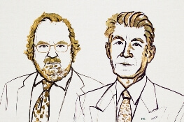
Two immunologists have been awarded the Nobel Prize in Physiology or Medicine for discoveries that represent a “paradigmatic shift in the fight against cancer,” the Nobel committee said.
James P. Allison, PhD, of MD Anderson Cancer Center, and Tasuku Honjo, MD, PhD, of Kyoto University, shared the prize for their discovery of cancer therapies that work by inhibiting negative immune regulation.
Dr. Allison studied the protein CTLA-4 found on T cells, which acts as a T-cell brake, and Dr. Honjo discovered a protein on immune cells called PD-1 that also acts as a T-cell brake.
In addition to sharing the honor, the scientists will split the 9 million Swedish kronor ($1.01 million) that comes with the prize.
Drs. Allison and Honjo, working in parallel, pursued different strategies for inhibiting the brakes on the immune system. Both strategies produced effective checkpoint inhibitors in the treatment of cancer.
James P. Allison
Dr. Allison was one of several scientists during the 1990s who noticed that CTLA-4 functions as a brake on T cells. Unlike other scientists, however, he set out to investigate whether blocking CTLA-4 with an antibody he had already developed could release the brake on the immune system.
The antibody had “spectacular” effects in curing mice with cancer. Despite little interest from the pharmaceutical industry, Dr. Allison continued efforts to develop the antibody therapy for humans.
The antibody turned out to be ipilimumab, which was approved in 2011 by the U.S. Food and Drug Administration (FDA) for the treatment of advanced melanoma.
Tasuko Honjo
A few years prior to Dr. Allison’s finding, Dr. Honjo discovered PD-1 and set out to determine its function. PD-1 also operates as a T-cell brake, but it uses a different mechanism than does CTLA-4.
Dr. Honjo and others demonstrated in animal experiments that PD-1 blockade could be an effective anticancer therapy. Over the years he demonstrated the efficacy of targeting PD-1 in different types of human cancers.
The first two PD-1 checkpoint inhibitors—pembrolizumab and nivolumab—were approved by the FDA in 2014 for the treatment of melanoma.
Nivolumab is also approved to treat classical Hodgkin lymphoma (HL), non-small cell lung cancer (NSCLC), small cell lung cancer, squamous cell carcinoma of the head and neck, colorectal cancer, hepatocellular carcinoma, renal cell carcinoma, urothelial carcinoma, and microsatellite instability-high or mismatch repair deficient colorectal cancer.
Pembrolizumab is also approved to treat primary mediastinal large B-cell lymphoma, advanced NSCLC, classical HL, advanced gastric cancer, advanced cervical cancer, head and neck squamous cell cancer, advanced urothelial bladder cancer, and microsatellite instability-high cancer.
And targeting both CTLA-4 and PD-1 in combination therapy together may prove to be even more effective in eliminating cancer cells than either strategy alone, as is being demonstrated in patients with melanoma.
The Nobel organization wrote in a press release, “Checkpoint therapy has now revolutionized cancer treatment and has fundamentally changed the way we view how cancer can be managed.”
Brentuximab improves survival in older HL patients

Older patients with untreated Hodgkin lymphoma (HL) can achieve significantly improved survival by adding brentuximab vedotin to their treatment before and after standard chemotherapy, a recent study found.
In patients with low comorbidity scores, responses were even more robust, reported lead author Andrew M. Evens, DO, of the Rutgers Cancer Institute of New Jersey, and colleagues.
“Causes of poor outcomes for older patients with HL are not fully understood but have been attributed to a combination of factors, including presence of comorbidities, poorer performance status, disease and biological differences, inability to tolerate chemotherapy at the full dose, and increased treatment-related toxicities,” the authors wrote in the Journal of Clinical Oncology.
The primary goal of the study was to improve outcomes for untreated, older patients, a group that’s historically been a difficult-to-treat patient population.
The phase 2 trial included 48 HL patients with a median age of 69 (range, 60 – 88).
All patients underwent geriatric assessment for comorbidities and loss of activities of daily living.
Treatment consisted of two doses of brentuximab followed by six cycles of doxorubicin, vinblastine, and dacarbazine (AVD), then four more doses of brentuximab (consolidation doses).
The primary endpoint was complete remission at completion of AVD.
Secondary outcomes included overall response rate, 2-year progression-free survival, 2-year overall survival, and safety.
Just over half the patients (52%) completed all cycles of therapy, and almost three quarters (73%) received at least one consolidation dose of brentuximab.
Among the first 23 evaluable patients, both the complete remission rate and overall response rate were 96%. Intention-to-treat survival rates for all 48 patients were 84% for 2-year progression-free survival and 93% for 2-year overall survival.
Historical 2-year progression-free survival rates in similar older patients is poor, at 50%, so the progression-free survival rate of 84% in this study represents a significant improvement.
Of note, patients with fewer comorbidities and without loss of instrumental activities of daily living showed more robust responses.
Patients with Cumulative Illness Rating Scale for Geriatrics (CIRS-G) comorbidity scores of less than 10 had a 2-year progression-free survival rate of 100% versus 45% for those with higher scores.
Similarly, patients without loss of instrumental activities achieved a progression-free survival rate of 94% versus 25% for those who had lost some instrumental activities.
Grade 3 or 4 adverse events occurred in 42% of patients, with neutropenia being the most common (44%).
“This study represents among the best-reported outcomes to date for untreated older patients with HL,” the investigators concluded.
Seattle Genetics supported the investigator-initiated trial.
CAR T may have curative potential in Hodgkin lymphoma
NEW YORK – Although the data set is small and not yet mature, chimeric antigen receptor (CAR) T-cell therapy appears to be a promising approach for Hodgkin lymphoma, according to Philippe Armand, MD, PhD, of Dana-Farber/Brigham and Women’s Cancer Center and the Massachusetts General Hospital Cancer Center in Boston.
While based on a handful of patients, the data do suggest this approach may play a role by targeting CD30 or Epstein Barr virus (EBV), Dr. Armand said at the NCCN Annual Congress: Hematologic Malignancies.
“Most importantly perhaps, like its experience outside of Hodgkin lymphoma, it may really have curative potential, based on the long [complete response] rates that have been already exhibited,” he said.
Much of the published clinical experience to date is with CD30-directed CAR Ts, Dr. Armand said, noting that in Hodgkin lymphoma, results so far show promise for this particular approach.
In a recent phase 1 dose escalation study, nine patients with relapsed/refractory Hodgkin lymphoma or anaplastic large-cell lymphoma (ALCL) received infusions of autologous T cells modified to express CD30-specific CAR T cells encoding the CD28 costimulatory domain, with no conditioning regimen.
Out of seven relapsed Hodgkin lymphoma patients, one had a complete response (CR) lasting beyond 2.5 years following a second infusion. Another patient had a CR persisting almost 2 years, and three patients had transient stable disease. One of the two ALCL patients had a CR lasting 9 months after a fourth infusion.
No toxicities attributable to the therapy were seen, according to the investigators.
The CD30 CAR T cells are being evaluated with a conditioning regimen in the phase 1 RELY-30 trial. Preliminary results presented at the 2018 European Society for Blood and Marrow Transplantation meeting in Lisbon showed better expansion of CAR T cells and responses in three out of five patients, including two CRs, according to Dr. Armand.
A CD30-directed CAR T-cell therapy with a 4-1BB costimulatory domain has also been tested in a small group of Hodgkin patients with a response rate of 35% – including some CRs – with an apparent lower response rate in patients with extranodal involvement. Dr. Armand noted that those findings need to be validated in additional studies.
Among non CD-30 targeted products, a CD19 CAR-T approach has been tried in Hodgkin lymphoma, though preliminary results suggest only transient activity, according to the presenter.
One interesting approach has been the targeting of EBV, he added. Recently reported results showed that two doses of T cells with specificity for EBV-derived tumor antigens induced clinical responses in patients with EBV-positive Hodgkin lymphoma.
The cells were engineered to express dominant-negative TGF-beta receptor type 2, according to the report. “We know that TGF-beta provides a strong immunosuppressant signal in the tumor microenvironment,” Dr. Armand said, noting that some of the responses in the seven evaluable patients lasted 4 years or more.
Dr. Armand reported financial disclosures related to Adaptive Biotechnologies/Sequenta, Affimed, Bristol-Myers Squibb, Merck, and Roche.
EXPERT ANALYSIS FROM NCCN ANNUAL CONGRESS: HEMATOLOGIC MALIGNANCIES
Drug approved as part of frontline therapy for HL

The Japanese Ministry of Health, Labour and Welfare has approved brentuximab vedotin (Adcetris) in combination with doxorubicin, vinblastine, and dacarbazine as a frontline treatment option for CD30-positive Hodgkin lymphoma (HL).
The approval was based on the phase 3 ECHELON-1 trial.
Result from ECHELON-1 were presented at the 2017 ASH Annual Meeting and simultaneously published in The New England Journal of Medicine.
In this trial, researchers compared brentuximab vedotin plus doxorubicin, vinblastine, and dacarbazine (A+AVD) to doxorubicin, bleomycin, vinblastine, and dacarbazine (ABVD) as frontline treatment for 1334 patients with advanced HL.
The primary endpoint was modified progression-free survival (PFS), which was defined as time to progression, death, or evidence of non-complete response after completion of frontline therapy followed by subsequent anticancer therapy.
According to an independent review committee, A+AVD provided a significant improvement in modified PFS compared to ABVD. The hazard ratio was 0.77 (P=0.035), which corresponds to a 23% reduction in the risk of progression, death, or the need for additional anticancer therapy.
The 2-year modified PFS rate was 82.1% in the A+AVD arm and 77.2% in the ABVD arm.
There was no significant difference between the treatment arms when it came to response rates or overall survival.
The objective response rate was 86% in the A+AVD arm and 83% in the ABVD arm (P=0.12). The complete response rate was 73% and 70%, respectively (P=0.22).
The interim 2-year overall survival rate was 97% in the A+AVD arm and 95% in the ABVD arm (hazard ratio=0.72; P=0.19).
The overall incidence of adverse events (AEs) was 99% in the A+AVD arm and 98% in the ABVD arm. The incidence of grade 3 or higher AEs was 83% and 66%, respectively, and the incidence of serious AEs was 43% and 27%, respectively.
Neutropenia, febrile neutropenia, and peripheral neuropathy were more common with A+AVD, while pulmonary toxicity was more common with ABVD.
Brentuximab vedotin boosts elderly Hodgkin survival
For elderly patients with untreated Hodgkin lymphoma (HL), adding brentuximab vedotin (Bv) before and after standard chemotherapy significantly improved survival, a recent study found.
In patients with low comorbidity scores, responses were even more robust, reported Andrew M. Evens, DO, of the Rutgers Cancer Institute of New Jersey in New Brunswick and his colleagues.
“Causes of poor outcomes for older patients with HL are not fully understood but have been attributed to a combination of factors, including presence of comorbidities, poorer performance status, disease and biological differences, inability to tolerate chemotherapy at the full dose, and increased treatment-related toxicities,” the authors wrote in the Journal of Clinical Oncology.
The primary goal of the study was to improve outcomes for untreated, older patients, a group that’s historically been a difficult-to-treat patient population.
The phase 2 trial included 48 elderly patients (median age, 69 years) with Hodgkin lymphoma. All patients underwent geriatric assessment for comorbidities and loss of activities of daily living. Treatment consisted of two doses of Bv followed by six cycles of doxorubicin, vinblastine, and dacarbazine (AVD), then four more doses of Bv (consolidation doses). The primary endpoint was complete remission at completion of AVD. Secondary outcomes included overall response rate, 2-year progression-free survival, 2-year overall survival, and safety.
Just over half of the patients (52%) completed all cycles of therapy, and almost three-quarters (72%) received at least one consolidation dose of Bv.
Among the first 23 evaluable patients, both the complete remission rate and overall response rate were 96%. Intention-to-treat survival rates for all 48 patients were 84% for 2-year progression-free survival and 93% for 2-year overall survival.
Historical 2-year progression-free survival rates in similar elderly patients is poor, at 50%, so the progression-free survival rate of 84% in this study represents a significant improvement. Of note, patients with fewer comorbidities and without loss of instrumental activities of daily living showed more robust responses.
Patients with Cumulative Illness Rating Scale for Geriatrics (CIRS-G) comorbidity scores of less than 10 had a 2-year progression-free survival rate of 100% versus 45% for those with higher scores. Similarly, patients without loss of instrumental activities achieved a progression-free survival rate of 94%versus 25% for those who had lost some instrumental activities.
Grade 3 or 4 adverse events occurred in 42% of patients, with neutropenia being the most common (44%).
“This study represents among the best-reported outcomes to date for untreated older patients with HL,” the investigators concluded.
Seattle Genetics supported the investigator-initiated trial. Dr. Evens reported consulting or advisory relationships with Seattle Genetics and several other companies.
SOURCE: Evens AM et al. J Clin Oncol. 2018 Sep 4. doi: 10.1200/JCO.2018.79.0139
FROM JOURNAL OF CLINICAL ONCOLOGY
Key clinical point:
Major finding: Two-year PFS was 84% for patients treated additionally with Bv, compared with 50% historically for standard therapy.
Study details: A prospective phase 2 study involving 48 elderly patients with Hodgkin lymphoma.
Disclosures: Seattle Genetics supported the investigator-initiated trial. Dr. Evens reported consulting or advisory relationships with Seattle Genetics and several other companies.
Source: Evens AM et al. J Clin Oncol. 2018 Sep 4. doi: 10.1200/JCO.2018.79.0139.
Guidelines for proton therapy in mediastinal lymphomas
Proton therapy can help mitigate toxicity in adults with mediastinal lymphomas, but the treatment should only be used in patients expected to derive the most benefit, according to new guidelines from the International Lymphoma Radiation Oncology Group.
The guidelines note that proton therapy reduces the radiation dose to organs at risk in certain clinical presentations, such as when the mediastinal target is on both sides of the heart.
However, the advantages of proton therapy are not always clear in other situations, such as when the target spans the right side of the heart or when the target is above the heart with no axillary involvement.
“The limited availability of proton therapy calls for case selection based on a clear understanding of which cases will derive most benefit from proton therapy as compared to advanced photon techniques,” said guideline author Bouthaina Dabaja, MD, of the University of Texas MD Anderson Cancer Center in Houston, and her colleagues.
The group’s guidelines were published in Blood.
The guidelines note that proton therapy—like intensity-modulated radiotherapy and 3-dimensional conformal radiotherapy—presents an opportunity for more conformal dose distribution and better sparing of organs at risk.
Proton therapy can greatly benefit certain patients with mediastinal disease, including:
- Young female patients in whom proton therapy would reduce the breast dose and decrease the risk of secondary breast cancer
- Patients at high risk of radiation-related toxicity due to previous treatment
- Patients with disease spanning below the origin of the left main stem coronary artery that is anterior to, posterior to, or on the left side of the heart.
“The relation of disease to organs at risk determines the situations in which proton therapy is most beneficial,” the experts said in the guidelines.
However, the consideration of proton therapy needs to factor in the complexities of proton therapy planning, the need to manage uncertainties, and the “evolving nature of the technology,” which includes the development of pencil beam scanning.
While passive scattering proton therapy is the least complex delivery technique, it is challenging because beams can conform only to one side of the target. In contrast, active mode pencil beam scanning proton therapy potentially provides better conformality and sparing of organs at risk.
“Because treatment involves delivery of individual controlled spots, inhomogenous doses can be created deliberately,” the guideline authors said.
However, “motion management is of prime importance” with pencil beam scanning proton therapy, which is more sensitive to density changes in the beam path than is passive scattering proton therapy.
To that end, physicians should pay close attention to evaluating intrafractional movement, which is frequently tied to the breathing cycle.
Dr. Dabaja and her coauthors reported no funding or conflicts of interest.
Role of SES in childhood cancer survival disparities

Socioeconomic status (SES) may explain some racial/ethnic disparities in childhood cancer survival, according to new research.
The study showed that whites had a significant survival advantage over blacks and Hispanics for several childhood cancers.
SES significantly mediated the association between race/ethnicity and survival for acute lymphoblastic leukemia (ALL), acute myeloid leukemia (AML), neuroblastoma, and non-Hodgkin lymphoma (NHL).
Rebecca Kehm, PhD, of Columbia University in New York, New York, and her colleagues reported these findings in Cancer alongside a related editorial.
The researchers examined population-based cancer survival data from the Surveillance, Epidemiology, and End Results database.
The team collected information on 31,866 patients, ages 0 to 19, who were diagnosed with cancer between 2000 and 2011.
Survival differences by race/ethnicity
The researchers found that whites had a significant survival advantage over blacks for the cancers listed in the following table.
| Survival—black vs white | |||
| Cancer | Mortality hazard ratio | 95% confidence interval | P value |
| ALL | 1.43 | 1.15-1.77 | <0.01 |
| AML | 1.68 | 1.36-2.07 | <0.001 |
| Neuroblastoma | 1.38 | 1.08-1.75 | 0.01 |
| NHL | 1.53 | 1.14-2.07 | 0.01 |
| Hodgkin lymphoma | 1.66 | 1.06-2.60 | 0.03 |
| Astrocytoma | 1.95 | 1.57-2.43 | <0.001 |
| Non-astrocytoma CNS tumor | 1.53 | 1.25-1.88 | <0.001 |
| Non-rhabdomyosarcoma STS | 1.40 | 1.06-1.84 | 0.02 |
| Rhabdomyosarcoma | 1.44 | 1.10-1.88 | 0.01 |
In addition, whites had a significant survival advantage over Hispanics for the following cancers.
| Survival—Hispanic vs white | |||
| Cancer | Mortality hazard ratio | 95% confidence interval | P value |
| ALL | 1.63 | 1.43-1.86 | <0.001 |
| Neuroblastoma | 1.31 | 1.04-1.65 | 0.02 |
| NHL | 1.65 | 1.29-2.12 | <0.001 |
| Astrocytoma | 1.34 | 1.10-1.64 | <0.01 |
| Wilms tumor | 1.60 | 1.04-2.45 | 0.03 |
| Germ cell tumor | 1.63 | 1.19-2.24 | <0.01 |
Impact of SES
SES significantly mediated the association between race/ethnicity and survival for ALL, AML, neuroblastoma, and NHL but not for Hodgkin lymphoma or other cancers.
For black versus white patients, SES reduced the original association between race/ethnicity and survival by:
- 44% for ALL
- 28% for AML
- 49% for neuroblastoma
- 34% for NHL.
For Hispanics versus whites, SES reduced the original association between race/ethnicity and survival by:
- 31% for ALL
- 73% for AML
- 48% for neuroblastoma
- 28% for NHL.
“These findings provide insight for future intervention efforts aimed at closing the survival gap,” Dr Kehm said.
“For cancers in which socioeconomic status is a key factor in explaining racial and ethnic survival disparities, behavioral and supportive interventions that address social and economic barriers to effective care are warranted. However, for cancers in which survival is less influenced by socioeconomic status, more research is needed on underlying differences in tumor biology and drug processing.”
This research was supported by a grant from the National Institutes of Health, and the study’s authors made no disclosures.