User login
Updated CLL guidelines incorporate a decade of advances
include new and revised recommendations based on major advances in genomics, targeted therapies, and biomarkers that have occurred since the last iteration in 2008.
The guidelines are an update from a consensus document issued a decade ago by the International Workshop on CLL, focusing on the conduct of clinical trials in patients with CLL. The new guidelines are published in Blood.
Major changes or additions include:
Molecular genetics: The updated guidelines recognize the clinical importance of specific genomic alterations/mutations on response to standard chemotherapy or chemoimmunotherapy, including the 17p deletion and mutations in TP53.
“Therefore, the assessment of both del(17p) and TP53 mutation has prognostic and predictive value and should guide therapeutic decisions in routine practice. For clinical trials, it is recommended that molecular genetics be performed prior to treating a patient on protocol,” the guidelines state.
IGHV mutational status: The mutational status of immunoglobulin variable heavy chain (IGHV) genes has been demonstrated to offer important prognostic information, according to the guidelines authors led by Michael Hallek, MD of the University of Cologne, Germany.
Specifically, leukemia with IGHV genes without somatic mutations are associated with worse clinical outcomes, compared with leukemia with IGHV mutations. Patients with mutated IGHV and other prognostic factors such as favorable cytogenetics or minimal residual disease (MRD) negativity generally have excellent outcomes with a chemoimmunotherapy regimen consisting of fludarabine, cyclophosphamide, and rituximab, the authors noted.
Biomarkers: The guidelines call for standardization and use in prospective clinical trials of assays for serum markers such as soluble CD23, thymidine kinase, and beta-2-microglobulin. These markers have been shown in several studies to be associated with overall survival or progression-free survival, and of these markers, beta-2-microglobulin “has retained independent prognostic value in several multiparameter scores,” the guidelines state.
The authors also tip their hats to recently developed or improved prognostic scores, especially the CLL International Prognostic Index (CLL-IPI), which incorporates clinical stage, age, IGHV mutational status, beta-2-microglobulin, and del(17p) and/or TP53 mutations.
Organ function assessment: Not new, but improved in the current version of the guidelines, are recommendations for evaluation of splenomegaly, hepatomegaly, and lymphadenopathy in response assessment. These recommendations were harmonized with the relevant sections of the updated lymphoma response guidelines.
Continuous therapy: The guidelines panel recommends assessment of response duration during continuous therapy with oral agents and after the end of therapy, especially after chemotherapy or chemoimmunotherapy.
“Study protocols should provide detailed specifications of the planned time points for the assessment of the treatment response under continuous therapy. Response durations of less than six months are not considered clinically relevant,” the panel cautioned.
Response assessments for treatments with a maintenance phase should be performed at a minimum of 2 months after patients achieve their best responses.
MRD: The guidelines call for minimal residual disease (MRD) assessment in clinical trials aimed at maximizing remission depth, with emphasis on reporting the sensitivity of the MRD evaluation method used, and the type of tissue assessed.
Antiviral prophylaxis: The guidelines caution that because patients treated with anti-CD20 antibodies, such as rituximab or obinutuzumab, could have reactivation of hepatitis B virus (HBV) infections, patients should be tested for HBV serological status before starting on an anti-CD20 agent.
“Progressive multifocal leukoencephalopathy has been reported in a few CLL patients treated with anti-CD20 antibodies; therefore, infections with John Cunningham (JC) virus should be ruled out in situations of unclear neurological symptoms,” the panel recommended.
They note that patients younger than 65 treated with fludarabine-based therapy in the first line do not require routine monitoring or infection prophylaxis, due to the low reported incidence of infections in this group.
The authors reported having no financial disclosures related to the guidelines.
FROM BLOOD
FDA updates breast implant–associated lymphoma cases, risk
(BIA-ALCL), including nine deaths.
This figure includes all medical device reports received by the agency between 2011 and September 2017. The FDA recently provided an update on ALCL linked to breast implants and an estimate of lifetime risk of developing ALCL.
Based on available medical literature, the lifetime risk of developing BIA-ALCL for patients with textured breast implants ranges from 1 in 3,817 to 1 in 30,000, according to the update.
Of the 272 reports with data on surface type, 242 were textured implants and 30 were smooth implants. In addition, 413 reports include information on the implant fill type: 234 used silicone gel and 179 were saline filled.
“The FDA has been closely tracking the relationship between breast implants and a rare type of non-Hodgkin’s lymphoma since we first identified this possible association. We’ve been working to gather additional information to better characterize and quantify the risk so that patients and providers can have more informed discussions about breast implants,” said Binita Ashar, MD, director of the division of surgical devices in the FDA’s Center for Devices and Radiological Health. “As part of that effort, we are working to update and enhance the information we have on this association, including updating the total number of known cases of BIA-ALCL and the lifetime risk of developing BIA-ALCL as reported in medical literature.”
The possible association between breast implants and the development of anaplastic large cell lymphoma (ALCL) was first identified in 2011. At that time, there were not enough cases of to determine what factors increased a patient’s risk of developing the disease. As more information became available, the World Health Organization designated BIA-ALCL as a T-cell lymphoma that can develop following breast implants.
Ibrutinib linked to invasive fungal infections
The tyrosine kinase inhibitor ibrutinib (Imbruvica) may be associated with early-onset invasive fungal infections (IFI) in patients with hematologic malignancies, investigators caution.
French investigators identified 33 cases of invasive fungal infections occurring among patients who had been treated with ibrutinib as monotherapy or in combination with other agents. Of the 33 cases, 27 were invasive aspergillosis, and 40% of these were localized in the central nervous system. The findings were published in the journal Blood.
“IFI tend to occur within the first months of treatment and are infrequent thereafter. Whilst it seems difficult at this point to advocate for systematic antifungal prophylaxis in all patients, an increased awareness about the potential risk of IFI after initiating ibrutinib is warranted, especially when other predisposing factors are associated,” wrote David Ghez, MD, PhD, and colleagues at the Gustave Roussy Institute in Villejuif and other centers in France.
Although ibrutinib, an inhibitor of Bruton’s tyrosine kinase, is generally considered to be less immunosuppressive than other therapies, it was associated with five cases of Pneumocystis jirovecii pneumonia in patients with chronic lymphocytic leukemia (CLL) treated with ibrutinib monotherapy in a 2016 report (Blood. 2016;128:1940-3). Of these five patients, four were treatment naive, suggesting that ibrutinib itself could increase risk for invasive opportunistic infections, Dr. Ghez and his colleagues noted.
Based on this finding and on case reports of invasive infections in other patients being treated with ibrutinib, the authors conducted a retrospective survey of centers in the French Innovative Leukemia Organization CLL group.
They identified 33 cases, including 30 patients with CLL (15 of whom had deleterious 17p deletions), 1 with mantle cell lymphoma, and 2 with Waldenstrom macroglobulinemia.
Invasive aspergillosis accounted for 27 of the 33 cases, and 11 cases had CNS localization. There were four cases of disseminated cryptococcosis, and one each of mucormycosis and pneumocystis pneumonia.
The median time between the start of ibrutinib therapy and a diagnosis of invasive fungal infection was 3 months, with some cases occurring as early as 1 month, and others occurring 30 months out. However, the majority of cases – 28 – were diagnosed within 6 months of the start of therapy, including 20 that occurred within 3 months of ibrutinib initiation.
In 21 patients, the diagnosis of an invasive fungal infection led to drug discontinuation. In the remaining patients, the drug was either resumed after resolution of the IFI, or continued at a lower dose because of potential for interaction between ibrutinib and the antifungal agent voriconazole.
Dr. Ghez reported receiving a research grant from Janssen, and coauthor Loic Ysebaert, MD, PhD, reported consultancy fees from the company. All other authors declared no competing financial interests.
SOURCE: Ghez D et al., Blood. 2018 Feb 1. doi: 10.1182/blood-2017-11-818286.
FROM BLOOD
Key clinical point:
Major finding: Of 33 identified cases, 27 were invasive aspergillosis.
Study details: Retrospective review of case reports from 16 French centers.
Disclosures: Dr. Ghez reported receiving a research grant from Janssen, and coauthor Loic Ysebaert, MD, PhD, reported consultancy fees with the company. All other authors declared no competing financial interests.
Source: Ghez D et al. Blood. 2018 Feb 1. doi: 10.1182/blood-2017-11-818286.
Mycosis fungoides increases risk for second cancers
LA JOLLA, CALIF. – Patients with mycosis fungoides are at increased risk for developing other cancers and should be screened for second primary and hematologic malignancies, results of a cancer registry survey suggest.
A study of data on 6,196 patients included in 18 population-based cancer registries comprising the SEER-18 (Surveillance, Epidemiology, and End Results 18) database who were diagnosed and followed from 2000 to 2014 showed that 514 (8.3%) developed second cancers, compared with the 70.8 secondary malignancies that would be expected in the general population. This difference translated into a standardized incidence ratio (SIR) of 7.3, reported Amrita Goyal, MD, and Aleksandr Lazaryan, MD, PhD, of the University of Minnesota, Minneapolis.

Patients with MF have a 500% greater risk for developing a second solid malignancy and a 2700% greater likelihood of developing a second hematologic malignancy, she said.
The investigators hypothesized that MF predisposes patients to second malignancies because of its immunocompromising effects.
Dr. Goyal said that, although the SEER data set does not include information on disease stage for all patients, when they looked at a separate cohort of 173 University of Minnesota patients with MF, they saw that patients with higher-stage MF were significantly more likely to develop secondary malignancies than patients with lower-stage disease.
The investigators looked at the actual and expected cancer incidence rates for the SEER-18 population sample, and used data on age, sex, race, and calendar year to generate incidence estimates for the general population.
They found that 514 patients in the SEER-18 population developed a total of 170 second primary hematologic malignancies, for a SIR of 27.4, compared with the general population. The most common hematologic cancers were Hodgkin lymphoma (SIR 69.8) and non-Hodgkin lymphoma (SIR 46.5), and other second hematologic malignancies included multiple myeloma (SIR 4.5), chronic lymphocytic leukemia (SIR 9.1). and acute leukemias (SIR 8.1).
The most frequently occurring second solid tumors included cancers of the nose, nasal cavity, and middle ear (SIR 30.4); thyroid (SIR 16.1); brain (SIR 15.1); and breast (SIR 8.0).
Other solid tumors with an approximately 400%-500% higher incidence included cancers of the prostate, bladder, colon, and kidneys.
Dr. Goyal and Dr. Lazaryan recommend development of targeted cancer screening strategies for patients with MF.
The study was funded in part by an American Society of Hematology HONORS grant. The researchers reported having no conflicts of interest. The T-Cell Lymphoma Forum is held by Jonathan Wood & Associates, which is owned by the same company as this news organization.
SOURCE: Goyal A et al. TCLF 2018 Abstract EP18_2.
LA JOLLA, CALIF. – Patients with mycosis fungoides are at increased risk for developing other cancers and should be screened for second primary and hematologic malignancies, results of a cancer registry survey suggest.
A study of data on 6,196 patients included in 18 population-based cancer registries comprising the SEER-18 (Surveillance, Epidemiology, and End Results 18) database who were diagnosed and followed from 2000 to 2014 showed that 514 (8.3%) developed second cancers, compared with the 70.8 secondary malignancies that would be expected in the general population. This difference translated into a standardized incidence ratio (SIR) of 7.3, reported Amrita Goyal, MD, and Aleksandr Lazaryan, MD, PhD, of the University of Minnesota, Minneapolis.
Patients with MF have a 500% greater risk for developing a second solid malignancy and a 2700% greater likelihood of developing a second hematologic malignancy, she said.
The investigators hypothesized that MF predisposes patients to second malignancies because of its immunocompromising effects.
Dr. Goyal said that, although the SEER data set does not include information on disease stage for all patients, when they looked at a separate cohort of 173 University of Minnesota patients with MF, they saw that patients with higher-stage MF were significantly more likely to develop secondary malignancies than patients with lower-stage disease.
The investigators looked at the actual and expected cancer incidence rates for the SEER-18 population sample, and used data on age, sex, race, and calendar year to generate incidence estimates for the general population.
They found that 514 patients in the SEER-18 population developed a total of 170 second primary hematologic malignancies, for a SIR of 27.4, compared with the general population. The most common hematologic cancers were Hodgkin lymphoma (SIR 69.8) and non-Hodgkin lymphoma (SIR 46.5), and other second hematologic malignancies included multiple myeloma (SIR 4.5), chronic lymphocytic leukemia (SIR 9.1). and acute leukemias (SIR 8.1).
The most frequently occurring second solid tumors included cancers of the nose, nasal cavity, and middle ear (SIR 30.4); thyroid (SIR 16.1); brain (SIR 15.1); and breast (SIR 8.0).
Other solid tumors with an approximately 400%-500% higher incidence included cancers of the prostate, bladder, colon, and kidneys.
Dr. Goyal and Dr. Lazaryan recommend development of targeted cancer screening strategies for patients with MF.
The study was funded in part by an American Society of Hematology HONORS grant. The researchers reported having no conflicts of interest. The T-Cell Lymphoma Forum is held by Jonathan Wood & Associates, which is owned by the same company as this news organization.
SOURCE: Goyal A et al. TCLF 2018 Abstract EP18_2.
REPORTING FROM TCLF 2018
Key clinical point:
Major finding: Patients with MF have a 730% greater likelihood of developing a second primary hematologic malignancy.
Study details: A retrospective review of data on 6,196 patients in the SEER-18 database.
Disclosures: The study was funded in part by an American Society of Hematology HONORS grant. The researchers reported having no conflicts of interest.
Source: Goyal A et al. TCLF 2018 Abstract EP18_2.
High objective response rate, OS seen with ATA129 in PTLD
SALT LAKE CITY, UTAH – An allogeneic off-the-shelf Epstein-Barr virus–targeted cytotoxic T lymphocyte–cell product known as ATA129 (tabelecleucel), is associated with a high response rate and a low rate of serious adverse events in patients with posttransplant lymphoproliferative disorder (PTLD), according to interim findings from an ongoing multicenter study.
The objective response rate at a median of 3.3 months among patients who were treated with ATA129 and who had sufficient follow-up to assess response was 80% in six patients treated following hematopoietic cell transplantation (HCT), and 83% in six who were treated after solid organ transplant (SOT), Susan E. Prockop, MD, reported at the combined annual meetings of the Center for International Blood & Marrow Transplant Research and the American Society for Blood and Marrow Transplantation.
Study participants included those with or without underlying immune deficiency with Epstein-Barr virus (EBV)–positive PTLD, EBV-positive lymphoma, EBV-positive hemophagocytic lymphohistiocytosis, or EBV viremia, and they had to have measurable disease. All had adequate organ function and performance status. The overall median age of the cohort was 41 years, and among the transplant recipients the median age was 24.5 years. They received a median of 5 weeks of therapy (2.1 months among post-HCT patients and 12.9 months among post-SOT patients), she said.
Patients in the trial underwent the adoptive T cell therapy with partially human leukocyte antigen (HLA)–matched ATA129 that shared at least 2 of 10 HLA alleles at high resolution, including at least 1 through which ATA129 exerted cytotoxicity, or “HLA restriction,” Dr. Prockop said, noting that the product was licensed and obtained breakthrough designation in February 2015.
The ATA129 dose was 1.6-2 million T cells/kg infused on days 1, 8, and 15 of every 35-day cycle. Those without toxicity were eligible to receive additional cycles, and patients with progressive disease after one cycle were allowed to switch to an ATA129 product with a different HLA restriction, she noted.
Treatment-emergent adverse events occurred in 21 patients, including 17 who experienced grade 3 or greater adverse events or serious adverse events. Six were treatment related; one of those was grade 3 or greater, and five were considered serious adverse events. One patient had a grade 5 treatment-emergent adverse event (disease progression); two in the post-HCT group experienced graft-versus-host disease (GVHD), including one with grade 3 skin GVHD after sun exposure, which resolved with topical therapy; and one had grade 4 GVHD of the gastrointestinal tract and liver. One patient had a tumor flare that resolved, Dr. Prockop said.
“The most common safety events were GI disorders in seven patients, infections and infestations in five patients, and general disorders and administration site conditions in four,” she said. “No events have been categorized as drug reactions.”
PTLD, an EBV-driven lymphoproliferative disorder, is a life-threatening condition typically involving aggressive, clonal, diffuse large B cell lymphomas. Survival without therapy is a median of 31 days, she explained. Patients at high risk have a mortality rate of 72%, and these included those over age 30 years, those with GVHD at the time of diagnosis, and those with extranodal disease, three or more sites of disease involved, or central nervous system disease.
Although some patients respond to single-agent rituximab (Rituxan) therapy, those with rituximab-refractory disease have a median overall survival of 16-56 days, she said.
SOT recipients who develop indolent PTLD may respond to reduction of immunosuppression. Two-year risk-based survival in these patients is 88% with zero or one risk factors, and 0% with three or more risk factors, which include older age, poor performance status at diagnosis, high lactate dehydrogenase, CNS involvement, and short time from transplant to development of PTLD.
Rituximab monotherapy response rates are 76% in those with early lesions, and 47% in those with high-grade lesions, she said.
“Two-year overall survival in this patient population is 33%, reflecting their eligibility for multiagent chemotherapy, although this approach comes with significant morbidity,” she added, noting that patients failing rituximab experience increased chemotherapy-induced treatment-related mortality, compared with other lymphoma patients.
The benefit-risk profile observed in this multicenter trial is favorable with maximum response rates being reached after two cycles of therapy, and the findings confirm those from prior single-center studies, she said, noting that based on those earlier findings in patients treated with both primary and third-party donor EBV-cytotoxic T lymphocytes, the therapy is now an established National Comprehensive Cancer Network guideline therapeutic alternative for PTLD.
“Further evaluation in rituximab-refractory PTLD is ongoing in phase 3 registration trials,” she said.
Atara Biotherapeutics sponsored the trial. Dr. Prockop reported having no disclosures.
SOURCE: Prockop S et al. BMT Tandem Meetings Abstract 21.
REPORTING FROM THE 2018 BMT TANDEM MEETINGS
Key clinical point:
Major finding: Overall 1-year survival was 90.3%.
Study details: Interim results in 23 patients from a multicenter study.
Disclosures: Atara Biotherapeutics sponsored the trial. Dr. Prockop reported having no disclosures.
Source: Prockop S et al. BMT Tandem Meetings Abstract 21.
Triple therapy ups response in refractory mantle cell lymphoma
A combination of ibrutinib, lenalidomide, and rituximab produced an overall response rate of 76% at 17.8 months median follow-up among 50 adults with relapsed or refractory mantle cell lymphoma, according to an open-label, single-arm, phase 2 trial.
There were complete responses in 28 patients (56%) and partial responses in 10 (20%). Median progression-free survival was 16 months and median overall survival was 22 months. Similar proportions of patients, with and without TP53 mutations, had overall and complete responses, suggesting that triple therapy might be particularly useful in patients with high-risk genetic features.
“Our results provide preliminary evidence that the triplet combination of ibrutinib, lenalidomide, and rituximab is an active regimen in patients with relapsed or refractory mantle cell lymphoma, and should be evaluated in a prospective randomized controlled trial,” wrote Mats Jerkeman, MD, of Lund University, Sweden, and colleagues. The report was published in The Lancet Haematology.
“Addition of lenalidomide to ibrutinib and rituximab might increase the proportion of patients who have complete remission ... Previous studies reported complete responses in 44% of patients on ibrutinib and rituximab, in 36% of patients on rituximab and lenalidomide, and in 19% of patients on ibrutinib alone,” they wrote.
Treatment was divided into an induction phase of 12 cycles of 28 days with all three drugs and a maintenance phase with ibrutinib and rituximab only, given until disease progression or unacceptable toxicity. All the patients had previously been treated with at least one rituximab-containing regimen.
Janssen and Celgene funded the work. Dr. Jerkeman reported ties to Janssen and Celgene, as well as AbbVie and Gilead.
SOURCE: Jerkeman M et al. Lancet Haematol. 2018 Jan 29. doi: 10.1016/S2352-3026(18)30018-8.
FROM THE LANCET HAEMATOLOGY
Key clinical point:
Major finding: The overall response from for the combination of the three drugs was 76% at 17.8 months median follow-up.
Study details: An open-label, single-arm, phase 2 trial of 50 adults with relapsed/refractory MCL.
Disclosures: Janssen and Celgene funded the work. Dr. Jerkeman reported ties to Janssen and Celgene, as well as AbbVie and Gilead.
Source: Jerkeman M et al. Lancet Haematol. 2018 Jan 29. doi: 10.1016/S2352-3026(18)30018-8.
Background color a dermoscopic clue to cutaneous B-cell lymphoma
A salmon-colored background and prominent serpentine blood vessels are two characteristic features of primary cutaneous B-cell lymphoma (PCBCL) that can be identified dermoscopically and may aid diagnosis, researchers say.
In the January issue of the Journal of the European Academy of Dermatology and Venereology, researchers reported the results of a retrospective observational study using the dermoscopic images of 58 biopsy-confirmed primary cutaneous B-cell lymphoma lesions in 51 patients.
While all the lesions were nonpigmented, 46 (79.3%) of them showed salmon- or yellow- to orange- colored background areas. More than three-quarters of the lesions also featured prominent blood vessels (77.6%), the majority of which were serpentine in nature.
, while only 8.6% of the lesions showed neither feature.
Of the 58 lesions, the authors selected 17 to be evaluated by two dermoscopy experts who were blinded to the diagnosis. In 70.6% of these cases they included cutaneous B-cell lymphoma in the differential diagnosis, while other diagnoses included spider bite (58.8%), basal cell carcinoma (52.9%), amelanotic melanoma (47.1%), and scar/keloid (47.1%). Overall, the two experts did not agree on almost 30% of the suggested differential diagnoses.
“The presentation of cutaneous lymphomas in general and of PCBCLs in particular can be nonspecific, and a biopsy is essential for a definitive diagnosis,” wrote Shamir Geller, MD, of the dermatology service at Memorial Sloan Kettering Cancer Center, New York, and his coauthors.
The 58 PCBCLs analyzed were among 172 biopsy-proven PCBCL lesions in the study, which were newly diagnosed and whose pathology reports included the clinical differential diagnosis in the pathology requisition slip, in patients referred to the cancer center between 1992 and 2016. In only 16.3% of these cases, the clinician suspected cutaneous lymphoma. Skin malignancies were suspected in 54.7% of cases, with the leading diagnosis being basal cell carcinoma in 17.4% of cases. Basal cell carcinoma was considered in nearly one-third of lesions, particularly those on the head and neck.
Nonneoplastic conditions suspected by clinicians included cyst in 21.5% of cases, granulomatous processes in 15.7%, and infectious disease in 4.7%.
The authors commented that a low index of suspicion for skin lymphoma was seen regardless of the subtype or site.
“While dermoscopy offers a bridge between the naked eye examination and the histopathological appearance, cutaneous lymphoma is diagnosed on a cellular level using histopathology, immunohistochemistry and molecular studies,” they wrote. “Therefore, dermoscopy may serve as an ancillary tool in PCBCL; however, it cannot be diagnostic.”
The study was supported in part by the National Institutes of Health/National Cancer Institute Cancer Center. Dr. Geller is a recipient of a grant from the American Physicians and Friends For Medicine in Israel. No conflicts of interest were declared.
SOURCE: Geller S et al. J Eur Acad Dermatol Venereol. 2018 Jan;32(1):53-6.
FROM THE JOURNAL OF THE EUROPEAN ACADEMY OF DERMATOLOGY AND VENEREOLOGY
Key clinical point: A salmon-colored background and prominent serpentine blood vessels are two characteristic dermoscopic features of primary cutaneous B-cell lymphoma (PCBCL).
Major finding: Nearly 80% of PCBCLs had a salmon-colored background on dermoscopy.
Data source: A retrospective observational study that analyzed 172 biopsy-proven PCBCLs, including 58 PCBCL dermoscopic images.
Disclosures: The study was supported by the NIH/NCI Cancer Center. The lead author received a grant from the American Physicians and Friends for Medicine in Israel. No conflicts of interest were declared.
Source: Geller S et al. J Eur Acad Dermatol Venereol. 2018 Jan;32(1):53-6.
VIDEO: Practice changers out of ASH 2017
ATLANTA – There were a lot of new data presented during the annual meeting of the American Society of Hematology. But what findings could actually change the way you practice?
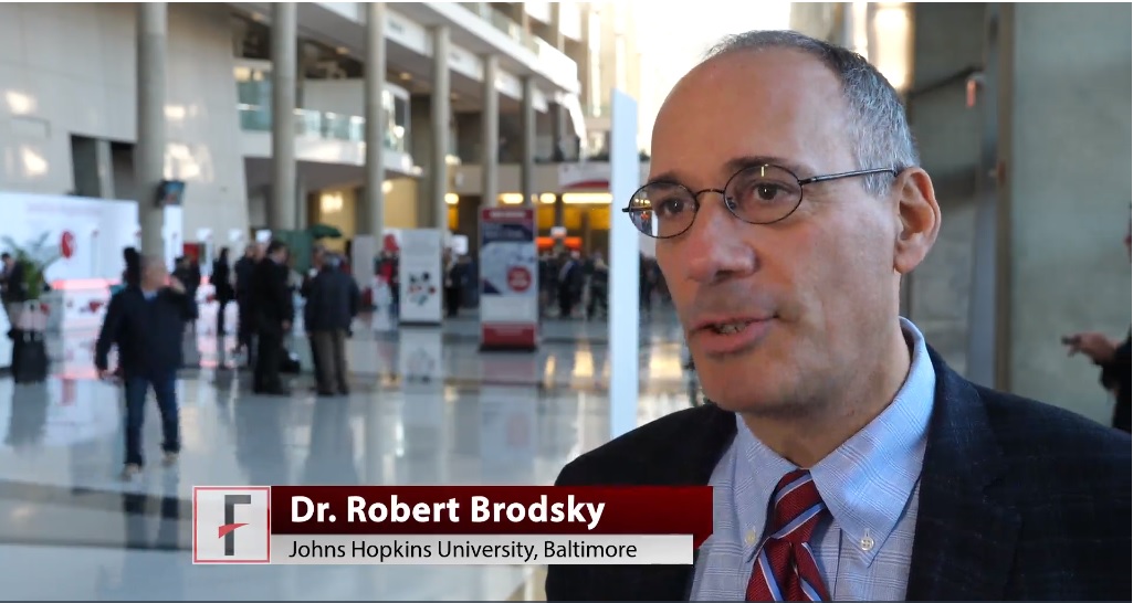
Robert A. Brodsky, MD, director of the division of hematology at Johns Hopkins University in Baltimore and the moderator for the late-breaking abstract session at ASH, highlighted results from two studies.
Data from the MURANO trial showed robust results for a combination of venetoclax and rituximab in patients with relapsed/refractory chronic lymphocytic leukemia (CLL). At a median follow-up of 23.8 months, median progression-free survival -had not been reached in patients randomized to venetoclax/rituximab, while patients who received bendamustine plus rituximab had a median PFS of 17 months.
The based on the data presented, Dr. Brodsky said.
Another “enormously exciting and practice-changing” finding is that direct oral anticoagulants can be used safely in patients with cancer, Dr. Brodsky said in an interview.
In a randomized, open-label study, 12 months of daily treatment with edoxaban was noninferior to standard subcutaneous therapy with dalteparin for treatment of venous thromboembolism in cancer patients.
The video associated with this article is no longer available on this site. Please view all of our videos on the MDedge YouTube channel
ATLANTA – There were a lot of new data presented during the annual meeting of the American Society of Hematology. But what findings could actually change the way you practice?
Robert A. Brodsky, MD, director of the division of hematology at Johns Hopkins University in Baltimore and the moderator for the late-breaking abstract session at ASH, highlighted results from two studies.
Data from the MURANO trial showed robust results for a combination of venetoclax and rituximab in patients with relapsed/refractory chronic lymphocytic leukemia (CLL). At a median follow-up of 23.8 months, median progression-free survival -had not been reached in patients randomized to venetoclax/rituximab, while patients who received bendamustine plus rituximab had a median PFS of 17 months.
The based on the data presented, Dr. Brodsky said.
Another “enormously exciting and practice-changing” finding is that direct oral anticoagulants can be used safely in patients with cancer, Dr. Brodsky said in an interview.
In a randomized, open-label study, 12 months of daily treatment with edoxaban was noninferior to standard subcutaneous therapy with dalteparin for treatment of venous thromboembolism in cancer patients.
The video associated with this article is no longer available on this site. Please view all of our videos on the MDedge YouTube channel
REPORTING FROM ASH 2017
CLL drug in limited supply outside U.S.
Ofatumumab (Arzerra), a monoclonal antibody treatment for chronic lymphocytic leukemia, will soon be available outside the United States through compassionate use programs only. The drug will continue to be widely available in the United States.
Novartis announced in January that it would begin limiting the availability of the drug outside of the United States and would work with regulatory authorities to set up compassionate use programs for patients who are currently being treated with the drug. Patients who use these programs will receive the drug for free.
The decision was driven by the surge in CLL drugs that have become available over the last 5 years, according to Novartis.
The decision to pull the drug from international markets will not affect its use in ongoing clinical trials, particularly two phase 3 studies in relapsing multiple sclerosis and indolent non-Hodgkin lymphoma.
AUDIO: Immunotherapy’s role in NHL
ATLANTA – The use of immune checkpoint blockade is increasingly becoming standard therapy in Hodgkin lymphoma, but this approach has so far garnered mixed results in non-Hodgkin lymphoma, Stephen Ansell, MD, PhD, said at the annual meeting of the American Society of Hematology.
In an interview, Dr. Ansell, professor of medicine and chair of the lymphoma group at the Mayo Clinic, Rochester, Minn., said responses have been variable with promising results from immune checkpoint inhibitors in primary mediastinal large B-cell lymphoma, some NK/T-cell lymphomas, and primary CNS lymphoma. However, responses have been modest in low-grade lymphoma.
Dr. Ansell, who chaired a session at ASH 2017 on immunotherapy’s expanding role in non-Hodgkin lymphoma, said one of the major challenges of using immune checkpoint blockade in non-Hodgkin lymphoma is the complicated biology. For example, there are a lot of regulatory T cells that actually inhibit the immune response, and many of the T cells that are present within the tumor have an exhausted phenotype and are poorly functioning. Additionally, some of the cytokines that would seem to be stimulating the immune system can, over time, slowly produce T-cell exhaustion.
“Sort of like too much of a good thing ends up being a bad thing,” he said.
These are the issues that are fueling research today, Dr. Ansell said. Going forward he said he expects to see more combination approaches to therapy, such as using an agonistic positive signal plus the blocking of an inhibitory signal with chemotherapy.
Dr. Ansell reported that Mayo Clinic receives clinical trial support from Merck, Bristol-Myers Squibb, Seattle Genetics, Trillium, and Affimed.
EXPERT ANALYSIS FROM ASH 2017