User login
CNS involvement predicts relapse but not survival in ARL, study shows
CHICAGO—Investigators have found evidence to suggest that identifying central nervous system (CNS) involvement at diagnosis does not impact overall survival for patients with AIDS-related lymphoma (ARL).
The research showed that ARL patients with CNS involvement at diagnosis were nearly 3 times as likely as their peers to have CNS relapse during cancer treatment.
But there was no significant difference between the 2 groups with regard to survival.
This may be due to the low overall incidence of CNS relapse, the use of insufficient treatments, and/or inadequate methods for identifying patients with CNS involvement, according to the investigators.
Stefan K. Barta, MD, of the Fox Chase Cancer Center in Philadelphia, Pennsylvania, and his colleagues conducted this research and presented the results at the 2014 ASCO Annual Meeting (abstract 8570).
In 2013, Dr Barta led the assembly of a database containing medical data from more than 1500 patients newly diagnosed with ARL who participated in clinical trials in Europe and the US from 1990 through 2010.
In the new study, he and his colleagues used the database to identify 880 patients with ARL whose data included complete information on CNS involvement at diagnosis and CNS relapse.
The team set out to find associations between CNS relapse and patient characteristics, including age, sex, CD4 count, lymphoma subtype, treatment history with combination antiretroviral therapies (cART), rituximab use, and the type of initial chemotherapy.
All of the patients had received either intrathecal therapy for CNS involvement or intrathecal prophylaxis with single-agent or 3-agent regimens. Sixty-nine percent of patients (n=607) had received cART, and 31% (n=276) had received rituximab-containing induction chemoimmunotherapy.
CNS involvement was identified in 13% of patients at diagnosis, including 27% of patients with Burkitt lymphoma or Burkitt-like lymphoma and 6% of patients with diffuse large B-cell lymphoma.
There was no difference in the prevalence of baseline CNS involvement between patients treated before and after the introduction of cART (13% each).
In all, 5.3% of patients experienced CNS relapse at a median of 4.2 months after diagnosis (range, 0.3-19.3 months). This included 12% of patients diagnosed with CNS involvement at baseline and 4% of patients who were not.
The median overall survival after CNS relapse was 1.6 months (range, 0-86.4 months). There was no significant difference in overall survival between patients with CNS involvement at diagnosis and those without it. The hazard ratio was 0.85 (P=0.32).
Multivariate analysis showed the only baseline characteristic significantly associated with the frequency of CNS relapse was CNS involvement, with a hazard ratio of 2.9 (P=0.01). None of the treatments had a significant impact on CNS relapse.
Dr Barta said these results suggest that current treatments are insufficient, and the approaches used to identify CNS involvement may be missing many patients who are at risk of CNS relapse.
“A lot of patients who relapsed probably had undetected CNS involvement at diagnosis,” he said. “We want to figure out if there are better strategies to identify patients at risk.” ![]()
VIDEO: Team approach is best for successful oncofertility clinic
PHILADELPHIA – Whether you want to build an entire oncofertility clinic or just elevate the level of care your current facility offers, an integrated approach to care is key, according to Dr. Leslie A. Appiah, director of oncofertility at the University of Kentucky Medical Center, Lexington.
In a video interview at the annual meeting of the North American Society for Pediatric and Adolescent Gynecology, Dr. Appiah offers suggestions for how oncologists, gynecologists, reproductive endocrinologists, nurse managers, social workers, and others – even when they’re with other institutions – can work together to offer care that adheres to position statements on fertility rights for cancer patients from the American Society of Clinical Oncology and other associations.
Dr. Appiah also discusses how to achieve fluid communication so that fertility can be preserved without delaying cancer treatments, and best practices for oncofertility concerns in the pediatric setting.
On Twitter @whitneymcknight
The video associated with this article is no longer available on this site. Please view all of our videos on the MDedge YouTube channel
AT THE NASPAG ANNUAL MEETING
Novel agent shows promising activity in heavily pretreated NHL

©ASCO/Rodney White
CHICAGO—The novel, oral selective inhibitor of nuclear transport known as selinexor (KPT-330) can safely be given as monotherapy to patients with heavily pretreated non-Hodgkin lymphoma (NHL), according to a presentation at the 2014 ASCO Annual Meeting.
“Selinexor has favorable pharmacokinetic and pharmacodynamic characteristics,” said presenter Martin Gutierrez, MD, of the John Theurer Cancer Center at Hackensack University Medical Center in New Jersey.
Selinexor is a slowly reversible, selective inhibitor of nuclear transport that inhibits XPO1, which is elevated in NHL, chronic lymphocytic leukemia (CLL), and other malignances.
“It shows single-agent anti-tumor activity across all NHL types, with durable cancer control of more than 9 months [and] marked activity across germ cell B (GCB), non-germ cell B, and double-hit diffuse large B-cell lymphoma (DLBCL),” Dr Gutierrez said.
He provided an update of the ongoing phase 1 dose-escalation study at the meeting as abstract 8518.
The study now includes 51 patients, half of whom have NHL. Their median age is 60 years.
The patients received selinexor across 8 dose levels, ranging from 3 mg/m2 to 60 mg/m2. Dosing at 60 mg/m2 twice weekly is ongoing, and the maximum-tolerated dose has not been reached.
Among the 43 evaluable patients, the disease control rate was 74%, the overall response rate was 28%, and the complete response rate was 5%.
“All patients who had their disease controlled had a reduction in lymph nodes and some degree of activity across all dose levels,” Dr Gutierrez said. “GCB and non-GCB patients responded similarly.”
The length of response was up to 632 days in the DLBCL group.
Most adverse events were gastrointestinal in nature, and most of them were grade 1 or 2. The most common adverse events were nausea, anorexia, and fatigue.
The investigators found a higher incidence of side effects in the first treatment cycle. The dosing schedule was interrupted and reduced to maintain steady state levels.
“The results suggest that an intermittent dosing schedule optimally induces a steady state with maximal induction of XPO1 mRNA,” Dr Gutierrez said.
There were 3 dose-limiting toxicities, including 1 multiple myeloma patient with grade 4 thrombocytopenia, 1 follicular lymphoma patient with grade 4 thrombocytopenia, and 1 CLL patient with grade 2 fatigue.
ASCO discussant Owen O’Connor, MD, from Columbia University Medical Center in New York, commented, “These clinical data are interesting with a provocative target. I applaud the investigators for doing a trial across a diversity of B- and T-cell lymphomas . . . . The results suggest a potential effect in a rare subset of lymphoma patients that have little treatment options.”
Frontline trials of selinexor are planned, including patients with Richter transformation and follicular lymphoma.
Selinexor recently received orphan drug status from the US Food and Drug Administration for the treatment of DLBCL. ![]()
Lenalidomide plus R-CHOP tackles thorny DLBCL subtype
CHICAGO – Lenalidomide in combination with R-CHOP chemotherapy induced responses in 98% of patients with newly diagnosed diffuse large B-cell lymphoma in a phase II study.
Among the 60 evaluable patients, 80% had complete responses, and 18% had partial responses.
Moreover, progression-free and overall survival were increased with lenalidomide (Revlimid) in the activated B-cell (ABC) or non–germinal center B-cell (non-GCB) subtype, which is associated with a poor outcome when treated with standard R-CHOP monotherapy.
"It appears the addition of lenalidomide to R-CHOP may ameliorate the negative effect of the non-GCB phenotype on outcome," Dr. Grzegorz Nowakowski said at the annual meeting of the American Society of Clinical Oncology.
About 40% of patients with diffuse large B-cell lymphoma (DLBCL) given R-CHOP every 21 days will relapse or develop refractory disease. For those who do, the response rate for single-agent lenalidomide is about 30%, he said.
The study involved 64 patients with untreated stages II-V CD20-positive DLBCL who received oral lenalidomide 25 mg on days 1-10 with standard-dose R-CHOP (cyclophosphamide, hydroxydaunorubicin, vincristine, and prednisone) chemotherapy every 21 days for six cycles (R2CHOP).
All patients received pegfilgrastim on day 2 of each cycle and aspirin prophylaxis throughout.
At baseline, 25% of patients were older than age 70 years, 60% had stage IV disease, and 52% were International Prognostic Index high intermediate or high risk.
The DLBCL subtype was germinal-center B-cell (GCB) in 51% and non-GCB in 34%, with 16% unknown.
Efficacy was examined by DLBCL subtype and against a matched case-control cohort of 87 consecutive, contemporary DLBCL cases in the Mayo Clinic Lymphoma Database who were treated with R-CHOP alone.
At 2 years, progression-free survival (PFS) and overall survival rates in the R2CHOP group were 59% and 78%, compared with rates of 52% and 65% in the R-CHOP case-matched controls, said Dr. Nowakowski of the Mayo Clinic, Rochester, Minn.
When the case controls were examined by molecular subtype, there was a significant difference between non-GCB and GCB controls treated with standard R-CHOP alone for progression-free survival (28% vs. 64%; P = .00029) and overall survival (46% vs. 74%; P = .000036).
In contrast, non-GCB and GCB treated with R2CHOP with lenalidomide had similar rates of progression (60% vs. 59%; P = 0.83) and overall survival at 2 years (83% vs. 75%; P = .61), he reported.
Similar results were recently seen in elderly DLBCL patients in the phase II Italian REAL07 trial (Lancet Oncol. 2014 Jun;15:730-7), said Dr. Nowakowski and invited discussant Dr. Andre Goy, chief of lymphoma and chair and director, John Theurer Cancer Center, Hackensack (N.J.) University Medical Center.
Lenalidomide offers "very promising activity with R2CHOP, particularly in the non-GC subtype," Dr. Goy said "These data will need to be confirmed in ongoing randomized studies, but might offer a new platform in combination/maintenance post–R-CHOP, particularly in elderly patients because the ABC percentage increases with age and they tend to have a worse outcome."
Dr. Goy observed that lenalidomide increased the incidence of grade 3 and 4 neutropenia and thrombocytopenia, but this did not translate into more infections, serious adverse events, or treatment delays.
Grade 3 febrile neutropenia developed in six patients, grade 4 sepsis in one, hemorrhage in one, and thrombosis in one, Dr. Nowakowski said. One patient died of perforation/sepsis unrelated to treatment.
The ongoing phase II ECOG 1412 trial is utilizing gene expression profiling of DLBCL subtype in its evaluation of the efficacy of R2CHOP vs. R-CHOP, he said.
The study was supported by Celgene. Dr. Nowakowski reported having no financial disclosures. Dr. Goy reported serving in a consultant or advisory role with Celgene and Pharmacyclics and receiving honoraria from Janssen Oncology and Millennium Pharmaceuticals.
AT THE ASCO ANNUAL MEETING 2014
Key clinical point: DLBCL patients with the molecular subtype non-GCB may benefit from the addition of lenalidomide to R-CHOP chemotherapy.
Major finding: Non-GCB and GCB subtypes treated with R-CHOP plus lenalidomide had similar rates of progression (60% vs. 59%; P = .83) and overall survival at 2 years (83% vs. 75%; P = .61)
Data source: A phase II study in 64 newly diagnosed diffuse large B-cell lymphoma patients.
Disclosures: The study was supported by Celgene. Dr. Nowakowski reported having no financial disclosures. Dr. Goy reported serving in a consultant or advisory role with Celgene and Pharmacyclics and receiving honoraria from Janssen Oncology and Millennium Pharmaceuticals.
PET-CT accurately predicts survival of follicular lymphoma patients
CHICAGO – For patients with follicular lymphoma, positron emission tomography–computed tomography performed at the end of induction therapy is strongly predictive of both progression-free and overall survival, a retrospective analysis showed.
The pooled analysis of data on 246 PET-CT scans performed following chemoimmunotherapy in three clinical trials showed that patients with 18-fluorodeoxyglucose (FDG) uptake of 4 or greater on a 5-point scale had a fourfold higher risk for disease progression, compared with patients who became PET negative after induction, reported Dr. Judith Trotman of the University of Sydney (Australia).
At 4.5 years of follow-up, median progression-free survival (PFS) was 16.9 months for patients with a PET uptake of 4 or greater on the 5-point Deauville scale for postinduction response assessment, vs. 74 months for PET-negative patients.
Overall survival at 4.5 years for patients with a higher uptake of FDG PET was 87%, compared with 97% for patients who were PET negative after induction, Dr. Trotman reported at the American Society of Clinical Oncology annual meeting.
"We argue that for the patients who remain PET positive, follicular lymphoma is no longer an indolent histology," Dr. Trotman said.
The study results also showed that conventional CT assessment provides only limited additional value, and that "PET-CT applying the 5-point scale should be the new gold standard for therapeutic response assessment in this lymphoma," she said.
Not so indolent
Although the natural history of follicular lymphoma is a generally indolent course, approximately 15% of patients will die within 5 years of diagnosis, and high-risk scores on the Follicular Lymphoma International Prognostic Index (FLIPI) or its revision (FLIPI2) are not sufficient for predicting which patients are at greatest risk for death, Dr. Trotman said.
Three recent clinical trials reported that positron emission tomography assessment after first-line rituximab-based chemotherapy has good predictive value in patients with high tumor burden follicular lymphoma. Dr. Trotman and his colleagues conducted a pooled analysis of data from the trials with independent review of PET-CT scans to come up with more precise survival estimates and identify the best cutoff for survival using the Deauville scale for response assessment of FDG-avid lymphoma.
The trials were the PRIMA (Primary Rituximab and Maintenance) study of 122 patients, the FOLL05 randomized trial of the Fondazione Italiana Linfomi in 202 patients, and the PET Folliculaire trial in 106 patients.
The Deauville 5-point scale for FDG-avid lymphoma uses the following criteria:
1. No uptake.
2. Uptake less than or equal to mediastinum.
3. Uptake greater than mediastinum but less than or equal to liver.
4. Uptake moderately higher than liver.
5. Uptake markedly higher than liver and/or new lesions.
Dr. Trotman and his colleagues looked at cutoffs of 3 and higher and 4 and higher to see whether they were predictive of prognosis. Reviews of concordance with the trial results, performed by three independent reviewers, showed that a cutoff of 3 or higher had moderate concordance, while a score of 4 or higher had substantial agreement with results.
They then evaluated PET results to see whether they could sharpen the prognostic ability, and found that both cutoffs predicted PFS, but because of the higher concordance score, they chose to focus on the 4+ cutoff.
They found that the hazard ratio (HR) for progression with a score of 4 or greater was 3.9 (P less than .0001), and the hazard ratio for death was 6.7 (P = .0002). Median overall survival in patients with scores of 4 or greater was 79 months, vs. not reached for PET-negative patients.
In multivariate analyses, factors associated with PFS included PET-positive scores of 4 or greater (HR, 3.1; P less than .0001), stable or progressive disease vs. complete responses or complete responses unconfirmed (CR/CRu; HR, 3.7; P = .0013), and partial responses (PRs) vs. CR/CRu (HR, 1.6; P = .04).
Factors associated with OS were PET score and stable/progressive disease vs. CR/CRu (HR, 5.3; P = .05).
"I hope that we can now move on: postinduction PET-CT is a platform for response–adapted therapy, because while achieving PET negativity can better reassure our patients, particularly those otherwise in CRu or PR, the inferior survival of those who remain PET positive compels us to study such PET-response–adapted approaches," Dr. Trotman said.
The invited discussant, Dr. Christopher Flowers of the department of hematology and medical oncology at Emory University, Atlanta, noted that in their analysis, Dr. Trotman and his colleagues included only those scans that were of sufficient quality for central review, and that slightly more than half of all patients had PET scans.
"I think it’s important to try and understand how the PET-available cohort compared to the other clinical trial characteristics of patients to understand whether or not this PET population is a unique population, and [whether] the behavior characteristics may be different from what you might expect from a broader population of patients," he said.
It will be important to see whether the findings will hold up in patients treated with emerging regimens, such as rituximab and bendamustine, and other combinations now in clinical trials, he said.
Dr. Trotman reported having no relevant relationships to disclose. Dr. Flowers disclosed uncompensated consultation from several companies, and receiving research funding from Gilead Sciences, Janssen Pharmaceuticals, Millennium, and Spectrum Pharmaceuticals.
AT THE ASCO ANNUAL MEETING 2014
Key clinical point: For patients with follicular lymphoma, a PET-CT performed at the end of induction therapy is predictive of survival.
Major finding: A PET-CT cutoff score of 4 out of 5 on the Deauville lymphoma-response assessment scale was strongly predictive of both progression-free and overall survival of follicular lymphoma.
Data source: A retrospective analysis of prospectively collected data on 246 patients in three clinical trials.
Disclosures: Dr. Trotman reported having no relevant relationships to disclose. Dr. Flowers disclosed uncompensated consultation from several companies, and receiving research funding from Gilead Sciences, Janssen Pharmaceuticals, Millennium, and Spectrum Pharmaceuticals.
Method reveals new targets of p53

Credit: A.T. Tikhonenko
A novel sequencing technique has allowed researchers to identify direct targets of p53, providing new insight into this gene’s anticancer activity.
The research, published in eLife, revealed nearly 200 genes that were directly regulated by p53, and many of these had never been identified before.
The study’s authors said this work lays the foundation for investigations into which of these genes are necessary for p53’s cancer-killing effects and how cancer cells evade these genes.
The researchers noted that all cancers must deal with p53’s antitumor effects. Generally, there are 2 ways they do this: by mutating p53 directly or by producing the protein MDM2, which inhibits p53 function. With the current study, the team explored the second strategy.
“MDM2 inhibitors, which are through phase 1 human trials, effectively activate p53 but manage to kill only about 1 in 20 tumors,” said study author Joaquín Espinosa, PhD, of the University of Colorado in Boulder.
“The question is why. What else is happening in these cancer cells that allow them to evade p53?”
According to the researchers, the answer is in the downstream effects of p53. The gene sets in motion a cascade of events that lead to cancer cell destruction. But it has been unclear exactly which other genes are directly activated by p53.
To identify genetic targets of p53, Dr Espinosa and his colleagues used a technique called Global Run-On Sequencing (GRO-Seq). Unlike other methods, GRO-Seq measures new RNA being created, not overall RNA levels.
“Many teams around the world have been getting cancer cells, treating them with MDM2 inhibitors, and waiting hours and hours to see what genes turn on, and then, only imprecisely,” Dr Espinosa said. “GRO-Seq lets us do it in minutes, and the discoveries are massive.”
The technique generates a large quantity of data because it requires counting tens of thousands of RNA molecules before and after p53 activation. So this research required designing algorithms to sort through the data, as well as a computational biologist driving a supercomputer.
But the researchers were able to pinpoint new genes directly regulated by p53. And they believe this could aid the future development of cancer-fighting strategies. ![]()
Screening catches breast cancer early in HL survivors
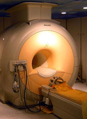
Results of a new study indicate that MRI and mammography can detect invasive breast tumors at very early stages in female survivors of Hodgkin lymphoma (HL).
Researchers said the findings underscore the need for at-risk childhood HL survivors and their physicians to be aware of screening guidelines.
The guidelines recommend survivors undergo breast MRI screening beginning at age 25 or 8 years after they received chest radiation, whichever is later.
“Female survivors of childhood HL who had chest radiation should speak with their family doctor about after-care assessment and breast cancer screening,” said David Hodgson, MD, of Princess Margaret Cancer Centre in Toronto, Canada.
“We estimate that 75% of women who are at high risk because of prior radiotherapy to the chest are not being screened. So my hope is that this new evidence will encourage these survivors to discuss early screening with their doctors.”
Dr Hodgson and his colleagues reported this evidence in Cancer.
The researchers evaluated the results of breast MRI and mammography screening among 96 female survivors of childhood HL who had been treated with chest radiotherapy.
The median patient age at first screening was 30 years, and the median number of MRI screening rounds was 3. Ten breast cancers—half of them invasive tumors—were diagnosed in 9 women during 363 person-years follow up.
The median age at breast cancer diagnosis was 39 years (range, 24 to 43 years), and the median latency period between HL diagnosis and age at breast cancer diagnoses was 21 years (range, 12 to 27 years).
“This illustrates the young age at which these cancers can occur,” Dr Hodgson said. “For some of these women, if they had been screened starting at age 40 or 50, like average-risk women, it would have been too late.”
MRI alone detected tumors with 80% sensitivity and 93.5% specificity. Mammography alone detected tumors with 70% sensitivity and 95% specificity. And both modalities combined detected tumors with 100% sensitivity and 88.6% specificity. All invasive tumors were detected by MRI.
In other words, of the 10 breast tumors, 5 were visible on both MRI and mammogram, 3 were visible only on MRI, and 2 were detected via mammogram alone (but were non-invasive). The median size of invasive tumors size was 8 mm (range, 3-15 mm), and none had spread to the lymph nodes.
The researchers noted that these results are substantially better than prior studies using only mammographic screening in young patients.
Dr Hodgson also pointed out that, because MRI screening is so much more sensitive to small changes in the appearance of the breast tissue than mammography, up to a third of patients may be called back for further testing of suspicious findings. But many of these are benign or not clinically significant and, therefore, require no treatment. ![]()
Group finds a way to target MDSCs

Researchers say they’ve found a way to target myeloid-derived suppressor cells (MDSCs) while sparing other immune cells.
In preclinical experiments, the team showed they could deplete MDSCs—and shrink tumors—using peptide antibodies.
These “peptibodies” wiped out MDSCs in the blood, spleen, and tumor cells of mice without binding to other white blood cells or dendritic cells.
The researchers described this work in Nature Medicine.
“We’ve known about [MDSCs] blocking immune response for a decade but haven’t been able to shut them down for lack of an identified target,” said the paper’s senior author, Larry Kwak, MD, PhD, of The University of Texas MD Anderson Cancer Center in Houston.
“This is the first demonstration of a molecule on these cells that allows us to make an antibody—in this case, a peptide—to bind to them and get rid of them. It’s a brand new immunotherapy target.”
Dr Kwak and his colleagues began this research by applying a peptide phage library to MDSCs, which allowed for a mass screening of candidate peptides that bind to the surface of MDSCs. This revealed 2 peptides, labeled G3 and H6, that bound only to MDSCs.
The researchers fused the 2 peptides to a portion of mouse immune globulin to generate experimental peptibodies called pep-G3 and pep-H6. Both peptibodies bound to both types of MDSCs—monocytic and granulocytic cells.
Dr Kwak and his colleagues then tested the peptibodies in 2 mouse models of thymic tumors, as well as models of melanoma and lymphoma. The team compared pep-G3 and pep-H6 to a control peptibody and an antibody against Gr-1.
Both pep-G3 and pep-H6 depleted monocytic and granulocytic MDSCs in the blood and spleens of all mice. But the Gr-1 antibody only worked against granulocytic MDSCs.
To see whether MDSC depletion would impede tumor growth, the researchers administered the peptibodies to mice with thymic tumors every other day for 2 weeks.
Mice treated with either pep-G3 or pep-H6 had tumors that were about half the size and weight of those in mice treated with the control peptibody or the Gr-1 antibody.
Lastly, the researchers analyzed surface proteins on the MDSCs and found that S100A9 and S100A8 are the likely binding targets for pep-G3 and pep-H6.
Dr Kwak and his colleagues said they are now working to extend these findings to human MDSCs. ![]()
Mutation causes ibrutinib resistance in CLL

Credit: Rhoda Baer
Researchers say they have identified a source of drug resistance in chronic lymphocytic leukemia (CLL).
In a letter to The New England Journal of Medicine, the team described how a mutation in Bruton’s tyrosine kinase (BTK) triggers resistance to ibrutinib, a drug that treats CLL by inhibiting BTK.
The researchers discovered this point mutation in a CLL patient enrolled in a clinical trial. The patient initially responded well to ibrutinib but stopped responding after almost 20 months.
“In a way, we are repeating, at a faster pace, the story of Gleevec [imatinib],” said study author Y. Lynn Wang, MD, PhD, of the University of Chicago in Illinois.
“That story began with development of an effective drug with few side effects, but, in many patients, the leukemia eventually found a way around it after long-term use. So researchers developed second-line drugs to overcome resistance.”
The ibrutinib study began in 2010 at Weill Cornell Medical College in New York, one of several sites for a phase 1 trial of ibrutinib. The researchers recruited 16 patients with CLL whose disease had progressed or relapsed despite multiple treatments.
Dr Wang arranged to track the progress of each patient’s leukemic cells before and during treatment and to correlate any cellular or molecular changes with each patient’s disease activity.
One of the 16 patients in the trial seemed to be unusual. This 61-year-old woman was diagnosed in 2000 at age 49. She had unsuccessfully received several different treatments before entering the study.
Within 18 months of starting ibrutinib, she showed significant improvement. At about 20 months, however, she started to decline, developing a respiratory infection that did not improve with treatment. By 21 months, it was clear she was having a relapse. The clinical team increased her dose, with no discernable effect.
Dr Wang’s team quickly began analyzing her blood samples, looking for changes that occurred between the period when she was responding well to ibrutinib and after she began to relapse.
Because complete gene sequencing would be time consuming, Dr Wang asked a graduate student working on the project, Menu Setty from Memorial Sloan-Kettering in New York, to first focus on 3 proteins that were likely candidates. One of the candidates was BTK.
And sure enough, Setty discovered a tiny but consistent change in BTK in about 90% of post-relapse cells. It was a thymidine-to-adenine mutation at nucleotide 1634 of the BTK complementary DNA, leading to a substitution of serine for cysteine at residue 481 (C481S).
When the researchers later analyzed the entire set of the patient’s genes, they found no other genetic changes that correlated with the patient’s clinical course. BTK made perfect sense as the cause for drug resistance, the researchers noted, as it’s the primary target of ibrutinib binding, and it plays a central role in rapid cell proliferation.
Dr Wang and her colleagues used structural and biochemical measures to confirm that the C481S mutation made CLL cells resistant to ibrutinib. The studies indicated that ibrutinib was 500 times less likely to bind to mutant BTK.
In an attempt to save the patient, the researchers tested alternative kinase inhibitors against the patient’s leukemic cells in the lab.
They found some kinase inhibitors remained effective against ibrutinib-resistant cells. (These studies are described in a separate manuscript that has been submitted for publication.) Unfortunately, despite this effort, the patient passed away a few weeks later, due to sepsis.
The researchers noted that the C481S mutation is one of several mechanisms that underlie resistance to ibrutinib, but this research highlights the mutation’s role in disease development and drug resistance.
Understanding the molecular and cellular mechanisms of resistance is the first step toward monitoring, early detection, and development of novel strategies to overcome drug resistance. ![]()
Wealth appears to affect distribution of cancer types
Credit: Rhoda Baer
Results of a large study suggest certain malignanices are more concentrated in areas with high levels of poverty, while other cancer types arise more often in wealthy regions.
The research, conducted using data from nearly 3 million US cancer cases, also indicated that areas with higher poverty levels tended to have a lower cancer incidence but higher mortality rate than areas with lower poverty levels.
Researchers reported these findings in the journal Cancer.
Francis Boscoe, PhD, of the New York State Cancer Registry in Albany, and his colleagues conducted this research to evaluate the role of socioeconomics in cancer incidence and mortality.
So the team compared individuals living in areas with the highest poverty levels to those living in areas with the lowest poverty levels.
The researchers collected information on nearly 3 million cancer cases diagnosed between 2005 and 2009 in16 states, plus Los Angeles (an area covering 42% of the US population). Cases were divided into 1 of 4 groupings based on the poverty rate of the residential census tract at the time of diagnosis.
For all malignancies combined, there was a negligible association between cancer incidence and poverty. However, 32 of 39 cancer types showed a significant association with poverty.
Fourteen cancers were positively associated with poverty, and 18 were negatively associated. Myeloma, Hodgkin lymphoma, and non-Hodgkin lymphoma (NHL) were among the negatively associated malignancies.
The cancers most significantly associated with higher poverty levels were Kaposi sarcoma and cancers of the larynx, cervix, penis, and liver. The cancers most significantly associated with lower poverty levels were melanoma, thyroid cancer, other non-epithelial skin cancers, and testis cancer.
Overall, male malignancy rates were more sensitive to poverty level than female rates. And, in general, the race-specific cancer incidence rates differed, but the poverty gradients were similar.
For example, there was a roughly 20-fold difference in rates of melanoma between white and black individuals, but the relationship between poverty and incidence remained regardless of race.
This was not true for NHL, however. Among black individuals, the incidence of NHL increased as the poverty level increased. But among white individuals, the incidence of NHL decreased as the poverty level increased.
The researchers pointed out that—for all cancer sites, races, and sexes combined—the difference in risk between the greatest and lowest poverty category was less than 2%.
“At first glance, the effects seem to cancel one another out,” Dr Boscoe said. “But the cancers more associated with poverty have lower incidence and higher mortality, and those associated with wealth have higher incidence and lower mortality. When it comes to cancer, the poor are more likely to die of the disease, while the affluent are more likely to die with the disease.”
Dr Boscoe noted that recent gains in technology have made it much easier to link patient addresses with neighborhood characteristics, therefore making it possible to incorporate socioeconomic status into cancer surveillance.
“Our hope is that our paper will illustrate the value and necessity of doing this routinely in the future,” he concluded. ![]()

Credit: Rhoda Baer
Results of a large study suggest certain malignanices are more concentrated in areas with high levels of poverty, while other cancer types arise more often in wealthy regions.
The research, conducted using data from nearly 3 million US cancer cases, also indicated that areas with higher poverty levels tended to have a lower cancer incidence but higher mortality rate than areas with lower poverty levels.
Researchers reported these findings in the journal Cancer.
Francis Boscoe, PhD, of the New York State Cancer Registry in Albany, and his colleagues conducted this research to evaluate the role of socioeconomics in cancer incidence and mortality.
So the team compared individuals living in areas with the highest poverty levels to those living in areas with the lowest poverty levels.
The researchers collected information on nearly 3 million cancer cases diagnosed between 2005 and 2009 in16 states, plus Los Angeles (an area covering 42% of the US population). Cases were divided into 1 of 4 groupings based on the poverty rate of the residential census tract at the time of diagnosis.
For all malignancies combined, there was a negligible association between cancer incidence and poverty. However, 32 of 39 cancer types showed a significant association with poverty.
Fourteen cancers were positively associated with poverty, and 18 were negatively associated. Myeloma, Hodgkin lymphoma, and non-Hodgkin lymphoma (NHL) were among the negatively associated malignancies.
The cancers most significantly associated with higher poverty levels were Kaposi sarcoma and cancers of the larynx, cervix, penis, and liver. The cancers most significantly associated with lower poverty levels were melanoma, thyroid cancer, other non-epithelial skin cancers, and testis cancer.
Overall, male malignancy rates were more sensitive to poverty level than female rates. And, in general, the race-specific cancer incidence rates differed, but the poverty gradients were similar.
For example, there was a roughly 20-fold difference in rates of melanoma between white and black individuals, but the relationship between poverty and incidence remained regardless of race.
This was not true for NHL, however. Among black individuals, the incidence of NHL increased as the poverty level increased. But among white individuals, the incidence of NHL decreased as the poverty level increased.
The researchers pointed out that—for all cancer sites, races, and sexes combined—the difference in risk between the greatest and lowest poverty category was less than 2%.
“At first glance, the effects seem to cancel one another out,” Dr Boscoe said. “But the cancers more associated with poverty have lower incidence and higher mortality, and those associated with wealth have higher incidence and lower mortality. When it comes to cancer, the poor are more likely to die of the disease, while the affluent are more likely to die with the disease.”
Dr Boscoe noted that recent gains in technology have made it much easier to link patient addresses with neighborhood characteristics, therefore making it possible to incorporate socioeconomic status into cancer surveillance.
“Our hope is that our paper will illustrate the value and necessity of doing this routinely in the future,” he concluded.
