User login
Chemo regimen can be ‘highly effective’ against ENKTL
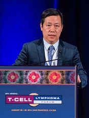
Photo by Larry Young
SAN FRANCISCO—A 3-agent chemotherapy regimen can be “highly effective” in patients with extranodal natural killer/T-cell lymphoma (ENKTL), according to researchers.
In a single-center study, this regimen—pegaspargase, gemcitabine, and oxaliplatin (P-GEMOX)—followed by extensive involved-field radiotherapy (EIFRT) produced high rates of long-term overall survival (OS) and progression-free survival (PFS) in newly diagnosed patients with stage I/II ENKTL.
P-GEMOX also proved effective—though to a much lesser degree—in advanced, relapsed, or refractory ENKTL, and these patients appeared to benefit from autologous stem cell transplant (auto-SCT) as consolidation.
Toxicity associated with P-GEMOX was mild to moderate and tolerable, according to Hui-Qiang Huang, MD, PhD, of State Key Laboratory of Oncology in Southern China, Guangzhou, China.
Dr Huang presented these results at the 8th Annual T-cell Lymphoma Forum.
Newly diagnosed patients
Dr Huang and his colleagues studied 56 patients newly diagnosed with stage I/II, nasal-type ENKTL. Most patients were younger than 60 years of age (80.4%, n=45).
About 79% (n=44) had an ECOG status of 0, and 21.4% (n=12) had a status of 1. About 61% (n=34) had stage I disease, and 39.3% (n=22) had stage II.
All patients received P-GEMOX—gemcitabine at 1000 mg/m2 on days 1 and 8, oxaliplatin at 150 mg/m2 on day 1, and pegaspargase at 2000 U/m2 on day 1. Doses could be adjusted in the event of toxicity.
The regimen was repeated every 3 weeks for a maximum of 4 cycles. Patients then underwent EIFRT—56 Gy in 28 fractions over 4 weeks.
The overall response rate (ORR) after P-GEMOX was 89.3% (50/56). Thirty-five patients achieved a complete response (CR), 15 had a partial response (PR), and 4 had stable disease (SD).
After EIFRT, the ORR increased to 94.6% (53/56). Fifty patients had a CR, 3 had a PR, and 1 had SD.
The median follow-up was 35.2 months (range, 10.6-51.4). Six patients relapsed, and the median time to relapse was 6.2 months.
Five patients died of disease progression. The median time to death was 10.9 months after the completion of EIFRT.
The 4-year OS rate was 90.7±4.0%, and the 4-year PFS rate was 89.1±4.2%.
OS and PFS were superior in patients with stage I disease as compared to stage II (P=0.056 and 0.023, respectively). And OS and PFS were superior in patients who responded to P-GEMOX (P=0.004 and 0.001, respectively).
There were no treatment-related deaths. The most common toxicities (occurring in more than 50% of patients) after P-GEMOX were neutropenia (80.3%), thrombocytopenia (55.3%), and hypoproteinemia (75.0%).
The most common grade 3/4 toxicities (occurring in more than 10% of patients) were granulocytosis (23.2%), thrombocytopenia (19.6%), and hypoproteinemia (10.7%).
Advanced & relapsed/refractory patients
Dr Huang and his colleagues also studied 60 patients with newly diagnosed, stage III/IV ENKTL (25%, n=15), relapsed ENKTL (21.7%, n=19), or refractory disease (43.3%, n=26). Seventy percent of these patients (n=42) had nasal-type ENKTL.
Most patients were younger than 60 years of age (91.7%, n=55). About 73% (n=44) had an ECOG status of 0-1, and 26.7% (n=16) had a status of 2. Fifteen percent of patients (n=9) had stage I disease, 16.7% (n=10) had stage II, 35% (n=21) had stage III, and 33.3% (n=20) had stage IV.
The patients received the same P-GEMOX regimen as the newly diagnosed, stage I/II patients, but they did not receive EIFRT, and responders could undergo auto-SCT.
For the whole cohort, the ORR after P-GEMOX was 70% (42/60). Twenty-one patients had a CR, 21 had a PR, and 9 had SD.
In the newly diagnosed patients, the ORR was 80% (12/15). Four patients had a CR, 8 had a PR, and 2 had SD. In the relapsed/refractory patients, the ORR was 66.7% (30/45). Seventeen patients had a CR, 13 had a PR, and 7 had SD.
The 4-year OS was 43.0±7.3%, and the 4-year PFS was 36.5±6.9%.
There was no significant difference in OS or PFS between the newly diagnosed and relapsed/refractory patients (P=0.653 and 0.825, respectively). However, there was a significant difference in PFS and OS between responders and non-responders (P<0.001 for both).
There was a difference in 3-year OS between patients who went on to auto-SCT and those did not, although it did not reach statistical significance (P=0.08). Eleven patients who achieved a CR went on to auto-SCT.
There were no treatment-related deaths. The most common toxicities (occurring in more than 50% of patients) after P-GEMOX were neutropenia (85%), hypoproteinemia (88.3%), anemia (71.6%), fibrinogen decrease (68.3%), and anorexia (53.3%).
The most common grade 3/4 toxicities (occurring in more than 10% of patients) were neutropenia (31.6%), hypoproteinemia (13.3%), and thrombocytopenia (11.7%).
Dr Huang said this research suggests P-GEMOX can be effective for patients with newly diagnosed or previously treated ENKTL. The next step is to investigate which novel agents could be added to the regimen to improve its efficacy. ![]()
Dual inhibitor could treat ATLL
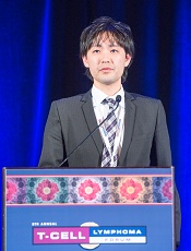
Photo by Larry Young
SAN FRANCISCO—Preclinical research suggests a compound that inhibits both EZH1 and EZH2 could be effective against adult T-cell leukemia/lymphoma (ATLL).
The compound, known as OR-S1, has demonstrated activity against ATLL in vitro and in vivo.
Researchers said OR-S1 reversed epigenetic disruption in ATLL cells, selectively eliminated both ATLL cells and cells infected with human T-cell leukemia virus type I (HTLV-1), and inhibited tumor growth in mouse models of ATLL.
Based on these results, the researchers are planning a phase 1 study of the compound.
Makoto Yamagishi, PhD, of The University of Tokyo in Japan, described the preclinical research with OR-S1 and discussed the rationale for developing the compound at the 8th Annual T-cell Lymphoma Forum. The work was carried out in collaboration with Daiichi Sankyo Co., Ltd.
“We do not precisely understand the molecular mechanism of ATLL development, including genetic and epigenetic abnormalities,” Dr Yamagishi noted.
To gain some insight, he and his colleagues performed microRNA profiling, gene expression profiling, and histone methylation/epigenetic factor profiling on cells from ATLL patients and CD4+ T cells from healthy donors.
The team found that PRC2 factors were significantly upregulated in ATLL. EZH2 was the most upregulated histone methyltransferase, but ATLL cells did not have active mutations in the EZH2 gene. Dr Yamagishi said this suggests EZH2 upregulation is critical for the ATLL-specific epigenome.
“At long last, we determined the epigenetic pattern of ATLL,” he said. “ATLL cells showed specific and significant reprogramming of the epigenome, especially H3K27me3 gain. We found abnormal H3K27me3 change in half of genes, and gain was dominant.”
“But, interestingly, the methylated genes are specific in ATLL and do not overlap with other EZH2-dependent cell types, such as embryonic stem cells and diffuse large B-cell lymphoma cells. So ATLL has a very unique epigenome.”
Further investigation revealed that both EZH1 and EZH2 contribute to ATLL-specific epigenetic deregulation. More than 80% of H3K27me3 accumulated genes are occupied by EZH1 and/or EZH2.
So the researchers decided to examine the effects of knocking down EZH1 and EZH2 in ATLL cells.
Compared with knockdown of either gene alone, double knockdown synergistically influenced target gene expression. It led to complete dysfunction of the Polycomb family and had a significant impact on ATLL cell survival.
The researchers also found that EZH1 depletion enhanced ATLL cells’ sensitivity to the EZH2 inhibitor GSK126.
So the team decided to develop a dual EZH1/EZH2 inhibitor. They created OR-S1, which showed “strong activity” against EZH1 and EZH2 but none of the other histone methyltransferases tested.
In in vitro experiments, OR-S1 completely removed H3K27me3 and significantly reduced cell growth in the ATLL-derived cell line TL-Om1.
The drug also reduced cell viability in primary ATLL cells. All 15 samples tested proved sensitive to OR-S1. In addition, OR-S1 treatment selectively removed HTLV-1-infected cells from samples taken from 16 asymptomatic carriers.
Finally, OR-S1 proved active in mice. The drug prevented engraftment of ATLL cells in immunocompromised mice. All 6 OR-S1-treated mice were alive and tumor-free at 49 days, whereas 5 of 6 control mice had died (P=0.0041).
In mice treated after ATLL cell engraftment, OR-S1 reduced tumor growth without causing notable weight loss.
“Synthetic lethality by targeting EZH1 and EZH2 is promising [for ATLL],” Dr Yamagishi said. “Toxicity tests suggest the EZH1/2 dual inhibitor may be sufficient for clinical use, so we are now planning a phase 1 study.” ![]()
Immunotherapy proves active against MF, SS
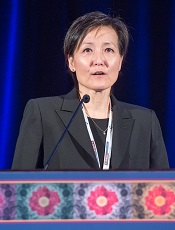
Photo by Larry Young
SAN FRANCISCO—The PD-1-blocking antibody pembrolizumab can produce “significant objective clinical responses” in patients with relapsed or refractory cutaneous T-cell lymphoma, according to researchers.
The drug elicited partial responses in 33% of patients enrolled in a phase 2 study. Half of the responders had mycosis fungoides (MF), and half had Sézary syndrome (SS).
All responses are ongoing, and a few patients with stable disease remain on treatment, so they may convert to partial responses, according to Youn Kim, MD, of Stanford University School of Medicine in California.
Dr Kim presented this research at the 8th Annual T-cell Lymphoma Forum.
The study was conducted by the Cancer Immunotherapy Trials Network (CITN) and supported by the National Cancer Institute and Merck, the company developing pembrolizumab.
“There’s good rationale for immune checkpoint blockade in [MF and SS],” Dr Kim said. “There’s systemic and local immune impairment in MF and Sézary, and there’s mounting evidence that T-cell immunity is critical for meaningful antitumor response.”
“[T]umor-infiltrating CD8+ T cells [have been] associated with improved survival, and therapies which augment T-cell function are effective in [MF and SS]. PD-1 and PD-L1 are very well expressed in the tissue and blood, [and] there’s good genomic evidence of immune evasion in [MF and SS].”
With all this in mind, Dr Kim and her colleagues conducted their phase 2 trial of pembrolizumab in 24 patients with relapsed or refractory MF/SS. Patients were excluded if they had central nervous system disease, autoimmune disease, immunodeficiency, or had received immunosuppressive therapy within 7 days.
The patients’ median age was 67 (range, 44-85), and most were male (75%). Thirty-eight percent of patients (n=9) had MF, and 62% (n=15) had SS. Twelve percent (n=3) had large-cell transformation.
Most patients had Stage IVA disease (62%, n=15), followed by IIIB (13%, n=3), IIIA (13%, n=3), IIB (8%, n=2), and IB (4%, n=1). The median number of prior systemic therapies was 4 (range, 1-10).
Treatment and response
Patients received pembrolizumab at 2 mg/kg intravenously every 3 weeks and were allowed to continue therapy for up to 2 years. Dr Kim noted that patients could continue treatment even after the initial documentation of progressive disease (PD) due to the possibility of immune-mediated flare reactions.
“So it’s the investigator’s decision to allow treatment beyond the initial PD,” she said. “However, if there’s confirmation of PD, those people will be removed.”
The median follow-up was 21 weeks (range, 7-39). Eight patients responded to treatment (according to global response criteria), all of which were partial responses. Four of the responders had MF, and 4 had SS. Responses occurred across all disease stages except IB.
“The range of prior therapies varied in the responders,” Dr Kim noted. “People think [patients tend to respond to] immunotherapy [if they are only] mildly [pre-]treated, but that was not the case. Heavily treated patients had great responses to pembrolizumab.”
All responses are ongoing, with a median duration of 13+ weeks (range, 3+ to 30+). The median time to response was 11 weeks (range, 8-22).
Dr Kim noted that 1 responder discontinued treatment because of a severe adverse event, but this patient remains in response without having received subsequent treatment.
The median best mSWAT (modified Skin Weighted Assessment Tool) reduction was 16%. Two patients had near-complete responses in the skin, and 2 patients with stable disease in the skin continue to improve.
Eleven of the 15 SS patients had measurable Sézary burden pre-treatment. And 3 of these patients had a greater than 50% reduction in Sézary count after treatment.
Four patients with stable disease are still on treatment. And at 20 weeks, 75% of patients are progression-free, according to Kaplan-Meier estimates.
Adverse events
Drug-related adverse events occurring at least twice included skin eruptions (21%, n=5), anemia (13%, n=3), decrease in white blood cell count (8%, n=2), elevated liver tests (8%, n=2), diarrhea (8%, n=2), fever (8%, n=2), and face edema (8%, n=2).
Grade 3/4 adverse events included skin eruptions (8%, n=2), anemia (8%, n=2), elevated liver tests (4%, n=1), and face edema (4%, n=1). Skin eruptions (of all grades) included exfoliative dermatitis (n=2), immune-mediated skin flare (n=2), and excessive peeling/edema (n=1).
There were no drug-related serious adverse events. The cause of the aforementioned serious adverse event (which prompted the responding patient to discontinue treatment) could not be determined.
There were 4 patients who did not report any adverse events, regardless of attribution.
In closing, Dr Kim said it is important to conduct biomarker correlative studies to understand the tumor escape mechanisms and enrich the response population.
She and her colleagues at CITN are now exploring the use of pembrolizumab in combination therapy. They are considering combining the drug with interferon-gamma, interleukin-12, low-dose total skin radiation, intratumoral ipilimumab, or Toll-like receptor agonists. ![]()
Mutations may impact response to HDACis in PTCL-NOS
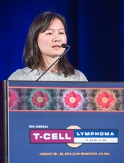
Photo by Larry Young
SAN FRANCISCO—Preclinical research has revealed mutations that may affect the performance of histone deacetylase inhibitors (HDACis) in patients with peripheral T-cell lymphoma-not otherwise specified (PTCL-NOS).
The researchers identified histone-modifying gene mutations in patients with PTCL-NOS and found evidence to suggest these mutations confer shorter survival.
The team also conducted experiments in Jurkat cells showing that certain HDACis could counteract loss-of-function mutations, while others could not.
Meng-Meng Ji, MD, PhD, of the Shanghai Institute of Hematology in China, presented this research at the 8th Annual T-cell Lymphoma Forum.
Dr Ji and her colleagues first performed targeted sequencing in tumor samples from 105 patients newly diagnosed with PTCL-NOS.
The team discovered 62 mutations of “important” lymphoma-associated histone-modifying genes in 31 patients. They found mutations in MLL2 (n=21), TET2 (n=17), EP300 (n=8), CREBBP (n=8), and SETD2 (n=6).
Clinical data revealed a significant difference in overall survival between patients who had these mutations and those who did not (P=0.0038).
Because most of the mutations they identified are loss-of-function mutations, Dr Ji and her colleagues wanted to determine whether HDACis could restore the expression of the mutated genes. So they tested 4 HDACis—valproic acid, vorinostat, romidepsin, and chidamide—in Jurkat cells.
All 4 HDACis upregulated expression of EP300 and CREBBP. However, only romidepsin and chidamide upregulated MLL2, and only valproic acid and vorinostat upregulated SETD2. Dr Ji said there was no obvious change in TET2 expression with any of the HDACis.
The researchers then took a closer look at the EP300, MLL2, and SETD2 mutations. They found that most EP300 mutations were located on the HAT domain. MLL2 mutations could be found in a variety of locations, but some were located on the SET domain. And most SETD2 mutations were located on the SET domain.
Based on the crystal structure of each gene, the team found that EP300 mutations on the HAT domain and both MLL2 mutations and SETD2 mutations located on the SET domain induce loss of function.
So the researchers constructed a mutant for EP300 (p.H1377R), MLL2 (p.V5389M), and SETD2 (p.R1598_) and transfected Jurkat cells with each mutant.
The mutants reduced gene expression significantly when compared to wild-type cells.
Like in the previous experiments, all 4 HDACis could restore the expression of EP300. But only romidepsin and chidamide could restore MLL2 expression, and only valproic acid and vorinostat could restore SETD2 expression.
In addition, all 4 HDACis restored H3K18 hypoacetylation, which was inhibited in Jurkat cells transfected with the EP300 mutant.
Romidepsin and chidamide restored H3K4me3 expression, which was inhibited by the MLL2 mutant. And valproic acid and vorinostat restored H3K36me3 expression, which was inhibited by the SETD2 mutant.
“HDACis targeted differently histone H3 acetylation or methylation modulated by the mutations, suggesting their distinct therapeutic efficiency in PTCL-NOS,” Dr Ji noted.
She said she and her colleagues are continuing this research in samples from patients with PTCL-NOS. ![]()
Lenalidomide shows promise for treating ATLL
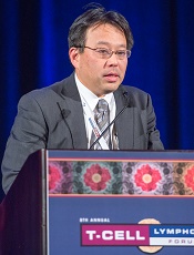
Photo by Larry Young
SAN FRANCISCO—Results of a phase 2 trial suggest lenalidomide may be a treatment option for patients with relapsed adult T-cell leukemia-lymphoma (ATLL).
Lenalidomide produced a 42% overall response rate (ORR) in this trial, and patients had a “favorable” median overall survival, according to Kisato Nosaka, MD, PhD, of the National Center for Global Health and Medicine in Tokyo, Japan.
He noted, however, that the survival data are still immature and may have been confounded by subsequent therapies.
Dr Nosaka presented these results at the 8th Annual T-cell Lymphoma Forum. The study was sponsored by Celgene K.K.
The trial included 26 Japanese patients with relapsed ATLL. Fifty-eight percent of patients had the acute subtype, 27% had the lymphoma subtype, and 15% had the unfavorable chronic subtype.
The patients’ median age was 68.5 (range, 53-81), and 65% of patients were 65 or older. Fifty percent of patients had an ECOG performance status of 0, 35% had a status of 1, and 15% had a status of 2.
Most patients had a low-risk simplified ATL-PI score (65%), but 35% had an intermediate-risk score. Fifteen percent had bone marrow involvement.
The median number of prior treatment regimens was 2 (range, 1-4). Forty-two percent of patients had prior mogamulizumab, and 35% had received LSG15 or modified LSG15.
Patients received lenalidomide at 25 mg per day, given continuously until disease progression or intolerability.
Safety
The median duration of treatment was 3.7 months (range, 0.4 to 18.3 months). There were no deaths during treatment or for 28 days after.
Nine patients (35%) experienced serious adverse events (AEs), but only 1 serious AE occurred in more than 1 patient. Two patients had serious thrombocytopenia.
The most frequent AEs were thrombocytopenia (77%), neutropenia (73%), lymphopenia (69%), and increased C-reactive protein (42%). The most frequent grade 3 or higher AEs were neutropenia (65%), leukopenia (38%), and lymphopenia (38%).
Response and survival
The median follow-up was 3.9 months. The ORR was 42%, including 5 complete responses/unconfirmed complete responses and 6 partial responses. Eight patients (31%) had stable disease, and 7 (27%) progressed.
Dr Nosaka noted that responses occurred in all disease subtypes and at all disease sites. The ORR was 33% (5/15) for the acute subtype, 50% (2/4) for the unfavorable chronic subtype, and 57% (4/7) for the lymphoma subtype.
The ORR was 31% (5/16) at the target lesion, 60% (6/10) in the peripheral blood, and 75% (6/8) for PGA (Physician’s Global Assessment of Clinical Condition, used to assess skin lesions).
Dr Nosaka also pointed out that the ORR was higher in patients who did not receive prior mogamulizumab (60%) than in patients who did (18%). However, he said the number of patients was too small for a definitive conclusion to be reached.
Similarly, the ORR was higher for patients in the low-risk simplified ATL-PI risk group than for those in the intermediate-risk group—53% and 22%, respectively.
Among the 11 responders, the median duration of response was not reached (range, 0.5 months to not reached). The mean duration of response was 5.2 months (range, 0 to 16.6 months).
The median progression-free survival was 3.8 months (range, 1.9 months to not reached). The median overall survival was 20.3 months (range, 9.1 months to not reached).
The overall survival was longer for patients in the low-risk ATL-PI group than the intermediate-risk group—not reached and 10.1 months, respectively (P=0.03).
In closing, Dr Nosaka said these results support lenalidomide as a possible treatment option for patients with relapsed/recurrent ATLL. ![]()
Study suggests chidamide could treat rel/ref CTCL too

Photo by Larry Young
SAN FRANCISCO—The oral histone deacetylase inhibitor chidamide can elicit responses in patients with relapsed or refractory cutaneous T-cell Lymphoma (CTCL), a new study suggests.
Chidamide has already demonstrated efficacy against relapsed or refractory peripheral T-cell lymphoma and has been approved for this indication in China.
Now, results of a phase 2 trial suggest chidamide might be a feasible treatment option for relapsed or refractory CTCL as well.
Yuankai Shi, MD, PhD, of the Chinese Academy of Medical Science & Peking Union Medical College in Beijing, China, discussed this trial at the 8th Annual T-cell Lymphoma Forum. The trial is sponsored by Chipscreen Biosciences Ltd.
He presented results observed in 50 patients with relapsed/refractory CTCL. They had a median age of 47 (range, 26-75), and half were male. Most patients had stage II disease (44%), 20% percent had stage III, and 18% each had stage I and IV. The median time from diagnosis was 2 years.
Patients were randomized to receive chidamide at 30 mg twice a week for 2 weeks out of a 3-week cycle (n=12), 4 weeks out of a 6-week cycle (n=13), or 30 mg twice a week without a drug-free holiday (n=25).
The objective response rate was 32% for the entire cohort, 33% for the 3-week cycle arm, 23% for the 6-week cycle arm, and 36% for the successive dosing arm.
There was 1 complete response, and it occurred in the successive dosing arm. There were 15 partial responses—4 in the 3-week arm, 3 in the 6-week arm, and 9 in the successive dosing arm.
The median duration of response was 92 days overall (range, 78-106), 50 days in the 3-week arm (range, 26-130), 92 days in the 6-week arm (range, 84-99), and 169 days in the successive dosing arm (range, 58-279).
The median progression-free survival was 85 days overall (range, 78-92), 84 days in the 3-week arm (range, 43-126), 81 days in the 6-week arm (range, 39-222), and 88 days in the successive dosing arm (range, 58-261).
Dr Shi noted that the major toxicities associated with chidamide were hematologic and gastrointestinal in nature, and they were controllable.
The incidence of adverse events (AEs) was 83% overall, 92% in the 3-week arm, 85% in the 6-week arm, and 77% in the successive dosing arm. The incidence of grade 3 or higher AEs was 23%, 23%, 38%, and 15%, respectively.
The most common AEs were thrombocytopenia (33%, 39%, 23%, and 35%, respectively), leucopenia (29%, 54%, 31%, and 15%, respectively), fatigue (17%, 23%, 23%, and 12%, respectively), nausea (13%, 23%, 8%, and 12%, respectively), diarrhea (10%, 8%, 8%, and 12%, respectively), fever (8%, 0%, 15%, and 8%, respectively), and anemia (8%, 8%, 15%, and 4%, respectively).
There were 2 serious AEs. One patient in the 3-week arm was hospitalized for fever and lung infection, and 1 patient in the successive dosing arm was hospitalized for hyperglycemia.
Dr Shi said these results suggest chidamide is effective and tolerable for patients with relapsed/refractory CTCL. And, based on the overall profiles of the 3 dosing regimens, successive dosing of chidamide at 30 mg twice a week is recommended. ![]()
NICE issues draft guideline for NHL

a cancer patient
Photo courtesy of NCI/
Mathews Media Group
The National Institute for Health and Care Excellence (NICE) has issued a draft guideline for the diagnosis and management of non-Hodgkin lymphoma (NHL).
The guideline, which is open for consultation, covers adults and young people who are referred to secondary care with suspected NHL or who have newly diagnosed or relapsed NHL.
It contains recommendations for the management of 6 different NHL subtypes—diffuse large B-cell lymphoma, mantle cell lymphoma, follicular lymphoma, MALT lymphoma, Burkitt lymphoma, and peripheral T-cell lymphoma.
The guideline considers which method of biopsy is most appropriate, which diagnostic test most suitable, how the stage of disease is best assessed, and what treatment is likely to be most effective.
It also proposes recommendations for how best to support patients who complete their treatment. These include the provision of end-of-treatment summaries to be discussed with the patient and an increase in education on the possible relapse/late side-effects of their treatment.
“This draft guideline is now open for consultation,” said Mark Baker, director of the Centre of Clinical Practice at NICE.
“We want to hear from patients and all those who provide care for people with non-Hodgkin’s lymphoma in the NHS [National Health Service] so that we can produce a guideline which will support everyone who diagnoses, treats, and has to live with this disease.”
The consultation closes on March 11, 2016, with the final guideline expected in the summer. ![]()

a cancer patient
Photo courtesy of NCI/
Mathews Media Group
The National Institute for Health and Care Excellence (NICE) has issued a draft guideline for the diagnosis and management of non-Hodgkin lymphoma (NHL).
The guideline, which is open for consultation, covers adults and young people who are referred to secondary care with suspected NHL or who have newly diagnosed or relapsed NHL.
It contains recommendations for the management of 6 different NHL subtypes—diffuse large B-cell lymphoma, mantle cell lymphoma, follicular lymphoma, MALT lymphoma, Burkitt lymphoma, and peripheral T-cell lymphoma.
The guideline considers which method of biopsy is most appropriate, which diagnostic test most suitable, how the stage of disease is best assessed, and what treatment is likely to be most effective.
It also proposes recommendations for how best to support patients who complete their treatment. These include the provision of end-of-treatment summaries to be discussed with the patient and an increase in education on the possible relapse/late side-effects of their treatment.
“This draft guideline is now open for consultation,” said Mark Baker, director of the Centre of Clinical Practice at NICE.
“We want to hear from patients and all those who provide care for people with non-Hodgkin’s lymphoma in the NHS [National Health Service] so that we can produce a guideline which will support everyone who diagnoses, treats, and has to live with this disease.”
The consultation closes on March 11, 2016, with the final guideline expected in the summer.
New insight into CLL development

Photo by Graham Colm
New research indicates that chronic lymphocytic leukemia (CLL) can develop during nearly any stage of B-cell maturation.
However, CLL that arises from more progressive maturation stages responds better to therapy.
The study also suggests that most methylation events that were previously thought to be tumor-specific are normally present in non-malignant B cells.
These findings were published in Nature Genetics.
Christoph Plass, PhD, of the German Cancer Research Center (DKFZ) in Heidelberg, and his colleagues conducted this study to determine which development stage of B cells marks the origin of B-cell CLL.
The team took blood samples from 268 CLL patients, separated the blood cells using specific B-cell maturation markers and analyzed the methylation patterns of each individual maturation stage.
The investigators were surprised to find that CLL can develop from almost all maturation stages. They also found that maturation was associated with “increasingly favorable clinical outcomes.”
In addition, methylation patterns that were previously regarded as cancer-specific actually reflect the characteristic patterns of the development stages at the moment of cancerous transformation.
The investigators found that the cell “freezes” this methylation pattern, and this is followed by only a few changes that are truly cancer-specific.
The team said they used advanced bioinformatic methods to calculate the small percentage of cancer-specific methylation patterns from the wealth of maturation-related variations.
“Up until recently, it was technically impossible to study the various maturation stages in such detail as we have done,” Dr Plass said. “It took the advanced sequencing technology and the powerful bioinformatic methods that we have available now to make such a detailed comparison possible.”
The investigators said their findings differ from those of prior studies because, with the current study, they compared CLL cells with the whole pool of B-cell maturation stages.
“All differences found were attributed to cancer,” Dr Plass said, adding that some previous works on the cancer epigenome will need to be re-interpreted in the light of the current results.
Next, Dr Plass and his colleagues want to examine other cancer types to determine whether methylation patterns that are thought to be cancer-specific also arise from the normal cellular maturation program. In particular, they plan to study other hematologic malignancies and prostate cancer.
Drug granted another breakthrough designation for CLL

The US Food and Drug Administration (FDA) has granted breakthrough therapy designation to the BCL-2 inhibitor venetoclax when given with rituximab to treat patients with relapsed or refractory chronic lymphocytic leukemia (CLL).
Venetoclax already had breakthrough designation from the FDA as single-agent treatment for patients with relapsed or refractory CLL and 17p deletion.
The drug was granted priority review for this indication as well.
Breakthrough therapy designation is designed to accelerate the development and review of medicines that demonstrate early clinical evidence of a substantial improvement over current treatment options for serious diseases.
The latest breakthrough designation for venetoclax is supported by a phase 2 study of the drug in combination with rituximab in patients with relapsed/refractory CLL. Results from this trial were presented at the 2015 ASH Annual Meeting (abstract 325).
Another phase 2 trial presented at that meeting (abstract LBA-6) showed that single-agent venetoclax is effective against CLL as well.
The drug has also proven active against other hematologic malignancies, including acute myeloid lekemia and multiple myeloma.
However, venetoclax has been shown to pose a risk of tumor lysis syndrome (TLS). In fact, TLS-related deaths temporarily halted enrollment in trials of venetoclax. But researchers discovered ways to reduce the risk of TLS, and the trials continued.
Venetoclax is being developed by AbbVie in partnership with Genentech and Roche.
FDA approves maintenance therapy for CLL
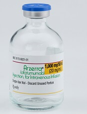
Photo courtesy of GSK
The US Food and Drug Administration (FDA) has approved the use of ofatumumab (Arzerra) as maintenance therapy for patients with chronic lymphocytic leukemia (CLL).
The drug can now be given for an extended period to patients who are in complete or partial response after receiving at least 2 lines of therapy for recurrent or progressive CLL.
Ofatumumab is also FDA-approved as a single agent to treat CLL that is refractory to fludarabine and alemtuzumab.
And the drug is approved for use in combination with chlorambucil to treat previously untreated patients with CLL for whom fludarabine-based therapy is considered inappropriate.
The FDA granted the new approval for ofatumumab based on an interim analysis of the PROLONG study. The results suggested that ofatumumab maintenance can improve progression-free survival (PFS) in CLL patients when compared to observation.
Ofatumumab is marketed as Arzerra under a collaboration agreement between Genmab and Novartis. For more details on ofatumumab, see the full prescribing information.
PROLONG trial
The PROLONG trial was designed to compare ofatumumab maintenance to no further treatment in patients with a complete or partial response after second- or third-line treatment for CLL. Interim results of the study were presented at ASH 2014.
These results—in 474 patients—suggested that ofatumumab can significantly improve PFS. The median PFS was about 29 months in patients who received ofatumumab and about 15 months for patients who did not receive maintenance therapy (P<0.0001).
There was no significant difference in the median overall survival, which was not reached in either treatment arm.
The researchers said there were no unexpected safety findings. The most common adverse events (≥10%) were infusion reactions, neutropenia, and upper respiratory tract infection.