User login
Noninvasive prenatal testing brings new options, opportunities, questions
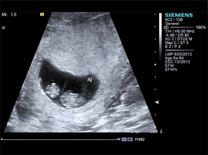
Noninvasive prenatal testing is moving from bench to bedside at a dizzying pace, and while this rapid integration into clinical practice is raising important clinical and ethical questions, it also is creating exciting new opportunities – such as the potential for antenatal treatment of Down syndrome.
In an editorial in Prenatal Diagnosis, Dr. Lyn S. Chitty and Dr. Diana W. Bianchi note that "we are in the midst of a paradigm shift in the way that prenatal screening and diagnosis are performed around the world."
The shift is occurring at a pace unprecedented in the history of prenatal care, and the available tests, which measure cell free fetal DNA (cfDNA) in the maternal blood and currently are used to detect trisomy 21, 18, and 13, as well as sex chromosome aneuploidies, provide a "readily accessible and generally safer option for prenatal testing than can be offered from 10 until 40 weeks of gestation," wrote Dr. Chitty of Great Ormond Street and University College London hospitals and Dr. Bianchi, the Natalie V. Zucker Professor of Pediatrics, Obstetrics, and Gynecology of Tufts University, Boston (Prenatal Diagnosis 2013;33:511-13).
October will mark 2 years since the commercial debut of the first noninvasive prenatal test (NIPT) for aneuploidy in the United States – MaterniT21 (Sequenom, San Diego), a laboratory-developed test that detects increases in the amount of chromosomal 21, 18, and 13 material in maternal blood.
"This was followed by multiple publications, several professional society recommendations, and a logarithmic uptake in the number of tests ordered. The reason why noninvasive prenatal testing (NIPT) represents a paradigm shift is that it changes the algorithm of screening followed by invasive testing that has been in practice worldwide for the last 30 years," they said.
Rather than undergoing combined ultrasound screening and serum screening (the "combined test"), followed by more invasive chorionic villus sampling or amniocentesis to rule out aneuploidy for a positive combined test, more women now have the option of a simple blood test. The negative predictive value of NIPT is high, so fewer women are subjected to the invasive procedures, which increase the risk for miscarriage and other adverse outcomes.
The NIPT options are expanding quickly; since MaterniT21 became available, three other companies launched similar tests: Harmony (Ariosa Diagnostics, San Jose, Calif.), verifi (Verinata Health, Redwood City, Calif.), and – most recently – Panorama (Natera, San Carlos, Calif.).
None are approved by the Food and Drug Administration, but all are performed in Clinical Laboratory Improvement Amendments (CLIA)–approved laboratories as laboratory-developed tests.
The tests are typically administered any time after 10 weeks’. gestation because that is generally when an adequate amount of fetal DNA is present in the maternal serum, Dr. Bianchi, also a geneticist and executive director of the Mother Infant Research Institute at Tufts, explained during an NIPT symposium at the annual meeting of the American College of Obstetricians and Gynecologists in New Orleans.
Some of the tests use massively parallel sequencing, which can be used to "look at everything," including sex chromosome aneuploidies, although most laboratories use software that masks all but the results of interest. Other tests use more targeted sequencing to focus on the chromosomes of interest.
Studies suggest that NIPT is at least 99% accurate for detecting trisomy 21 and trisomy 18, and between 79% and 92% accurate for trisomy 13, with a false positive rate of less than 1% for each, according to a 2012 fact sheet developed by the National Coalition for Health Professional Education in Genetics and the National Society of Genetic Counselors.
Serum screening, by comparison, has an 80%-95% detection rate for trisomy 21 and trisomy 18, with false-positive rates of 3% -5%. The rates for trisomy 13 are uncertain, according to the fact sheet.
Most studies to date have included mainly women with singleton pregnancies at high risk of trisomy 21 due to advanced maternal age, an abnormal serum screen, a personal or family history of aneuploidy, or an abnormal ultrasound, and as a result most current recommendations support screening only in this population.
A December 2012 opinion from the American College of Obstetricians and Gynecologists Committee on Genetics and the Society for Maternal-Fetal Medicine Publications Committee, for example, notes that NIPT "offers tremendous potential as a screening tool for fetal aneuploidy," but that it should be offered, after pretest counseling, only to women with high-risk singleton pregnancies. Cell-free fetal DNA testing has not been sufficiently evaluated in those at low risk or with multiple gestations, the committee said (Obstet. Gynecol. 2012;120:1532-4).
In January 2013, the National Society of Genetic Counselors released a similar position statement (J. Genet. Counsel. 2013 [doi: 10.1007/s10897-012-9564-0]).
New evidence, however, suggests that NIPT also is feasible in low-risk pregnancies.
For example, two studies in the July issue of Ultrasound in Obstetrics and Gynecology indicate that both routine and contingent implementation of NIPT for first trimester trisomy screening are feasible.
In one of the studies, Dr. M.M. Gil of King’s College Hospital, London, and his colleagues demonstrated that in 1,005 singleton pregnancies, routine NIPT for trisomies 21, 18, and 13 at 10 weeks’ gestation had a lower false positive rate than did the combined test performed at 12 weeks (0.1% vs. 3.4%), although abnormal results required confirmation using invasive testing.
"This study has shown that routine screening for trisomies by cfDNA testing at 10 weeks is feasible, allowing diagnosis of aneuploidies and the option of pregnancy termination within the first trimester," the investigators concluded (Ultrasound Obstet. Gynecol. 2013;42:34-40).
In the second study, which involved women with singleton pregnancies who underwent screening between March 2006 and May 2012, Dr. K.H. Nicolaides, also of King’s College Hospital, London, and his colleagues demonstrated that effective first trimester screening for Down syndrome can be achieved – with a 98% detection rate and an invasive testing rate below 0.5% – using contingent screening (Ultrasound Obstet. Gynecol. 2013;42:41-50).
"The results demonstrate that in contingent screening, a detection rate of 98% in fetuses with trisomy 21, at an overall invasive testing rate less than 0.5%, could be achieved by offering the cfDNA test to about 36%, 21%, and 11% of cases identified by first-line screening using the combined test alone, using the combined test with the addition of serum placental growth factor and alpha-fetoprotein, and using the combined test with the addition of placental growth factor, alpha-fetoprotein, and ductus venosus pulsatility index for veins, respectively," they said. "Screening for trisomy 21 by cfDNA testing contingent on the results of an expanded combined test would retain the advantages of the current method of screening, but with a simultaneous major increase in detection rate and decrease in the rate of invasive testing," they concluded.
Data also suggest that NIPT can be used successfully in twin pregnancies, albeit with the caveat that deeper sequencing may be necessary in these pregnancies, Dr. Bianchi noted.
These findings suggest that even wider implementation of NIPT is likely, she said, adding that there are major concerns about such implementation – not the least of which is fitting adequate pre- and posttest counseling into busy practices.
"It is not straightforward – there are multiple issues that need to be discussed," she said.
Questions also remain about the performance of the tests in normal- vs. high-risk pregnancies, she said.
Recently, a Blue Cross Blue Shield Technology Evaluation Center report concluded that NIPT meets the company’s criteria for both normal- and high-risk pregnancies, Dr. Bianchi noted.
"We’ll be hearing more about this in the coming year. Everyone is looking for more data," she said.
The results thus far – with more than 100,000 tests performed – undoubtedly have been encouraging. Since the integration of these tests into clinical practice began, many medical centers already are witnessing significant declines in the number of invasive procedures being performed for aneuploidy, Dr. Bianchi said, noting that such procedures have decreased by 34% in the first year at Tufts Medical Center, where NIPT is routinely offered as an option to high-risk women, and as an alternative to invasive procedures in average-risk women who test positive on traditional screening. Invasive procedures are strongly encouraged in those with aneuploidy detected on NIPT.
As a result of these outcomes, every aspect of the current standard of care is being questioned, Dr. Chitty and Dr. Bianchi wrote in their editorial. "For example, do we still need to measure maternal serum biomarkers, and what is the place of nuchal translucency measurement?" they asked.
The use of NIPT and the rapid advances taking place in the field, also raise important ethical questions.
Even before Sequenom introduced the MaterniT21 test to the market, the ethical implications of NIPT were being weighed. In a 2009 paper, Dr. Antina de Jong of Maastricht (the Netherlands) University and colleagues noted that although the introduction of NIPT would have some "ethically favorable consequences," such as the absence of iatrogenic miscarriage resulting from a test, earlier reassurance of a healthy fetus, a longer period for decision-making and the possibility of an early abortion, "which may be physically and psychologically less burdening and ethically less problematic because of presumed lower mortal fetal status," the possibility that NIPT would eventually include testing for a broader scope of abnormalities complicates the ethical issues.
Notwithstanding the potential benefits with respect to earlier decision making, one concern is the "normalization and trivialization of early selective abortion," they said, adding that "more generalized use of noninvasive testing could facilitate selective terminations of pregnancy in a range of conditions hitherto not diagnosed prenatally and where the arguments for and against termination may not have received sufficient scrutiny."
Also, testing for a broader scope of abnormalities has important implications regarding informed consent, they said (Eur. J. Hum. Genet. 2009 [doi:10.1038/ejhg.2009.203]).
"Informed consent will become far more challenging – if attainable at all. Moreover, should [NIPT] testing become available for a wider range of disorders including late-onset disease, this may lead to the same ethical difficulties as with regard to wide range testing of newborns, in which the dominant view is that the child’s right not to know should be respected," the authors wrote. "It is difficult to see how this respect can be upheld when, after broadening prenatal testing, children will be born with a positive test result for a serious late-onset disease."
Another facet of NIPT that is rife with ethical implications is noninvasive fetal whole genome sequencing, which was shown in a recent proof-of-concept study to be possible using only parental DNA samples and plasma. That study is revisited from a clinical standpoint in an article published in the same issue of Prenatal Diagnosis along with the editorial by Dr. Chitty and Dr. Bianchi and a number of other related articles and studies. (To give readers an overview of "this rapidly changing field," both the June and July issues of the journal are dedicated to NIPT, said Dr. Bianchi, the journal’s editor-in-chief).
"If noninvasive determination of the entire fetal genomic sequence becomes clinically available, there will be a significant increase in the number of ethical issues that arise," Dr. Chitty and Dr. Bianchi noted.
"You can imagine, it’s hard enough now to counsel about serum screening results for Down syndrome. Imagine if you had to deal with the entire human genome. But it’s coming – it’s coming, so we have to figure out ... what’s to be communicated to expectant couples," Dr. Bianchi said, adding that she hopes much of the groundwork for dealing with these issues will be laid in adult and pediatric populations before they have to be considered for fetuses.
While these advances bring with them ethical concerns, they also open doors to exciting opportunities, she said.
"The challenge now is to translate this technology into practice that is accessible to all pregnant women and in an ethical way that preserves informed parental choice, while not increasing overall costs to the health care system," Dr. Chitty and Dr. Bianchi said.
Dr. Bianchi is chair of the clinical advisory board for Verinata Health Inc., and has received honorarium and research funding from the company. She is editor-in-chief of the Journal of Prenatal Diagnosis, and has received honorarium from the company. Dr. Nicolaides’ and Dr. Gil’s studies were funded by grants from the Fetal Medicine Foundation. Dr. de Jong’s research was supported by the Centre for Society and Genomics in the Netherlands, funded by the Netherlands Genomics Initiative, and the Netherlands Organisation for Scientific Research Zonmw Prevention Fund.
New drugs approved in 2012
In 2012, the Food and Drug Administration approved 39 new molecular entities (i.e., drugs). This was the highest number of approvals in the 2003 to 2012 period. Of the 39, 2 have not yet come onto the market, 3 are highly unlikely to be used in women of reproductive age, and 1 is a four-drug combination for HIV-1 infection.
The remaining 33 agents are classified as anorexiant (1), anticoagulant (1), anticonvulsant (1), antidote (1), antilipemic (1), antineoplastic (11), antituberculosis (1), dermatologic (1), endocrine/metabolic (3), gastrointestinal (4), hematologic (1), immunologic (2), impotence (1), ophthalmic (2), respiratory (1), and urinary tract agent (1).
There is no reported human pregnancy experience for any of these agents. Consequently, the potential risk to the embryo and/or fetus must be estimated based on the indication, mechanism of action, other drugs with a similar mechanism, route of administration, molecular weight, elimination half-life, and animal reproduction data. Some of these drugs have been included in the quarterly updates to the 9th edition of my book "Drugs in Pregnancy and Lactation." The remainder will appear in the 10th edition, scheduled to be released in the spring of 2014.
Lorcaserin (Belviq) is an anorexiant. Because weight loss in pregnancy usually offers no benefit to a pregnant woman, the manufacturer classifies the drug as contraindicated in pregnancy.
A new anticoagulant, apixaban (Eliquis), is used to reduce the risk of stroke in patients with atrial fibrillation. The animal data suggest low risk and, when combined with the indication, the drug should not be withheld because of pregnancy.
The animal data for the anticonvulsant perampanel (Fycompa) suggest risk, but the absence of human pregnancy experience prevents a full assessment of the embryo-fetal risk. If the drug is used in pregnancy, physicians are encouraged to recommend that their patients enroll in the North American Antiepileptic Drug Pregnancy Registry by calling 1-888-233-2334.
The antidote, glucarpidase (Voraxaze), is used to treat toxic levels of methotrexate. Because methotrexate is contraindicated in pregnancy, it appears that there will be few opportunities for use of this drug in a pregnant woman. The same can be said for the antilipemic agent, lomitapide (Juxtapid). Decreasing the levels of lipids and cholesterol offers no benefit in pregnancy. The manufacturer classifies the agent as contraindicated because of the toxicity observed in three animal species.
Among the new antineoplastic agents approved, six are kinase inhibitors. Of these, five are in the subclass of tyrosine kinase inhibitors (trade name; indication): Axitinib (Inlyta; renal cancer), bosutinib (Bosulif; leukemia), cabozantinib (Cometriq; thyroid cancer), ponatinib (Iclusig; leukemia), and ziv-aflibercept (Zaltrap; colorectal cancer). The sixth kinase inhibitor is a multikinase inhibitor: regorafenib (Stivarga; colorectal cancer).
Other new antineoplastics are the antiandrogen enzalutamide (Xtandi; prostate cancer but could be used for other cancers), the proteasome inhibitor carfilzomib (Kyprolis; multiple myeloma), and the protein synthesis inhibitor omacetaxine (Synribo; leukemia). Pertuzumab (Perjeta; breast cancer) is a monoclonal antibody that is given in combination with trastuzumab and docetaxel. Vismodegib (Erivedge; basal cell cancer) is a miscellaneous antineoplastic that has been associated with amenorrhea in clinical trials. Animal data suggest risk of embryo-fetal harm.
All of the above antineoplastics are contraindicated in pregnancy because their mechanisms suggest the potential for embryo-fetal harm. However, if a drug is the best choice for a woman with a severe or a potentially fatal disease, it should not be withheld because the maternal benefit should far outweigh the unknown embryo-fetal risk.
Bedaquiline (Sirturo) is a new antituberculosis agent that can be used in pregnancy because of the low risk in animal studies and its indication. The dermatologic agent, ingenol mebutate (Picato) is used topically for actinic keratosis and appears to be compatible in pregnancy, because blood levels of the drug and two of its metabolites were below the lower limit of quantification (0.1 ng/mL).
The three endocrine/metabolic agents are ivacaftor (Kalydeco; cystic fibrosis), taliglucerase alfa (Elelyso; Gaucher disease), and pasireotide (Signifor; Cushing’s disease). The animal data for ivacaftor suggest low risk. Taliglucerase alfa appears to be compatible in pregnancy because it might reduce the risk of spontaneous abortion and bleeding complications. However, based on animal data, if a pregnant woman takes pasireotide, she should be informed of the potential risk, including abortion, to her embryo and/or fetus.
There are four gastrointestinal agents: crofelemer (Fulyzaq; antidiarrheal), linaclotide (Linzess; laxative), teduglutide (Gattex; short bowel syndrome), and the combination of sodium picosulfate, magnesium oxide, and citric acid (Prepopik; osmotic laxative). All appear to be compatible in pregnancy because of limited, if any, absorption (crofelemer, linaclotide, and sodium picosulfate) or because it is an analogue of a naturally occurring peptide (teduglutide).
The animal data for the hematologic agent, peginesatide (Omontys; anemia in patients with chronic renal disease) suggest risk. The drug has a high molecular weightthat should limit its passage across the placenta, but it still might cross in the third trimester. Because it stimulates erythropoiesis in human red blood cell precursors, it could do the same in the fetus if it crosses.
The two immunologic drugs are teriflunomide (Aubagio; multiple sclerosis) and tofacitinib (Xeljanz; rheumatoid arthritis). Teriflunomide, the principal active metabolite of leflunomide and responsible for leflunomide’s activity, is contraindicated in pregnancy. If a woman conceives while taking this drug, an 11-day procedure for accelerated elimination is recommended because it takes an average of 8 months (and it may be as long as 2 years) to reach plasma concentrations that are considered minimal risk (see package insert for procedure). The animal data for tofacitinib suggest risk. It is contraindicated if combined with methotrexate.
At first glance, it does not appear that the vasodilator avanafil (Stendra; impotence agent) will be used in pregnancy. However, a similar agent, sildenafil, has been used for the treatment of pulmonary arterial hypertension, a high-risk condition in pregnancy. Based on its indication, mechanism of action, and low-risk animal data, avanafil can be used in pregnancy if indicated.
Aclidinium bromide (Tudorza Pressair; bronchodilator) is a respiratory agent used in chronic obstructive pulmonary disease, chronic bronchitis, and emphysema. The animal data suggest low risk. The low plasma concentrations suggest that the drug represents a low, if any, risk in pregnancy.
The two ophthalmic preparations are ocriplasmin (Jetrea; symptomatic vitreomacular adhesions) and tafluprost (Zioptan; glaucoma). Both are probably compatible in pregnancy because of the undetectable or very low systemic concentrations.
The animal data for mirabegron (Myrbetriq; antispasmodic for overactive bladder) suggest low risk. It is an adrenergic agonist that increases bladder capacity. Although there are no human data, it probably can be used in pregnancy, but avoiding the first trimester should be considered.
As with pregnancy, there are no reports involving the use of the above drugs during breast-feeding. When there are little or no human data, the risk to a nursing infant can be estimated by considering several factors: indication, duration of therapy, molecular weight, plasma protein binding, elimination half-life, presence of the drug in the systemic circulation, and the most common adverse effects observed in adults.
Using these criteria for the drugs discussed above, there are only 10 that are probably compatible with breast-feeding: glucarpidase, ingenol mebutate, sodium picosulfate, taliglucerase alfa, crofelemer, linaclotide, peginesatide, aclidinium bromide, tafluprost, and ocriplasmin. The remaining drugs are either contraindicated (most of the antineoplastics and tofacitinib if combined with methotrexate) or may cause toxicity in a nursing infant.
Mr. Briggs is a pharmacist clinical specialist at the outpatient clinics of Memorial Care Center for Women at Miller Children’s Hospital in Long Beach, Calif.; clinical professor of pharmacy at the University of California, San Francisco; and adjunct professor of pharmacy at the University of Southern California, Los Angeles, and Washington State University, Spokane. He also is coauthor of "Drugs in Pregnancy and Lactation," and coeditor of "Diseases, Complications, and Drug Therapy in Obstetrics." He had no relevant financial disclosures. Contact him at [email protected].

Mothers’ postpartum concerns predict failure to meet breastfeeding goals
First-time mothers’ widely reported breastfeeding problems or concerns predicted the likelihood that they would stop breastfeeding or give their infants formula within the first 2 months after giving birth, a study showed.
The 92% of mothers with any concerns at 3 days post partum were nine times more likely to stop breastfeeding before 2 months post partum, primarily because of infant feeding difficulties or concerns about milk quantity.
"Breastfeeding problems were a nearly universal experience in this cohort of first-time mothers," reported Erin A. Wagner of Cincinnati Children’s Hospital Medical Center and her colleagues in Pediatrics (2013 Sept. 23 [doi:10.1542/peds.2013-0724]). The concerns were "highly prevalent, persistent, and associated with not meeting breastfeeding goals," the investigators said.

Meanwhile, the lack of association between early breastfeeding cessation and prenatal breastfeeding concerns (as opposed to postpartum concerns) implies that the women’s failure to meet breastfeeding goals "do not appear to be simply the ‘self-fulfillment’ of anticipated problems," the researchers added.
The investigators conducted 2,946 interviews, starting with 532 primiparous expectant mothers, about half of whom (49%) were younger than 25. Just over a quarter (27%) were older than 30. Approximately half had private insurance and half had public insurance.
Ms. Wagner and her colleagues then followed up post partum with 447 of the participants, beginning within 24 hours of delivery and conducting additional interviews on days 3, 7, 14, 30, and 60 days post partum. After those lost to follow-up, 418 mothers comprised the final sample of participants with infant feeding information at 2 months post partum.
From that final sample, 47% of the 354 mothers who intended to feed their babies only breast milk for at least 2 months ended up feeding any formula to their child between 30 and 60 days post partum. Among the 406 mothers who intended to breastfeed for at least 2 months, 21% stopped breastfeeding by 60 days post partum.
To qualify the women’s breastfeeding concerns, the researchers categorized the women’s 4,179 open-ended answers involving concerns into nine main categories with a total of 49 subcategories. The most prevalent concern on delivery day was infant feeding difficulties, reported by 44% of the mothers. Three days post partum, 54% of mothers reported infant feeding difficulties, 42% reported breastfeeding pain, and 42% reported concerns about milk quantity. Infant feeding difficulties included latch problems, sleepy infants, nipple confusion or infant feeding refusal, fussy or frustrated infants, poor infant feeding, and problems with the baby’s length or frequency of breastfeeding sessions.
After taking into account women’s education and their breastfeeding intentions in prenatal interviews, the researchers identified two breastfeeding concerns that contributed most to early cessation of breastfeeding. Thirty-two percent of those who stopped were estimated to have continued if not for infant feeding difficulties reported 7 days post partum (population attributable risk (PAR) = 32%).
"Most notably, the predominant subcategories at day 7 contributing to stopping breastfeeding under the infant feeding difficulty main category were ‘fussy or frustrated at the breast,’ ‘infant refusing to breastfeed/nipple confusion,’ and ‘problems with latch,’ " the researchers wrote. The second highest PAR was 23% for reporting concerns about milk quantity 2 weeks post partum. Accounting for the women’s age, ethnicity, health insurance status, or prenatal perceptions about breastfeeding ability did not significantly alter these findings.
Although 79% of the mothers reported at least one breastfeeding concern during prenatal interviews, the peak of concerns occurred on the third postpartum day, with 92% of women reporting at least one concern. The peak for reporting pain while breastfeeding occurred 1 week post partum with 47% of mothers.
Concerns about breastfeeding during the first week post partum appeared to contribute the most toward women’s using formula or stopping breastfeeding by 2 months post partum. Mothers reporting any breastfeeding concerns at 3 days after giving birth were three times more likely to feed their child formula between 1 and 2 months post partum (ARR = 3.3; 95% CI, 1.7-15.0). They were nine times more likely to stop breastfeeding within 60 days post partum (ARR 9.2, 95% CI, 3.0 to infinity).
The researchers identified 34 outliers who reported no breastfeeding concerns three days post partum; all but one of these women continued breastfeeding past 60 days post partum. These women were more likely to be younger than 30, to be Hispanic, to have confidence prenatally of their ability to breastfeed, to have an unmedicated vaginal delivery, and to report having strong breastfeeding support.
The researchers noted that the high levels of concerns reported in the first week postpartum might have been most strongly associated with adverse outcomes because the "day 3 and day 7 interviews captured a time when there is often a gap between hospital and community lactation support resources." The results "reinforce the recommendation of the American Academy of Pediatrics that all breastfed newborns receive an evaluation by a provider knowledgeable in lactation management within 2 to 3 days post discharge," the investigators said.
One limitation cited by the investigators is that the study’s generalizability might be limited by how similar breastfeeding norms and support rates are in other communities, compared with the study population.
The study was funded by the National Institutes of Health and the Perinatal Institute of Cincinnati Children’s Hospital Medical Center. Laurie A. Nommsen-Rivers, Ph.D., received a stipend for a lecture at the 2012 National WIC Association meeting. The other three authors reported no relevant financial disclosures.
First-time mothers’ widely reported breastfeeding problems or concerns predicted the likelihood that they would stop breastfeeding or give their infants formula within the first 2 months after giving birth, a study showed.
The 92% of mothers with any concerns at 3 days post partum were nine times more likely to stop breastfeeding before 2 months post partum, primarily because of infant feeding difficulties or concerns about milk quantity.
"Breastfeeding problems were a nearly universal experience in this cohort of first-time mothers," reported Erin A. Wagner of Cincinnati Children’s Hospital Medical Center and her colleagues in Pediatrics (2013 Sept. 23 [doi:10.1542/peds.2013-0724]). The concerns were "highly prevalent, persistent, and associated with not meeting breastfeeding goals," the investigators said.
Meanwhile, the lack of association between early breastfeeding cessation and prenatal breastfeeding concerns (as opposed to postpartum concerns) implies that the women’s failure to meet breastfeeding goals "do not appear to be simply the ‘self-fulfillment’ of anticipated problems," the researchers added.
The investigators conducted 2,946 interviews, starting with 532 primiparous expectant mothers, about half of whom (49%) were younger than 25. Just over a quarter (27%) were older than 30. Approximately half had private insurance and half had public insurance.
Ms. Wagner and her colleagues then followed up post partum with 447 of the participants, beginning within 24 hours of delivery and conducting additional interviews on days 3, 7, 14, 30, and 60 days post partum. After those lost to follow-up, 418 mothers comprised the final sample of participants with infant feeding information at 2 months post partum.
From that final sample, 47% of the 354 mothers who intended to feed their babies only breast milk for at least 2 months ended up feeding any formula to their child between 30 and 60 days post partum. Among the 406 mothers who intended to breastfeed for at least 2 months, 21% stopped breastfeeding by 60 days post partum.
To qualify the women’s breastfeeding concerns, the researchers categorized the women’s 4,179 open-ended answers involving concerns into nine main categories with a total of 49 subcategories. The most prevalent concern on delivery day was infant feeding difficulties, reported by 44% of the mothers. Three days post partum, 54% of mothers reported infant feeding difficulties, 42% reported breastfeeding pain, and 42% reported concerns about milk quantity. Infant feeding difficulties included latch problems, sleepy infants, nipple confusion or infant feeding refusal, fussy or frustrated infants, poor infant feeding, and problems with the baby’s length or frequency of breastfeeding sessions.
After taking into account women’s education and their breastfeeding intentions in prenatal interviews, the researchers identified two breastfeeding concerns that contributed most to early cessation of breastfeeding. Thirty-two percent of those who stopped were estimated to have continued if not for infant feeding difficulties reported 7 days post partum (population attributable risk (PAR) = 32%).
"Most notably, the predominant subcategories at day 7 contributing to stopping breastfeeding under the infant feeding difficulty main category were ‘fussy or frustrated at the breast,’ ‘infant refusing to breastfeed/nipple confusion,’ and ‘problems with latch,’ " the researchers wrote. The second highest PAR was 23% for reporting concerns about milk quantity 2 weeks post partum. Accounting for the women’s age, ethnicity, health insurance status, or prenatal perceptions about breastfeeding ability did not significantly alter these findings.
Although 79% of the mothers reported at least one breastfeeding concern during prenatal interviews, the peak of concerns occurred on the third postpartum day, with 92% of women reporting at least one concern. The peak for reporting pain while breastfeeding occurred 1 week post partum with 47% of mothers.
Concerns about breastfeeding during the first week post partum appeared to contribute the most toward women’s using formula or stopping breastfeeding by 2 months post partum. Mothers reporting any breastfeeding concerns at 3 days after giving birth were three times more likely to feed their child formula between 1 and 2 months post partum (ARR = 3.3; 95% CI, 1.7-15.0). They were nine times more likely to stop breastfeeding within 60 days post partum (ARR 9.2, 95% CI, 3.0 to infinity).
The researchers identified 34 outliers who reported no breastfeeding concerns three days post partum; all but one of these women continued breastfeeding past 60 days post partum. These women were more likely to be younger than 30, to be Hispanic, to have confidence prenatally of their ability to breastfeed, to have an unmedicated vaginal delivery, and to report having strong breastfeeding support.
The researchers noted that the high levels of concerns reported in the first week postpartum might have been most strongly associated with adverse outcomes because the "day 3 and day 7 interviews captured a time when there is often a gap between hospital and community lactation support resources." The results "reinforce the recommendation of the American Academy of Pediatrics that all breastfed newborns receive an evaluation by a provider knowledgeable in lactation management within 2 to 3 days post discharge," the investigators said.
One limitation cited by the investigators is that the study’s generalizability might be limited by how similar breastfeeding norms and support rates are in other communities, compared with the study population.
The study was funded by the National Institutes of Health and the Perinatal Institute of Cincinnati Children’s Hospital Medical Center. Laurie A. Nommsen-Rivers, Ph.D., received a stipend for a lecture at the 2012 National WIC Association meeting. The other three authors reported no relevant financial disclosures.
FROM PEDIATRICS
Major finding: The 92% of women who reported breastfeeding concerns at 3 days post partum were nine times more likely to stop breastfeeding within 60 days (adjusted relative risk, 9.2; 95% CI, 3.0 to infinity), with 54% reporting infant feeding problems, 42% reporting breastfeeding pain, and 42% reporting milk quantity concerns.
Data source: An analysis of 2,946 interviews with 532 primiparas through University of California Davis Medical Center, with prenatal interviews and follow-up interviews at 0, 3, 7, 14, 30, and 60 days post partum.
Disclosures: The study was funded by the National Institutes of Health and the Perinatal Institute of Cincinnati Children’s Hospital Medical Center. Laurie A. Nommsen-Rivers, Ph.D., one of the researchers, received a stipend for a lecture at the 2012 National WIC Association meeting. The other three authors reported no relevant financial disclosures.

How I counsel patients regarding delayed cord clamping

Has the evidence tipped in favor of delayed cord clamping?
In December 2012, the American College of Obstetricians and Gynecologists
(ACOG) published a Committee Opinion on the timing of umbilical cord clamping after birth, but it found insufficient evidence to recommend early or delayed clamping.1
In a Cochrane review published earlier this year, McDonald and colleagues reviewed 15 trials and 3,911 mother-infant pairs, exploring the primary outcomes of severe maternal postpartum hemorrhage (≥1,000 mL), maternal death, and severe maternal morbidity and neonatal death associated with early versus delayed clamping. They also analyzed a number of secondary outcomes. None of the primary outcomes reached statistical significance.
In a statement from the World Health Organization (WHO) included in the Cochrane review, it was recommended that “the cord should not be clamped earlier than necessary”; the WHO graded this as a “weak recommendation”
based on “low-quality” evidence.
With such underwhelming evidence, I would guess that the average clinician does not feel very motivated to change his or her practice, if that practice involves early clamping.
One limitation of the Cochrane findings
In the studies included in the Cochrane review, there was marked heterogeneity in the definition of delayed cord clamping, which ranged
from 1 minute after delivery to the complete cessation of cord pulsation (~5 minutes). Some of the studies even used alternate times (2 minutes, 3 minutes, and so on).
Delayed clamping improved neonatal hemoglobin status
Among the secondary outcomes assessed in this review was an improvement in neonatal hemoglobin concentration and overall iron stores associated with delayed clamping—but this benefit came at the expense of a higher incidence of neonatal jaundice requiring phototherapy. As a result, the investigators
concluded that delayed cord clamping should be performed when there is ready
access to phototherapy.
Should we implement delayed clamping?
At this time, I am reluctant to recommend that we shift to delayed clamping. Here are my reasons:
- Data are lacking as to whether increased hemoglobin levels and iron stores in newborns improve outcomes—or provide any benefit. No long-term developmental outcome data were included in the Cochrane review.
- Although I am not a pediatrician, I am unaware of infant iron deficiency being a significant threat to public health in the developed world.
- The greater need for phototherapy in the delayed-clamping group should not be viewed as inconsequential.
- Iron supplementation is probably more readily available than phototherapy, especially in developing countries.
- In the minority of cases in which delayed clamping might be beneficial (eg, prematurity), it is not always feasible, as these infants may already be compromised. Anxious neonatologists generally want the newborn handed over to them for resuscitation as quickly as possible, generally frowning upon a delay of 3 to 5 minutes for the blood to move from the placenta to the infant.
What this evidence means for practice
I recommend that obstetric care providers continue their current practice until more detailed data emerge on the risks and benefits of delayed clamping. If a patient asks about the issue, we should counsel her about the risks and benefits of early versus delayed clamping and comply with her choice when there are no contraindications.
—John T. Repke, MD
Tell us what you think, at [email protected]. Please include your name and city and state.
Reference
1. American College of Obstetricians and Gynecologists. Committee Opinion #543: Timing of umbilical cord clamping after birth. Obstet Gynecol. 2012:120(6):1522–1526.

John T. Repke, MD
University Professor and Chairman, Department of Obstetrics and Gynecology, Penn State University College of Medicine, and Obstetrician-Gynecologist-in-Chief, Milton S. Hershey Medical Center, Hershey, Pennsylvania. Dr. Repke serves on the OBG Management Board of Editors.
The author reports no financial relationships relevant to this article.
In December 2012, the American College of Obstetricians and Gynecologists
(ACOG) published a Committee Opinion on the timing of umbilical cord clamping after birth, but it found insufficient evidence to recommend early or delayed clamping.1
In a Cochrane review published earlier this year, McDonald and colleagues reviewed 15 trials and 3,911 mother-infant pairs, exploring the primary outcomes of severe maternal postpartum hemorrhage (≥1,000 mL), maternal death, and severe maternal morbidity and neonatal death associated with early versus delayed clamping. They also analyzed a number of secondary outcomes. None of the primary outcomes reached statistical significance.
In a statement from the World Health Organization (WHO) included in the Cochrane review, it was recommended that “the cord should not be clamped earlier than necessary”; the WHO graded this as a “weak recommendation”
based on “low-quality” evidence.
With such underwhelming evidence, I would guess that the average clinician does not feel very motivated to change his or her practice, if that practice involves early clamping.
One limitation of the Cochrane findings
In the studies included in the Cochrane review, there was marked heterogeneity in the definition of delayed cord clamping, which ranged
from 1 minute after delivery to the complete cessation of cord pulsation (~5 minutes). Some of the studies even used alternate times (2 minutes, 3 minutes, and so on).
Delayed clamping improved neonatal hemoglobin status
Among the secondary outcomes assessed in this review was an improvement in neonatal hemoglobin concentration and overall iron stores associated with delayed clamping—but this benefit came at the expense of a higher incidence of neonatal jaundice requiring phototherapy. As a result, the investigators
concluded that delayed cord clamping should be performed when there is ready
access to phototherapy.
Should we implement delayed clamping?
At this time, I am reluctant to recommend that we shift to delayed clamping. Here are my reasons:
- Data are lacking as to whether increased hemoglobin levels and iron stores in newborns improve outcomes—or provide any benefit. No long-term developmental outcome data were included in the Cochrane review.
- Although I am not a pediatrician, I am unaware of infant iron deficiency being a significant threat to public health in the developed world.
- The greater need for phototherapy in the delayed-clamping group should not be viewed as inconsequential.
- Iron supplementation is probably more readily available than phototherapy, especially in developing countries.
- In the minority of cases in which delayed clamping might be beneficial (eg, prematurity), it is not always feasible, as these infants may already be compromised. Anxious neonatologists generally want the newborn handed over to them for resuscitation as quickly as possible, generally frowning upon a delay of 3 to 5 minutes for the blood to move from the placenta to the infant.
What this evidence means for practice
I recommend that obstetric care providers continue their current practice until more detailed data emerge on the risks and benefits of delayed clamping. If a patient asks about the issue, we should counsel her about the risks and benefits of early versus delayed clamping and comply with her choice when there are no contraindications.
—John T. Repke, MD
Tell us what you think, at [email protected]. Please include your name and city and state.
Reference
1. American College of Obstetricians and Gynecologists. Committee Opinion #543: Timing of umbilical cord clamping after birth. Obstet Gynecol. 2012:120(6):1522–1526.
Reference
1. American College of Obstetricians and Gynecologists. Committee Opinion #543: Timing of umbilical cord clamping after birth. Obstet Gynecol. 2012:120(6):1522–1526.

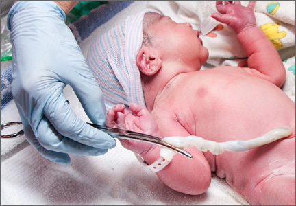
No increased birth defect risk found with oral fluconazole, with one exception
The overall risk of birth defects was not increased among infants whose mothers took oral fluconazole during the first trimester of pregnancy, in a large registry study that provides "largely reassuring" results regarding the teratogenicity of this drug, according to the authors.
In the study that included data on more than 7,000 pregnancies exposed to fluconazole in Denmark, fluconazole (largely at the most commonly prescribed 150-mg or 300-mg single doses) was also not associated with an increased risk of 14 of the 15 specific birth defects that have been associated with azole antifungal drugs in human or animal studies. But the risk of tetralogy of Fallot was increased by about threefold, an association that needs to be studied further, concluded Ditte Mølgaard-Nielsen and her associates in the department of epidemiology research, Statens Serum Institut, Copenhagen.

The risk of birth defects also was not increased among the smaller number of pregnancies with first-trimester exposure to two other oral azole antifungals, ketoconazole and itraconazole. The study is being published in the Aug. 29 issue of the New England Journal of Medicine (2013;369:830-9).
Most previous studies of the teratogenicity of fluconazole have involved case reports and have suggested that long-term use of fluconazole at high doses may be associated with an increased risk of certain birth defects – the basis of a Food and Drug Administration safety communication in 2011. But while a few epidemiologic studies of oral fluconazole, mostly at the 150-mg dose, have not found an association with birth defects, the studies were not large enough to investigate the risk with different doses or the risk of certain defects, the authors said.
In August 2011, the FDA described the possible association of oral fluconazole, at doses of 400-800 mg/day during the first trimester, and an increased risk of a rare but "distinct set of birth defects," based on case reports. The defects included brachycephaly, abnormal facies, abnormal calvarial development, cleft palate, femoral bowing, thin ribs and long bones, arthrogryposis, and congenital heart disease, which were also seen in animals. As a result, the FDA changed the pregnancy risk category of fluconazole from category C (adverse effects seen in animals but no adequate human data) to D (evidence of human fetal risk based on human data but the potential benefits may justify use in pregnant women with serious or life-threatening conditions) for all indications, except for the 150-mg single dose used for vaginal candidiasis. Because the available human data on that dose do not indicate there is an association with an increased risk of birth defects, the 150-mg dose used for vaginal candidiasis remained in category C.
The Danish study used data on all live-born infants in Denmark between January 1996 and March 2011 from the national birth registry, prescription data from the national prescription registry, and information on birth defects from a national patient registry. The most common cumulative doses of fluconazole were 150 mg (56%) and 300 mg (31%); the remaining percentage of patients were exposed to cumulative doses from 350 mg to 6,000 mg.
Among the 7,352 pregnancies exposed to fluconazole in the first trimester, there were 210 birth defects (2.86%), compared with 25,159 birth defects (2.6%) among 968,236 unexposed pregnancies.
When the investigators looked at 15 different birth defects that have been linked to in utero exposure to fluconazole and other azole antifungal agents, the risk was not significantly increased for defects that included craniosynostosis, cleft palate, cleft lip with or without cleft palate, limb defects, polydactyly, syndactyly, diaphragmatic hernia, heart defects overall, or ventricular septal defects.
However, there were 7 cases of tetralogy of Fallot (0.1%) among pregnancies exposed to fluconazole, compared with 287 cases (0.03%) among those not exposed – a threefold increased risk that was statistically significant. The cases were found in infants exposed to all three cumulative dose categories: four (0.10%) exposed to 150 mg, two (0.09%) exposed to 300 mg, and one (0.10%) exposed to the 350- to 6,000-mg dose range.
Although the increased risk of tetralogy of Fallot could be a chance finding, this was among the birth defects previously associated with exposure to fluconazole, which made the finding more reliable than if this had been the first time the association with this particular birth defect had been reported, the authors noted.
Among 687 pregnancies exposed to itraconazole and 72 pregnancies exposed to ketoconazole in the registry, there was no significantly increased risk of birth defects overall, or any clustering of malformations.
"Our finding that a 150-mg dose of fluconazole was not associated with an increased risk of birth defects overall confirms the results of previous studies; our study also adds to these safety data by reporting risk estimates for doses higher than 150 mg," the authors wrote. "Although fluconazole may confer an increased risk of tetralogy of Fallot, the absolute risk was small and the association needs to be confirmed," they added.
The study’s strengths include the nationwide cohort, the 1-year follow-up of the infants, the high degree of accuracy of the registry data on birth detects, and the use of prescription data, which eliminates the chance of recall bias, the authors said. The limitations included not being able to account for all confounding factors, such as ascertainment of maternal illnesses.
The study was funded by the Danish Medical Research Council. The authors reported having no relevant financial disclosures.
Fluconazole is marketed as Diflucan, ketoconazole is marketed as Nizoral, and itraconazole is marketed as Sporanox; all three are available in generic formulations.
The overall risk of birth defects was not increased among infants whose mothers took oral fluconazole during the first trimester of pregnancy, in a large registry study that provides "largely reassuring" results regarding the teratogenicity of this drug, according to the authors.
In the study that included data on more than 7,000 pregnancies exposed to fluconazole in Denmark, fluconazole (largely at the most commonly prescribed 150-mg or 300-mg single doses) was also not associated with an increased risk of 14 of the 15 specific birth defects that have been associated with azole antifungal drugs in human or animal studies. But the risk of tetralogy of Fallot was increased by about threefold, an association that needs to be studied further, concluded Ditte Mølgaard-Nielsen and her associates in the department of epidemiology research, Statens Serum Institut, Copenhagen.
The risk of birth defects also was not increased among the smaller number of pregnancies with first-trimester exposure to two other oral azole antifungals, ketoconazole and itraconazole. The study is being published in the Aug. 29 issue of the New England Journal of Medicine (2013;369:830-9).
Most previous studies of the teratogenicity of fluconazole have involved case reports and have suggested that long-term use of fluconazole at high doses may be associated with an increased risk of certain birth defects – the basis of a Food and Drug Administration safety communication in 2011. But while a few epidemiologic studies of oral fluconazole, mostly at the 150-mg dose, have not found an association with birth defects, the studies were not large enough to investigate the risk with different doses or the risk of certain defects, the authors said.
In August 2011, the FDA described the possible association of oral fluconazole, at doses of 400-800 mg/day during the first trimester, and an increased risk of a rare but "distinct set of birth defects," based on case reports. The defects included brachycephaly, abnormal facies, abnormal calvarial development, cleft palate, femoral bowing, thin ribs and long bones, arthrogryposis, and congenital heart disease, which were also seen in animals. As a result, the FDA changed the pregnancy risk category of fluconazole from category C (adverse effects seen in animals but no adequate human data) to D (evidence of human fetal risk based on human data but the potential benefits may justify use in pregnant women with serious or life-threatening conditions) for all indications, except for the 150-mg single dose used for vaginal candidiasis. Because the available human data on that dose do not indicate there is an association with an increased risk of birth defects, the 150-mg dose used for vaginal candidiasis remained in category C.
The Danish study used data on all live-born infants in Denmark between January 1996 and March 2011 from the national birth registry, prescription data from the national prescription registry, and information on birth defects from a national patient registry. The most common cumulative doses of fluconazole were 150 mg (56%) and 300 mg (31%); the remaining percentage of patients were exposed to cumulative doses from 350 mg to 6,000 mg.
Among the 7,352 pregnancies exposed to fluconazole in the first trimester, there were 210 birth defects (2.86%), compared with 25,159 birth defects (2.6%) among 968,236 unexposed pregnancies.
When the investigators looked at 15 different birth defects that have been linked to in utero exposure to fluconazole and other azole antifungal agents, the risk was not significantly increased for defects that included craniosynostosis, cleft palate, cleft lip with or without cleft palate, limb defects, polydactyly, syndactyly, diaphragmatic hernia, heart defects overall, or ventricular septal defects.
However, there were 7 cases of tetralogy of Fallot (0.1%) among pregnancies exposed to fluconazole, compared with 287 cases (0.03%) among those not exposed – a threefold increased risk that was statistically significant. The cases were found in infants exposed to all three cumulative dose categories: four (0.10%) exposed to 150 mg, two (0.09%) exposed to 300 mg, and one (0.10%) exposed to the 350- to 6,000-mg dose range.
Although the increased risk of tetralogy of Fallot could be a chance finding, this was among the birth defects previously associated with exposure to fluconazole, which made the finding more reliable than if this had been the first time the association with this particular birth defect had been reported, the authors noted.
Among 687 pregnancies exposed to itraconazole and 72 pregnancies exposed to ketoconazole in the registry, there was no significantly increased risk of birth defects overall, or any clustering of malformations.
"Our finding that a 150-mg dose of fluconazole was not associated with an increased risk of birth defects overall confirms the results of previous studies; our study also adds to these safety data by reporting risk estimates for doses higher than 150 mg," the authors wrote. "Although fluconazole may confer an increased risk of tetralogy of Fallot, the absolute risk was small and the association needs to be confirmed," they added.
The study’s strengths include the nationwide cohort, the 1-year follow-up of the infants, the high degree of accuracy of the registry data on birth detects, and the use of prescription data, which eliminates the chance of recall bias, the authors said. The limitations included not being able to account for all confounding factors, such as ascertainment of maternal illnesses.
The study was funded by the Danish Medical Research Council. The authors reported having no relevant financial disclosures.
Fluconazole is marketed as Diflucan, ketoconazole is marketed as Nizoral, and itraconazole is marketed as Sporanox; all three are available in generic formulations.
FROM THE NEW ENGLAND JOURNAL OF MEDICINE
Major finding: Exposure to oral fluconazole in the first trimester was not associated with an increased risk of birth defects overall, but was associated with a threefold increased risk in tetralogy of Fallot, although the absolute risk was small.
Data source: A registry-based cohort study comparing the risk of birth defects overall, and the risk of specific birth defects associated with first-trimester exposure to different doses of oral fluconazole, in 7,352 fluconazole-exposed pregnancies and 968,236 unexposed pregnancies.
Disclosures: The study was funded by the Danish Medical Research Council. The authors reported having no relevant financial disclosures.

Atypical antipsychotics during pregnancy – What do we know?
Currently, the second-generation "atypical" antipsychotics, such as olanzapine (Zyprexa), aripiprazole (Abilify), and risperidone (Risperdal), are used far more frequently than are the first-generation "typical" antipsychotics, such as haloperidol. This is largely because the newer agents are used for a wider spectrum of illnesses that include bipolar disorder, anxiety disorders, and obsessive compulsive disorder, in addition to chronic psychotic illnesses such as schizophrenia.
As a result, a large number of women of reproductive age are being treated with atypicals, and given the persistent rate of unplanned pregnancies in the United States and globally, many women treated with atypicals become pregnant every year. But compared with the amount of reproductive safety information available for many classes of psychiatric medicines such as selective serotonin reuptake inhibitors (SSRIs), the reproductive safety data on both atypical and typical antipsychotics are relatively sparse.
The data on the older typical antipsychotics – from small-case series and one meta-analysis published in 1996 – do not suggest an increased risk for major malformations associated with first trimester exposure to this class (Am. J. Psychiatry 1996;153:592-606).
Atypical antipsychotics also include medications such as clozapine (Clozaril), ziprasidone (Geodon), paliperidone (Invega), quetiapine (Seroquel), asenapine (Saphris), lurasidone (Latuda), and iloperidone (Fanapt). The reproductive safety data on these drugs as a class include a small prospective study of 110 pregnancies in women treated with atypical antipsychotics (olanzapine, risperidone, quetiapine, and clozapine) during pregnancy, which found no signal for teratogenicity. The rate of major malformations was 0.9% in this group, which was not significantly different from the rate in the nonexposed group and was not higher than the 1%-3% baseline risk in the general population (J. Clin. Psychiatry 2005; 66:444-9).
However, this was a small sample and the findings were considered preliminary.
Because of the considerable use of atypicals to treat psychiatric disorders, the field has been waiting for more substantial information regarding the risks associated with fetal exposure to these newer drugs. Prompted by the lack of such data, the National Pregnancy Registry for Atypical Antipsychotics at Massachusetts General Hospital was established in 2008.
Now, the data we have been waiting for, from the registry and other sources, are beginning to emerge, and are providing some reassuring – though still preliminary – information regarding the risk for major congenital malformation risks associated with first trimester exposure to these drugs.
Published in August, a prospective observational cohort study conducted by the Institute for Clinical Teratology and Drug Risk Assessment in Pregnancy in Germany compared the risk for major malformations and other adverse pregnancy outcomes among 561 women exposed to atypical antipsychotics, 284 women exposed to typical antipsychotics, and 1,122 pregnant women treated with medications known not to be harmful during pregnancy (the reference group) from January 1997 to March 2009.
After an effort to adjust for potential confounders, there was not a significant difference between the rates of major malformations, the primary endpoint, between those exposed to atypical antipsychotics (5.1%) or typical antipsychotics (4.2%) in the first trimester. The major malformation rate was higher among those exposed to typical antipsychotics than in the reference group, but the difference was not statistically significant. However, the major malformation rate was about twofold greater among those on atypicals compared to the reference group (odds ratio, 2.17), a significant difference. In the group exposed to atypicals, the most common major malformations were cardiovascular, and of the cardiac malformations, most (8 of 12) were atrial or ventricular septal defects (J. Clin. Psychopharmacol. 2013;33 453-62).
(The rate of symptoms suggesting "poor neonatal adaptation" were also significantly higher in the two antipsychotic-exposed groups.)
As the authors point out, cardiac septal defects are one of the most common congenital malformations, and this difference could be due to a detection bias, because women exposed to a medicine for which there are sparse reproductive safety data – particularly one in a relatively new class of drugs – may be more likely to get prenatal and postnatal diagnostic testing.
In the first presentation of the National Pregnancy Registry for Atypical Antipsychotics at an annual meeting in June, my coinvestigators and I concluded that atypical antipsychotics are unlikely to be major teratogens, like valproic acid. Based on 143 cases of first trimester exposure to atypicals, we described a risk with a very wide confidence interval with its outer bound being 5.5%. These data are considered preliminary at this point, and we plan to publish data based on a larger series with a nonexposed reference group in the next year.
Considering how widely the atypical antipsychotics are used across disease states by sexually active reproductive-age women, what can the clinician conclude based on the currently available reproductive safety data on atypical antipsychotics? Based on the 2005 study, the German study, and our report on the registry data, it is reasonable to conclude that second-generation antipsychotics are not major teratogens. They are not valproate (Depakote) or thalidomide. As I have noted in previous columns, when it comes to using psychiatric medicines, or most other medications for that case during pregnancy, there is no perfect decision and no decision is risk free. Given the importance of treating psychiatric illness during pregnancy and the evolving data that illustrate the adverse impact of untreated psychiatric illness on a wide range of neonatal and obstetrical outcomes – and on increasing the risk for postpartum psychiatric illness – one could consider a risk-benefit decision that places the use of atypical antipsychotics as not contraindicated.
The use of atypical antipsychotics to treat women who suffer from underlying psychiatric illness frequently makes the difference between substantial suffering or takes patients from partial remission to euthymia. In the end, precise quantification of the reproductive safety of atypical antipsychotics may be challenging given the frequency of polytherapy – whether an atypical is used as mood stabilizers for patients with bipolar or as an adjunct to antidepressants in a woman with depression. However, after 2 decades of clinical work and research with the population of women who take psychiatric medications and who are either planning to conceive or who are pregnant, I maintain that little should trump the very significant downside of active psychiatric disorder during pregnancy.
For information on enrolling in the National Pregnancy Registry for Atypical Antipsychotics, women and clinicians can call 866-961-2388.
Dr. Cohen is the director of the Center for Women's Mental Health at Massachusetts General Hospital in Boston, which provides information about pregnancy and mental health. He is the principal investigator on the National Pregnancy Registry for Atypical Antipsychotics, which is sponsored by multiple manufacturers of atypical antipsychotics. To comment, e-mail him at [email protected].

Pregnancy sepsis score identifies safe ED discharges
BERNALILLO, N.M. – A score of 6 or greater on a sepsis risk scale specifically for pregnant women had an adjusted odds ratio of 109 for ICU admission, with a 95% confidence interval of 18-661, according to its developers at Brown University in Providence, R.I.
Dubbed the Sepsis in Obstetrics Score (SOS), the scale is based on the Rapid Emergency Medicine Score and the SIRS/Sepsis criteria, but is adjusted to reflect the slightly higher heart rates, lower blood pressures, and elevated white counts associated with pregnancy. Scores range from 0-28, with 0 being normal.

The team applied the scoring system retrospectively to 850 pregnant or recently postpartum women who had gotten blood cultures or influenza swabs in the ED; both were used as surrogate markers for septic presentations.
Forty-eight women had scores of at least 6, and eight were admitted to the ICU. There was one ICU admission among the 802 women with scores less than 6.
In addition to the high odds ratio, which was adjusted for age, race, and body mass index, a score of at least 6 had a sensitivity of 88.9%, a specificity of 95.2%, a negative predictive value of 99.9%, and a positive predictive value of 16.7% for ICU admission within 48 hours of presentation.
"In this population in general, there is a low overall rate of serious morbidity and mortality, resulting in our low positive predictive value; it is, however, significantly higher than other disease severity scoring systems studied in this population," said lead investigator Dr. Catherine Albright, an ob.gyn. resident at Brown.
A score of at least 6 also was independently associated with telemetry unit admission, length of hospital stay, fetal tachycardia, and positive blood cultures; the latter were found in 30.8% of women who met the cutoff, but only 8.5% of women who did not. There was no significant difference in the percentage of women with positive flu swabs, about 14% in each group.
"None of the current disease severity scoring systems included pregnant women in their initial study populations. They uniformly overestimate morbidity and mortality in obstetric populations," whereas SOS "can reliably identify patients" who require ICU treatment, Dr. Albright said. The next step is prospective validation, she added.
However, during the question and answer period, an audience member pointed out that the researchers "used your own cohort to develop the cutoff, and then applied that cutoff to the same cohort, and got a high odds ratio; that’s really not surprising. I think your follow-up of applying this to a different cohort [will be] really important."
"Absolutely," Dr. Albright responded.
The score includes temperature, heart rate, blood pressure, respiratory rate, oxygen saturation, white blood cell count, percentage of immature neutrophils, and lactic acid concentration. With adjustment for pregnancy, a heart rate up to 120 bpm, a WBC count of about 6-17,000 cells/mcL, and a systolic blood pressure down to 90 mm Hg are considered to be in the normal range.
Among higher-scoring women, the most common diagnoses at presentation were pyelonephritis and endometritis. Those with scores below six were most commonly diagnosed with influenzalike illness, Dr. Albright reported at the Infectious Diseases Society for Obstetrics and Gynecology annual meeting
There were no significant differences between the two groups in the presence of hypertension, diabetes, HIV, and other comorbidities. The only significant demographic difference was age, with higher-scoring women a mean of 24 years old, vs. 26.3 in the lower-scoring group, probably a clinically irrelevant finding, she said.
Known or suspected ectopic pregnancies and multiple gestations were among the exclusion criteria.
Dr. Albright said she had no relevant financial disclosures.
BERNALILLO, N.M. – A score of 6 or greater on a sepsis risk scale specifically for pregnant women had an adjusted odds ratio of 109 for ICU admission, with a 95% confidence interval of 18-661, according to its developers at Brown University in Providence, R.I.
Dubbed the Sepsis in Obstetrics Score (SOS), the scale is based on the Rapid Emergency Medicine Score and the SIRS/Sepsis criteria, but is adjusted to reflect the slightly higher heart rates, lower blood pressures, and elevated white counts associated with pregnancy. Scores range from 0-28, with 0 being normal.
The team applied the scoring system retrospectively to 850 pregnant or recently postpartum women who had gotten blood cultures or influenza swabs in the ED; both were used as surrogate markers for septic presentations.
Forty-eight women had scores of at least 6, and eight were admitted to the ICU. There was one ICU admission among the 802 women with scores less than 6.
In addition to the high odds ratio, which was adjusted for age, race, and body mass index, a score of at least 6 had a sensitivity of 88.9%, a specificity of 95.2%, a negative predictive value of 99.9%, and a positive predictive value of 16.7% for ICU admission within 48 hours of presentation.
"In this population in general, there is a low overall rate of serious morbidity and mortality, resulting in our low positive predictive value; it is, however, significantly higher than other disease severity scoring systems studied in this population," said lead investigator Dr. Catherine Albright, an ob.gyn. resident at Brown.
A score of at least 6 also was independently associated with telemetry unit admission, length of hospital stay, fetal tachycardia, and positive blood cultures; the latter were found in 30.8% of women who met the cutoff, but only 8.5% of women who did not. There was no significant difference in the percentage of women with positive flu swabs, about 14% in each group.
"None of the current disease severity scoring systems included pregnant women in their initial study populations. They uniformly overestimate morbidity and mortality in obstetric populations," whereas SOS "can reliably identify patients" who require ICU treatment, Dr. Albright said. The next step is prospective validation, she added.
However, during the question and answer period, an audience member pointed out that the researchers "used your own cohort to develop the cutoff, and then applied that cutoff to the same cohort, and got a high odds ratio; that’s really not surprising. I think your follow-up of applying this to a different cohort [will be] really important."
"Absolutely," Dr. Albright responded.
The score includes temperature, heart rate, blood pressure, respiratory rate, oxygen saturation, white blood cell count, percentage of immature neutrophils, and lactic acid concentration. With adjustment for pregnancy, a heart rate up to 120 bpm, a WBC count of about 6-17,000 cells/mcL, and a systolic blood pressure down to 90 mm Hg are considered to be in the normal range.
Among higher-scoring women, the most common diagnoses at presentation were pyelonephritis and endometritis. Those with scores below six were most commonly diagnosed with influenzalike illness, Dr. Albright reported at the Infectious Diseases Society for Obstetrics and Gynecology annual meeting
There were no significant differences between the two groups in the presence of hypertension, diabetes, HIV, and other comorbidities. The only significant demographic difference was age, with higher-scoring women a mean of 24 years old, vs. 26.3 in the lower-scoring group, probably a clinically irrelevant finding, she said.
Known or suspected ectopic pregnancies and multiple gestations were among the exclusion criteria.
Dr. Albright said she had no relevant financial disclosures.
AT IDSOG
Major finding: A sepsis risk scoring system for pregnant women has a negative predictive value of 99.9% for not needing ICU admission.
Data Source: A retrospective application of the scale to 850 women.
Disclosures: Dr. Albright said she had no relevant financial disclosures.

Get umbilical artery systolic-to-diastolic ratio in intrauterine growth restriction
SAN FRANCISCO – An umbilical artery systolic-to-diastolic ratio of less than 3 as measured on weekly Doppler ultrasounds in a fetus with 30 weeks’ or more gestation and suspected intrauterine growth restriction suggests that the fetus probably is doing okay, Dr. Vickie A. Feldstein said.
That "ballpark guideline" is most helpful if physicians at your institution have agreed to use the umbilical artery systolic/diastolic (S/D) ratio as the parameter for assessing fetuses with intrauterine growth restriction (IUGR) and have agreed on which anatomical location is preferred for the ultrasound interrogation, so that there is some uniformity in how results are presented and interpreted, she said.
The recent Practice Bulletin No. 134 from the American College of Obstetricians and Gynecologists recommended in May 2013 that if the ultrasonographically estimated fetal weight is below the 10th percentile for gestational age, further evaluation should be considered, such as Doppler blood flow studies of the umbilical artery (Obstet. Gynecol. 2013;121:1122-33).
The medical literature describes several Doppler ultrasound parameters that could be used in suspected IUGR, including the umbilical artery S/D ratio, the resistance index, or the pulsatility index. They’re all about the same phenomenon, which is measuring resistance to perfusion in the placenta as reflected in the interrogation of the umbilical artery, Dr. Feldstein said at a meeting on antepartum and intrapartum management sponsored by the University of California, San Francisco.
"I think a report that includes all of them would make our heads spin," said Dr. Feldstein, professor of clinical radiology at the university. She and her colleagues use the S/D ratio, calculated by using calipers to measure the peak of systole on umbilical artery Doppler ultrasound and dividing that by the measure of end diastole.
"I don’t care how fast the flow actually is, I care about the character of the flow, the relative difference between systole and diastole," she said. The ratio reflects the status of placental circulation. It normally is high early in pregnancy and decreases as gestation advances, placental resistance decreases, and there is more forward flow during diastole.
If separate umbilical artery Doppler tracings yield discrepant S/D ratios, that may reflect normal variability or be due to changes in fetal heart rate. "A significant change in heart rate might change the S/D ratio quite a bit," she said. Or, an ultrasound filter set too low can produce noise in the tracing that might alter where you place the calipers.
The location along the umbilical cord that the sonographer samples also can affect measurements. The medical literature is full of suggestions about where to sample. Dr. Feldstein recommends sampling toward the placenta, if possible, which will reflect resistance to perfusion in the placenta.
"We’ve found the cord insertion, typically, so we know where to look," she said. "The farther away from the placenta you go, you’re adding resistance of the cord to your tracing."
She encouraged obstetricians to talk to the people who do Doppler at their institutions "to decide together how you want this done, how you want it reported, and what parameter you want used, so you don’t overwhelm yourselves with excess information."
If the S/D ratio is a bit above 3 in a third-trimester fetus with IUGR but there’s decent diastolic flow, "don’t sweat the small stuff," she suggested. As the S/D ratio goes higher and higher, however, placental insufficiency (and resistance) increases and forward flow decreases, and can become absent or even reversed end-diastolic flow.
An absence of diastolic flow is associated with a 60%-70% loss of vasculature, "a really significant abnormality in the placenta," she said. With reversed diastolic flow, the odds of perinatal death increase more than fivefold.
A recent study of 1,116 fetuses with IUGR showed that an abnormal umbilical artery Doppler ultrasound tracing (defined by pulsatility index or the absence or reversal of end-diastolic flow) was significantly associated with adverse outcomes irrespective of estimated fetal weight or abdominal circumference (Am. J. Obstet. Gynecol. 2013;208:e1-6 [doi: 10.1016/j.ajog.2013.02.007]).
When Dr. Feldstein sees an abnormal umbilical artery Doppler tracing, she samples the fetal middle cerebral artery by Doppler ultrasound. The middle cerebral artery S/D ratio should always be higher than the umbilical artery SD ratio, with a typical middle cerebral artery S/D ratio greater than 4 after 30 weeks’ gestation.
A fetus in trouble with IUGR will respond by lowering cerebral vascular resistance to maintain blood flow to its brain, decreasing the middle cerebral artery S/D ratio in what’s known as a "brain-sparing" wave form. Although there’s nothing that can be done for the fetus at this point, she said, it can be helpful to know that brain-sparing in growth-restricted fetuses was associated with increased risk for abnormal neurobehavioral outcomes in a controlled study of 126 preterm infants (Ultrasound Obstet. Gynecol. 2011;38:288-94).
"It’s a sign of distress," she said. Until recently, clinicians would follow that fetus carefully with various kinds of testing and watch for reverse diastolic flow in the umbilical artery, which is a sign of significant compromise, hypoxemia, and possible death. Today, a finding of brain-sparing next leads Dr. Feldstein to interrogate the ductus venosus in the liver, which is "the hardest to do, but it can be done," she said.
The ductus venosus flow should be phasic but continuous, in a pattern called the "a wave" reflecting forward, continuous flow even during right atrial contractions. If the flow reverses backward into the ductus venosus during right atrial contractions, that’s a sign of cardiac compromise, severe hypoxia, and right ventricular dysfunction, associated with high risk of morbidity and mortality.
Locating the ductus venosus for sampling can be tricky, in part because it is so close to hepatic veins, but if the ductus venosus flow is abnormal enough, there’s a shortcut that is easier to do: Sample the umbilical vein. Abnormal phasicity in umbilical vein pulsation may reflect ductus venosus flow reversal.
A previous study reported that the risk for perinatal mortality increased to nearly 6% with an elevated umbilical artery S/D ratio, to more than 11% with absent or reversed diastolic flow in the uterine artery, and to 39% with an abnormal ductus venosus wave form (Ultrasound Obstet. Gynecol. 2003;22:240-5). In a 2010 Cochrane Review of 18 studies that included more than 10,000 women with high-risk pregnancies, Doppler ultrasound was associated with a 29% reduction in the rate of perinatal deaths (1.2% with Doppler and 1.7% without); analysis showed that using Doppler on 203 high-risk pregnancies would avoid 1 perinatal death (Cochrane Database Syst. Rev. 2010 Jan. 20). "So, there’s a significant impact and not that much excess work," Dr. Feldstein said.
She reported having no financial disclosures.
[email protected]
On Twitter @sherryboschert
EXPERT ANALYSIS FROM A MEETING ON ANTEPARTUM AND INTRAPARTUM MANAGEMENT
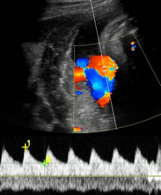
Identify monochorionic twins to guide pregnancy management
SAN FRANCISCO – The physician is responsible for knowing the chorionicity of a pregnant patient’s twins, so if you don’t offer diagnostic ultrasound or can’t make the diagnosis yourself, refer the patient to someone who can.
Knowing the chorionicity is essential because "it literally will define how you manage the pregnancy," Dr. Larry Rand said at a meeting on antepartum and intrapartum management sponsored by the University of California, San Francisco.

"This is a critical, critical concept," said Dr. Rand, director of perinatal services for the Fetal Treatment Program at the university. The chorionicity "should be the very first question that you ask yourself when you have a patient with twins."
You don’t need to be an expert on monochorionic twins; you just need to know if the patient is at high risk because of monochorionicity. Determining the chorionicity of twins before 14 weeks’ gestation is considered the standard of care, he stressed, and a physician who doesn’t know that twins are monochorionic could be liable if something goes wrong.
If the chorionicity is undetermined on your office ultrasound, refer the patient and request a chorionicity determination, he advised.
"This is one of the times that I have to say that ultrasound makes all the difference," Dr. Rand said. "The single most important ultrasound finding in the entire pregnancy is going to be the chorionicity."
A patient deserves to know whether her twins are monochorionic because she needs to be counseled appropriately about the risks. "It’s not the radiologist’s responsibility," he said. "The obstetrician is responsible for knowing the effect of chorionicity." Make sure to document that you have either determined the chorionicity yourself or have asked for an exam to assess chorionicity.
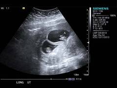
The rate of congenital anomalies in monochorionic twins, for example, is similar to the rate seen in diabetic mothers. This "changes the kind of tests you order and the things you see" compared with dichorionic twins, he said. Level II ultrasound examinations of anatomy that would be done in high-risk cases such as diabetic pregnancies, for example, should be done for monochorionic twins, whose elevated risk for anomalies may be due to an imperfect splitting of the single egg from which they came.
Compared with dichorionic twins, monochorionic twins also have increased risk for neurologic injury, including an eightfold increased incidence of cerebral palsy, and for preterm birth, spontaneous abortion, intrauterine growth restriction and growth discordance, intrauterine fetal demise, and twin-to-twin transfusion syndrome. Specialized centers can treat twin-to-twin transfusion syndrome with laser therapy if patients are diagnosed and referred in a timely manner – which is the reason for more-frequent screening of monochorionic twins. (See management recommendations below.)
Aterio-arterial (AA) anastomosis, a type of vascular connection within the placenta, can be detected on antenatal ultrasound. If present, the risk of twin-to-twin transfusion syndrome decreases 10-fold and the risk of intrauterine fetal demise decreases sevenfold. "It’s very helpful prognostically," he said.
Although AA anastomosis is not a new concept, it’s only recently that obstetrical imaging has evolved to identify it on antenatal ultrasound, which is "so helpful for counseling and management," said Dr. Rand, also the Lynne and Marc Benioff Endowed Chair in Maternal and Fetal Medicine at the university.
The major fear in monochorionic twins is intrauterine fetal demise of one twin, which leaves the surviving twin with a 10%-20% risk of death and a 20%-40% risk of long-term neurologic injury if the second twin survives. The difference even comes into play with genetic counseling. Aneuploidy risk in monochorionic twins is similar to risk in singletons (because they originated from a single egg). For invasive testing, one chorionic villus sampling covers both monochorionic twins (because there’s only one placenta to sample), but amniocentesis typically is performed on each amniotic sac. Some physicians sample just one amniotic sac for amniocentesis, thinking that it should cover both twins because they’re identical, but there are extremely rare cases of subtle differences that would not be detected by single-sac sampling, he said.

The other outcomes differ between monochorionic and dichorionic twins because the splitting of an egg can be imperfect; monochorionic twins share "plumbing" and vascular connections, and they have to share the resources of one placenta, Dr. Rand explained.
Before 14 weeks’ gestation, ultrasound is 99% sensitive and 100% specific for chorionicity, and it’s an easy assessment to do. In the second trimester, it gets trickier, which is why determining chorionicity early is so important, he said.
Dr. Rand gave several recommendations for management of monochorionic twins. In the first trimester, he said, determine the chorionicity, check the nuchal translucency, and assess fetal growth.
In the second trimester, conduct level II (high risk) anatomical surveys and echocardiograms to screen for fetal cardiac disease (as you would in a diabetic mother). Establish a minimum surveillance schedule of at least every 2 weeks for amniotic fluid between 16 and 28 weeks’ gestation, and every 4 weeks for fetal growth between 16 and 32 weeks’ gestation.
In the third trimester, continue the surveillance of amniotic fluid and fetal growth. Consider performing non–stress tests and Doppler ultrasounds. There’s debate about the optimal gestational age for delivery of monochorionic twins, between 36 and 38 weeks. "We’re not sure when to deliver," Dr. Rand said, but he recommends delivering monochorionic twins who’ve had issues with growth, fluid, or other aspects during pregnancy closer to 36 weeks and delivering twins with no issues closer to 38 weeks. "Monochorionicity is not, in and of itself, an indication for cesarean delivery," he added.
Approximately 75% of twins are dizygotic (from two separate eggs, each fertilized by sperm) and thus dichorionic. Among the 25% of twin pregnancies that are monozygotic (from one egg), the egg splits more slowly in 75% of cases, creating monochorionic twins that share a placenta and circulation. In the other 25% of monozygotic twins, the egg splits quickly, within 3 days, yielding two separate units and creating dichorionic twins, each with their own placenta.
Dr. Rand reported having no financial disclosures.
On Twitter @sherryboschert
SAN FRANCISCO – The physician is responsible for knowing the chorionicity of a pregnant patient’s twins, so if you don’t offer diagnostic ultrasound or can’t make the diagnosis yourself, refer the patient to someone who can.
Knowing the chorionicity is essential because "it literally will define how you manage the pregnancy," Dr. Larry Rand said at a meeting on antepartum and intrapartum management sponsored by the University of California, San Francisco.
"This is a critical, critical concept," said Dr. Rand, director of perinatal services for the Fetal Treatment Program at the university. The chorionicity "should be the very first question that you ask yourself when you have a patient with twins."
You don’t need to be an expert on monochorionic twins; you just need to know if the patient is at high risk because of monochorionicity. Determining the chorionicity of twins before 14 weeks’ gestation is considered the standard of care, he stressed, and a physician who doesn’t know that twins are monochorionic could be liable if something goes wrong.
If the chorionicity is undetermined on your office ultrasound, refer the patient and request a chorionicity determination, he advised.
"This is one of the times that I have to say that ultrasound makes all the difference," Dr. Rand said. "The single most important ultrasound finding in the entire pregnancy is going to be the chorionicity."
A patient deserves to know whether her twins are monochorionic because she needs to be counseled appropriately about the risks. "It’s not the radiologist’s responsibility," he said. "The obstetrician is responsible for knowing the effect of chorionicity." Make sure to document that you have either determined the chorionicity yourself or have asked for an exam to assess chorionicity.
The rate of congenital anomalies in monochorionic twins, for example, is similar to the rate seen in diabetic mothers. This "changes the kind of tests you order and the things you see" compared with dichorionic twins, he said. Level II ultrasound examinations of anatomy that would be done in high-risk cases such as diabetic pregnancies, for example, should be done for monochorionic twins, whose elevated risk for anomalies may be due to an imperfect splitting of the single egg from which they came.
Compared with dichorionic twins, monochorionic twins also have increased risk for neurologic injury, including an eightfold increased incidence of cerebral palsy, and for preterm birth, spontaneous abortion, intrauterine growth restriction and growth discordance, intrauterine fetal demise, and twin-to-twin transfusion syndrome. Specialized centers can treat twin-to-twin transfusion syndrome with laser therapy if patients are diagnosed and referred in a timely manner – which is the reason for more-frequent screening of monochorionic twins. (See management recommendations below.)
Aterio-arterial (AA) anastomosis, a type of vascular connection within the placenta, can be detected on antenatal ultrasound. If present, the risk of twin-to-twin transfusion syndrome decreases 10-fold and the risk of intrauterine fetal demise decreases sevenfold. "It’s very helpful prognostically," he said.
Although AA anastomosis is not a new concept, it’s only recently that obstetrical imaging has evolved to identify it on antenatal ultrasound, which is "so helpful for counseling and management," said Dr. Rand, also the Lynne and Marc Benioff Endowed Chair in Maternal and Fetal Medicine at the university.
The major fear in monochorionic twins is intrauterine fetal demise of one twin, which leaves the surviving twin with a 10%-20% risk of death and a 20%-40% risk of long-term neurologic injury if the second twin survives. The difference even comes into play with genetic counseling. Aneuploidy risk in monochorionic twins is similar to risk in singletons (because they originated from a single egg). For invasive testing, one chorionic villus sampling covers both monochorionic twins (because there’s only one placenta to sample), but amniocentesis typically is performed on each amniotic sac. Some physicians sample just one amniotic sac for amniocentesis, thinking that it should cover both twins because they’re identical, but there are extremely rare cases of subtle differences that would not be detected by single-sac sampling, he said.
The other outcomes differ between monochorionic and dichorionic twins because the splitting of an egg can be imperfect; monochorionic twins share "plumbing" and vascular connections, and they have to share the resources of one placenta, Dr. Rand explained.
Before 14 weeks’ gestation, ultrasound is 99% sensitive and 100% specific for chorionicity, and it’s an easy assessment to do. In the second trimester, it gets trickier, which is why determining chorionicity early is so important, he said.
Dr. Rand gave several recommendations for management of monochorionic twins. In the first trimester, he said, determine the chorionicity, check the nuchal translucency, and assess fetal growth.
In the second trimester, conduct level II (high risk) anatomical surveys and echocardiograms to screen for fetal cardiac disease (as you would in a diabetic mother). Establish a minimum surveillance schedule of at least every 2 weeks for amniotic fluid between 16 and 28 weeks’ gestation, and every 4 weeks for fetal growth between 16 and 32 weeks’ gestation.
In the third trimester, continue the surveillance of amniotic fluid and fetal growth. Consider performing non–stress tests and Doppler ultrasounds. There’s debate about the optimal gestational age for delivery of monochorionic twins, between 36 and 38 weeks. "We’re not sure when to deliver," Dr. Rand said, but he recommends delivering monochorionic twins who’ve had issues with growth, fluid, or other aspects during pregnancy closer to 36 weeks and delivering twins with no issues closer to 38 weeks. "Monochorionicity is not, in and of itself, an indication for cesarean delivery," he added.
Approximately 75% of twins are dizygotic (from two separate eggs, each fertilized by sperm) and thus dichorionic. Among the 25% of twin pregnancies that are monozygotic (from one egg), the egg splits more slowly in 75% of cases, creating monochorionic twins that share a placenta and circulation. In the other 25% of monozygotic twins, the egg splits quickly, within 3 days, yielding two separate units and creating dichorionic twins, each with their own placenta.
Dr. Rand reported having no financial disclosures.
On Twitter @sherryboschert
EXPERT ANALYSIS FROM A MEETING ON ANTEPARTUM AND INTRAPARTUM MANAGEMENT