User login
Mechanism of cGVHD response to ECP still unclear
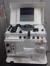
A prospective study did not reveal the mechanism driving response to extracorporeal photopheresis (ECP) in patients with chronic graft-versus-host disease (cGVHD).
However, researchers did find that responses occurred independent of risk factors, and results suggested that regulatory T cells (Tregs) are not the dominant mechanism of response to ECP.
Madan Jagasia, MBBS, of Vanderbilt University School of Medicine in Nashville, Tennessee, and his colleagues conducted this research and detailed the findings in Biology of Blood and Marrow Transplantation.
The study was funded by Therakos, Inc., a Mallinckrodt Pharmaceuticals Company.
The study included 77 patients with cGVHD. The median age at transplant was 49, 88% of patients were white, and 62% were male.
Patients had moderate (48%) or severe (52%) cGVHD. Sites of involvement included skin (86%), mouth (52%), gastrointestinal tract (29%), eye (62%), joint and fascia (51%), genital tract (11%), and lung (28%).
Patients had received a median of 2 (range, 0 to 7) prior lines of cGVHD therapy.
For this study, patients received 1689 ECP treatments, an average of 21.9 treatments per patient. The most common regimen was ECP twice a week for 4 weeks, then twice a week every 2 weeks for 8 weeks, then further tapering at the treating physician’s discretion.
Forty-eight patients (62.3%) completed all 6 months of ECP treatment. Reasons for early discontinuation included cGVHD progression (n=6), infection (n=4), cGVHD improvement (n=2), death from cGVHD-related cause (n=2), logistical issues (n=2), loss to follow-up (n=2), unknown reasons (n=4), and other various reasons (n=7) such as a finance issue and non-adherence.
Response
Provider-assessed response rates differed from response rates according to 2005 National Institutes of Health (NIH) consensus criteria.
According to providers, the response rate was 62.3% (48/77), with 14% of patients (n=11) achieving a complete response and 48% (n=37) attaining a partial response. Nineteen percent of patients (n=15) had stable disease, 14% (n=11) progressed, and 4% (n=3) did not have follow-up for cGVHD-related reasons.
Eight patients did not have enough data to assess NIH response. For the 69 evaluable patients, the NIH response rate was 43.5% (n=30), with 6% of patients (n=4) achieving a complete response and 38% (n=26) attaining a partial response. Fifteen percent of patients (n=10) had stable disease, 38% (n=26) progressed, and 4% (n=3) did not have follow-up for cGVHD-related reasons.
Risk factors
The researchers said there was no significant difference between responders and nonresponders when it came to age at treatment, platelet count, cGVHD severity, donor gender, and donor type.
For provider-assessed response, the median Karnofsky performance score at study entry was significantly higher in responders than nonresponders (90 vs 80; P=0.001). Responders also had a significantly shorter time from transplant to study entry compared to nonresponders (1.43 vs 2.06 years; P=0.051).
Similarly, according to NIH response, time from transplant to study entry was significantly shorter for responders than nonresponders (1.23 vs 1.92 years; P=0.04).
However, in a logistic regression model, time from transplant to study entry was not associated with provider-assessed or NIH response.
Tregs
The researchers found no significant difference in Treg percentages between responders and nonresponders.
For provider-assessed response, the baseline Treg frequency was 4.4% in responders and 4.8% in nonresponders (P=0.4). Treg percentages at the end of study were 4.2% and 5.5%, respectively (P=0.2). And the change in Treg frequency was 0.3% and 1.3%, respectively (P=0.3).
For NIH response, the baseline Treg frequency was 4.7% in responders and 4.4% in nonresponders (P=0.3). Treg percentages at the end of study were 4.4% and 4.7%, respectively (P=0.6). And the change in Treg percentages was 0.3% and 0.7%, respectively (P=0.4).
These findings run contrary to the researchers’ hypothesis that response to ECP would be associated with an increase in the percentage of Tregs.
The researchers did note that the number of Tregs varied between patients, and the team raised the possibility that the mechanism of Tregs in ECP is not visible by measuring cell abundance.
However, the researchers also said future studies should explore additional mechanisms of action for ECP and look particularly at other T-cell populations, dendritic cells, inhibitory cytokines, and proinflammatory cytokines.
OMS721 receives orphan designation for use in HSCT
The European Commission has granted orphan designation to OMS721 for use in the setting of hematopoietic stem cell transplant (HSCT).
OMS721 is a monoclonal antibody targeting MASP-2, the effector enzyme of the lectin pathway of the complement system.
Omeros Corporation is developing OMS721 as a treatment for HSCT-associated thrombotic microangiopathy (TMA) and other conditions.
OMS721 is currently under evaluation in a phase 2 trial (NCT02222545) of patients with HSCT-TMA, and Omeros released some results from this study in February.
Results were reported for 18 adults with HSCT-TMA persisting for at least 2 weeks following immunosuppressive regimen modification or more than 30 days post-transplant.
The patients received weekly OMS721 treatments for 4 to 8 weeks at the discretion of the investigator.
These patients had a significantly longer median overall survival than historical controls—347 days and 21 days, respectively (P<0.0001).
Omeros also reported that markers of TMA activity significantly improved following OMS721 treatment.
The mean platelet count increased from 18,100 x 106/mL at baseline to 52,300 x 106/mL (P=0.017). The mean LDH decreased from 591 U/L to 250 U/L (P<0.001). And the mean haptoglobin increased from 8 mg/dL to 141 mg/dL (P=0.003).
Mean creatinine remained stable—at approximately 120 μmol/L—but a majority of patients had co-existing conditions for which they were receiving nephrotoxic medications. These conditions included graft-versus-host disease, cytomegalovirus and human herpes virus 6 infections, prior sepsis, diffuse alveolar hemorrhage, and residual underlying malignancies.
The most commonly reported adverse events in this trial were diarrhea and neutropenia.
Four deaths occurred. One of these—due to acute renal and respiratory failure—was considered possibly related to OMS721.
The other deaths were due to progression of acute myeloid leukemia (n=1) and neutropenic sepsis (n=2).
About orphan designation
Orphan designation from the European Commission is available to companies developing products intended to treat a life-threatening or chronically debilitating condition that affects fewer than 5 in 10,000 people in the European Union.
The designation allows for financial and regulatory incentives that include 10 years of marketing exclusivity in the European Union after product approval, protocol assistance from the European Medicines Agency at reduced fees during the product development phase, and access to centralized marketing authorization.
Auto-HSCT linked to higher AML, MDS risk
Patients undergoing autologous hematopoietic stem cell transplant (auto-HSCT) for lymphoma or myeloma have an increased risk of acute myeloid leukemia (AML) and myelodysplastic syndromes (MDS), according to a retrospective study.
The study suggested these patients have 10 to 100 times the risk of AML or MDS as the general population.
The elevated risk also exceeds that of similar lymphoma and myeloma patients largely untreated with auto-HSCT.
Tomas Radivoyevitch, PhD, of the Cleveland Clinic Foundation in Ohio, and his colleagues reported these findings in Leukemia Research.
The investigators noted that exposure to DNA-damaging drugs and ionizing radiation—both used in auto-HSCT—is known to increase the risk of AML and MDS.
With this in mind, the team analyzed data on auto-HSCT recipients reported to the Center for International Blood and Marrow Transplant Research (CIBMTR).
Analyses were based on 9028 patients undergoing auto-HSCT from 1995 to 2010 for Hodgkin lymphoma (n=916), non-Hodgkin lymphoma (NHL, n=3546), or plasma cell myeloma (n=4566). Their median duration of follow-up was 90 months, 110 months, and 97 months, respectively.
Overall, 3.7% of the cohort developed AML or MDS after their transplant.
More aggressive transplant protocols increased the likelihood of this outcome. The risk of developing AML or MDS was higher for:
- Hodgkin lymphoma patients who received conditioning with total body radiation versus chemotherapy alone (hazard ratio [HR], 4.0)
- NHL patients who received conditioning with total body radiation (HR, 1.7) or with busulfan and melphalan or cyclophosphamide (HR, 1.8) versus the BEAM regimen (bischloroethylnitrosourea, etoposide, cytarabine, and melphalan)
- NHL or myeloma patients who received 3 or more lines of chemotherapy versus 1 line (HR, 1.9 for NHL and 1.8 for myeloma)
- NHL patients who underwent transplant in 2005 to 2010 versus 1995 to 1999 (HR, 2.1).
Patients reported to the Surveillance, Epidemiology and End Results database with the same lymphoma and myeloma diagnoses, few of whom underwent auto-HSCT, had risks of AML and MDS that were 5 to 10 times higher than the background level in the population.
However, the study auto-HSCT cohort had a risk of AML that was 10 to 50 times higher and a relative risk of MDS that was roughly 100 times higher than the background level.
“These increases may be related to exposure to high doses of DNA-damaging drugs given for [auto-HSCT], but this hypothesis can only be tested in a prospective study,” Dr Radivoyevitch and his coinvestigators wrote.
The reason for the greater elevation of MDS risk, compared with AML risk, is unknown.
“One possible explanation is that many cases of MDS evolve to AML, and that earlier diagnosis from increased post-transplant surveillance resulted in a deficiency of AML,” the investigators wrote. “A second is based on steeper MDS versus AML incidences versus age . . . and the possibility that transplantation recipient marrow ages (ie, marrow biological ages) are perhaps decades older than calendar ages.”
The study authors said they had no relevant conflicts of interest. The CIBMTR is supported by several US government agencies and numerous pharmaceutical companies.
FDA grants fast track designation to dilanubicel
The US Food and Drug Administration (FDA) has granted fast track designation to dilanubicel (NLA101) for use in patients with high-risk hematologic malignancies receiving an allogeneic cord blood transplant.
Dilanubicel is a universal-donor, ex-vivo-expanded hematopoietic stem and progenitor cell product.
It is intended to induce short-term hematopoiesis, which lasts until a patient’s immune system recovers.
However, dilanubicel may also produce long-term immunologic benefits and could potentially improve survival in transplant recipients, according to Nohla Therapeutics, the company developing the product.
Dilanubicel is manufactured ahead of time, cryopreserved, and intended for immediate off-the-shelf use.
Dilanubicel also has orphan drug designation from the FDA.
About fast track, orphan designations
The FDA’s fast track development program is designed to expedite clinical development and submission of applications for products with the potential to treat serious or life-threatening conditions and address unmet medical needs.
Fast track designation facilitates frequent interactions with the FDA review team, including meetings to discuss the product’s development plan and written communications about issues such as trial design and use of biomarkers.
Products that receive fast track designation may be eligible for accelerated approval and priority review if relevant criteria are met. Such products may also be eligible for rolling review, which allows a developer to submit individual sections of a product’s application for review as they are ready, rather than waiting until all sections are complete.
The FDA grants orphan designation to products intended to treat, diagnose, or prevent diseases/disorders that affect fewer than 200,000 people in the US. The designation provides incentives for sponsors to develop products for rare diseases. This may include tax credits toward the cost of clinical trials, prescription drug user fee waivers, and 7 years of market exclusivity if the product is approved.
Trials of dilanubicel
The fast track and orphan designations for dilanubicel were supported by data from a phase 2, single-center study. Results from this study were presented in a poster at the 23rd Congress of European Hematology Association (EHA) in June.
The trial included 15 patients with hematologic malignancies who underwent a cord blood transplant. Conditioning consisted of fludarabine (75 mg/m2), cyclophosphamide (120 mg/kg), and total body irradiation (13.2 Gy).
Patients received unmanipulated cord blood unit(s), followed 4 hours later by dilanubicel infusion. Prophylaxis for graft-vs-host disease (GVHD) was cyclosporine/mycophenolate mofetil.
The researchers compared outcomes in the 15 dilanubicel recipients to outcomes in a concurrent control cohort of 50 patients treated with the same transplant protocol, minus dilanubicel.
The time to neutrophil and platelet recovery were both significantly better in dilanubicel recipients than controls.
At day 100, the cumulative incidence of neutrophil recovery was 100% in dilanubicel recipients and 94% in controls (P=0.005). The median time to neutrophil recovery was 19 days (range, 9-31) and 25 days (range, 14-45), respectively.
The cumulative incidence of platelet recovery was 93% in dilanubicel recipients and 74% in controls (P=0.02). The median time to platelet recovery was 35 days (range, 21-86) and 48 days (range, 24-158), respectively.
At 100 days, there were no cases of grade 3-4 acute GVHD in dilanubicel recipients, but the incidence of grade 3-4 acute GVHD was 29% in the control group.
At 5 years, 27% of dilanubicel recipients had experienced chronic GVHD, compared to 38% of the control group.
There were no cases of transplant related mortality (TRM) in dilanubicel recipients, but the rate of TRM was 16% in the control group.
Two dilanubicel recipients (13%) relapsed post-transplant and subsequently died.
The 5-year disease-free survival rate was 87% in dilanubicel recipients and 66% in the control group. Overall survival rates were the same (87% and 66%, respectively).
Dilanubicel is currently under investigation in a phase 2b trial (NCT01690520) that has enrolled 160 patients with hematologic malignancies. The goal of the trial is to determine whether adding dilanubicel to standard donor cord blood transplant decreases the time to hematopoietic recovery, thereby reducing associated morbidities and mortality.
Another phase 2 trial, called LAUNCH (NCT03301597), is currently enrolling patients who have acute myeloid leukemia and chemotherapy-induced myelosuppression. The goals of this trial are to evaluate dilanubicel’s ability to reduce the rate of grade 3 or higher infections associated with chemotherapy-induced neutropenia and to identify the lowest effective cell dose of dilanubicel.
A new way to expand HSCs for UCB transplant
Researchers say they have discovered a new approach to expand hematopoietic stem cells (HSCs) from umbilical cord blood (UCB).
The team identified a protein, YTHDF2, that affects multiple targets and pathways involved in HSC self-renewal.
Experiments showed that reducing the function of YTHDF2 allowed UCB HSCs to expand.
The researchers therefore believe this approach could be used to improve UCB transplants.
“If we can expand cord adult stem cells, that could potentially decrease the number of cords needed per treatment,” said Linheng Li, PhD, of Stowers Institute for Medical Research in Kansas City, Missouri. “That’s a huge advantage.”
Dr Li and his colleagues conducted this research and described the work in Cell Research.
Past studies suggested that N6-methyladenosine (m6A) modulates the expression of a group of mRNAs that are critical for stem cell fate determination.
As the m6A reader YTHDF2 promotes targeted mRNA decay, Dr Li and his colleagues decided to target YTHDF2.
The researchers knocked out YTHDF2 function in a mouse model and observed an increase in functional HSCs. However, impairing YTHDF2 function did not alter lineage differentiation or lead to an increase in hematologic malignancies.
The researchers also knocked down YTHDF2 function in human UCB hematopoietic stem and progenitor cells. After 7 days of ex vivo culture, there was a roughly 14-fold increase in both the frequency and absolute number of HSCs with YTHDF2 knockdown (KD) cells compared to control cells.
When human UCB cells were transplanted into mice, there was a 9-fold increase in hematopoietic cell engraftment with YTHDF2 KD cells compared to control cells. In addition, the HSC frequency was about 4-fold higher in YTHDF2 KD cells.
The researchers transplanted bone marrow from primary recipient mice into sublethally irradiated secondary mice and found that, 12 weeks after transplant, human hematopoietic cell chimerism in the bone marrow was higher in YTHDF2 KD mice than in controls.
There was an 8-fold increase in competitive repopulating units from YTHDF2 KD cells compared to control cells.
As for why targeting YTHDF2 results in HSC expansion, the researchers found that YTHDF2 regulates HSC self-renewal gene expression by m6A-mediated mRNA decay.
The team discovered that m6A was enriched in mRNAs encoding transcription factors that are critical for stem cell self-renewal (such as GATA2, ETV6, STAT5, and TAL1). YTHDF2 recognizes these mRNAs and promotes their degradation.
“This work represents a path forward by demonstrating the ability to reliably expand adult stem cells from umbilical cord blood in the laboratory without terminally differentiating the cells into more mature and relatively short-lived blood cells,” said Joseph McGuirk, MD, a professor at the University of Kansas Health System who was not directly involved with this study.
“These findings represent a major advance in the field and have significant potential to improve the outcomes of thousands of children and adults who undergo umbilical cord blood transplantation every year.”
FDA advises against azithromycin use in allo-HSCT recipients
The US Food and Drug Administration (FDA) is warning against long-term use of azithromycin (Zithromax, Zmax) in patients who undergo allogeneic hematopoietic stem cell transplant (allo-HSCT).
Azithromycin has been used off-label as prophylaxis for bronchiolitis obliterans syndrome in these patients.
However, a trial published in JAMA last year suggested that long-term azithromycin use increases the risk of relapse and death in patients undergoing allo-HSCT as treatment for hematologic malignancies.
The FDA said it is reviewing additional data and will communicate its conclusions and recommendations when the review is complete.
In the meantime, the agency said healthcare professionals should not prescribe long-term azithromycin to allo-HSCT recipients for prophylaxis of bronchiolitis obliterans syndrome. However, patients should not stop taking azithromycin without first consulting their healthcare professionals.
Healthcare professionals and patients can report adverse events related to azithromycin to the FDA’s MedWatch program.
Pfizer, which markets Zithromax, has issued a Dear Healthcare Provider letter warning about the risks of relapse and death associated with long-term azithromycin use in allo-HSCT recipients.
The company said there is no evidence to suggest increased risks in other patient populations or when azithromycin is used for FDA-approved indications.
It isn’t clear why allo-HSCT recipients may have an increased risk of relapse/death with long-term azithromycin use. However, Pfizer said the available evidence raises questions about the safety of prophylactic azithromycin in this patient population, suggesting the risks outweigh the benefits.
The evidence comes from the ALLOZITHRO trial, which was published in JAMA in August 2017.
The trial included 480 patients who had undergone allo-HSCT for a hematologic malignancy. They were randomized to receive 250 mg of azithromycin (n=243) or placebo (n=237) 3 times a week for 2 years, beginning at the start of conditioning.
The trial was stopped about 13 months after enrollment was completed because there was an unexpected increase in the rate of relapse and death in patients taking azithromycin.
The 2-year cumulative incidence of relapse was 33.5% in the azithromycin group and 22.3% in the placebo group (unadjusted cause-specific hazard ratio [HR]=1.7, P=0.002).
The 2-year survival rate was 56.6% in the azithromycin group and 70.1% in the placebo group (adjusted HR=1.5, P=0.02).
The 2-year airflow decline-free survival rate was 32.8% in the azithromycin group and 41.3% in the placebo group (unadjusted HR=1.3, P=0.03).
And the incidence of bronchiolitis obliterans syndrome was 6% in the azithromycin group and 3% in the placebo group (P=0.08).
Kids may have higher risk of death long after allo-HSCT
Children may have an increased risk of premature death decades after allogeneic hematopoietic stem cell transplant (allo-HSCT), according to a study published in JAMA Oncology.
The leading causes of death in the patients studied were infection and chronic graft-vs-host disease (GVHD), patients’ primary disease, and subsequent cancers.
“This study shows that, while we are able to save the life of the child during their cancer treatment, we need to continue to provide proactive follow-up care with these types of patients throughout the rest of their life, as they are still an at-risk population,” said study author Smita Bhatia, MBBS, of the University of Alabama at Birmingham (UAB).
“The high intensity of therapeutic exposures at a young age lends itself to cause morbidities and organ compromise once they reach adulthood.”
Dr Bhatia and her colleagues conducted this retrospective study of children who underwent allo-HSCT between January 1, 1974, and December 31, 2010, and were followed until December 31, 2016.
The study included 1388 patients who lived 2 years or more after transplant. Their median age at transplant was 14.6 years (range, 0-21). The majority of patients were non-Hispanic white (70.7%), and most were male (59.7%).
Patients underwent allo-HSCT to treat acute lymphoblastic leukemia (ALL, 25.1%), acute myeloid leukemia (AML) or myelodysplastic syndromes (MDS, 23.5%), inborn errors of metabolism (13.8%), severe aplastic anemia (SAA, 10.6%), Fanconi anemia (8.3%), chronic myelogenous leukemia (CML, 6.5%), immune disorders (4%), sickle cell disease or thalassemia (1.9%), and other diseases.
Most patients had a related donor (57.9%), and most received a bone marrow transplant (73.4%).
The most common component of conditioning was cyclophosphamide (80.5%), followed by total body irradiation (TBI, 64.3%). About half of patients (49.8%) received both cyclophosphamide and TBI, and nearly a quarter (23.5%) received busulfan and cyclophosphamide.
Outcomes
The researchers found that allo-HSCT recipients had a 14.4-fold greater risk of premature death than the general population.
The team said the absolute excess risk of all-cause mortality was 12.0 per 1000 person-years, and relative mortality remained elevated 25 years or more after transplant (standardized mortality ratio, 2.9).
At a median follow-up of 14.9 years (range, 2.0 to 41.2), 295 patients had died. The 20-year overall survival rate was 79.3%.
The cause of death was available for 82.7% of patients (244/295), and some of these patients had more than 1 cause listed. Causes of death included:
- Infection and/or chronic GVHD—49.6%
- Primary disease—24.6%
- Subsequent malignant neoplasm—18.4%
- Cardiac disease—9.8%
- Pulmonary disease—7.8%
- External causes—2.9%
- Other causes—18.0%.
The hazard of all-cause late mortality was higher among patients who were older at transplant (hazard ratio [HR], 1.03; P=0.004) and those who had a high risk of relapse at transplant (HR, 1.95; P<0.001).
Compared to patients treated for ALL, the hazard of all-cause late mortality was lower among patients with AML/MDS (HR, 0.72; P=0.04), CML (HR, 0.53; P=0.02), Fanconi anemia (HR, 0.49; P=0.03), immune disorders (HR, 0.32; P=0.006), and SAA (HR, 0.33; P<0.001).
The hazard of all-cause late mortality was lower for patients who received conditioning with busulfan and cyclophosphamide (HR, 0.62; P=0.03) than for those who received TBI and cyclophosphamide.
Compared to patients treated for ALL, the hazard of relapse-related mortality was lower among patients with AML/MDS (HR, 0.39; P=0.01) and SAA (HR, 0.09; P=0.03), and the hazard of non-relapse mortality was lower for patients with SAA (HR, 0.36; P=0.004) and immune disorders (HR, 0.14; P=0.009).
The hazard of non-relapse mortality was higher for patients who were older at transplant (HR, 1.03; P=0.03), patients who received peripheral blood stem cells rather than bone marrow (HR, 2.39; P=0.01), and patients who had a high risk of relapse at transplant (HR, 2.05; P<0.001).
Orphan designation recommended for OMS721
The European Medicines Agency’s (EMA’s) Committee for Orphan Medicinal Products (COMP) has issued a positive opinion recommending orphan drug designation for OMS721 as a treatment for high-risk hematopoietic stem cell transplant-associated thrombotic microangiopathy (HSCT-TMA).
OMS721 is a monoclonal antibody targeting MASP-2, the effector enzyme of the lectin pathway of the complement system.
The COMP’s positive opinion of OMS721 for HSCT-TMA is expected to be adopted by the European Commission in August.
Orphan drug designation in Europe is available to companies developing products intended to treat a life-threatening or chronically debilitating condition that affects fewer than 5 in 10,000 people in the European Union (EU).
This designation allows for financial and regulatory incentives that include 10 years of marketing exclusivity in the EU after product approval, reduced EMA advisory, inspection and filing fees pre- and post-approval, and guaranteed access to centralized marketing authorization valid in all EU member states as well as in European Economic Area countries (ie, Iceland, Liechtenstein, and Norway).
Phase 2 trial
OMS721 is currently under evaluation in a phase 2 trial (NCT02222545). Omeros Corporation, the company developing OMS721, released some results from this study in February.
Results were reported for 18 adults with HSCT-TMA persisting for at least 2 weeks following immunosuppressive regimen modification or more than 30 days post-transplant. The patients received weekly OMS721 treatments for 4 to 8 weeks at the discretion of the investigator.
These patients had a significantly longer median overall survival than historical controls—347 days and 21 days, respectively (P<0.0001).
Omeros also reported that markers of TMA activity significantly improved following OMS721 treatment.
The mean platelet count increased from 18,100 x 106/mL at baseline to 52,300 x 106/mL (P=0.017). The mean LDH decreased from 591 U/L to 250 U/L (P<0.001). And the mean haptoglobin increased from 8 mg/dL to 141 mg/dL (P=0.003).
Mean creatinine remained stable—at approximately 120 μmol/L—but a majority of patients had co-existing conditions for which they were receiving nephrotoxic medications. These conditions included graft-versus-host disease, cytomegalovirus and human herpes virus 6 infections, prior sepsis, diffuse alveolar hemorrhage, and residual underlying malignancies.
The most commonly reported adverse events in this trial were diarrhea and neutropenia.
Four deaths occurred. One of these—due to acute renal and respiratory failure—was considered possibly related to OMS721.
The other deaths were due to progression of acute myeloid leukemia (n=1) and neutropenic sepsis (n=2).
Study could change treatment of MLSM7
New findings could help improve treatment of an inherited bone marrow disorder known as myelodysplasia and leukemia syndrome with monosomy 7 (MLSM7), according to researchers.
While studying families affected by MLSM7, researchers identified germline mutations in SAMD9L or SAMD9 in patients who had hematologic abnormalities, myelodysplastic syndromes (MDS), or acute myeloid leukemia (AML).
However, these mutations were also present in apparently healthy family members, and the researchers found that bone marrow monosomy 7 sometimes resolved without treatment.
The team recounted these findings in JCI Insight.
The researchers analyzed blood samples from 16 siblings in 5 families affected by MLSM7 and found they all carried germline mutations in SAMD9 or SAMD9L. In 3 of the 5 families, there were apparently healthy parents who also carried the mutations.
“Surprisingly, the health consequences of these mutations varied tremendously for reasons that must still be determined, but the findings are already affecting how we may choose to manage these patients,” said study author Jeffery Klco, MD, PhD, of St. Jude Children’s Research Hospital in Memphis, Tennessee.
Three of the 16 siblings developed AML and died of the disease or related complications. Two other siblings were diagnosed with MDS.
The remaining 11 siblings with the mutations were apparently healthy, although several had been treated for anemia and other conditions associated with low blood counts.
Some of these patients had a previous history of bone marrow monosomy 7 that spontaneously corrected over time. These patients, despite no therapy, appeared to have normal bone marrow function.
“This was an even greater surprise,” Dr Klco said. “The spontaneous recovery experienced by some children with the germline mutations suggests some patients with SAMD9 and SAMD9L mutations who were previously considered candidates for bone marrow transplantation may recover hematologic function on their own.”
Dr Klco and his colleagues have a theory that could explain the spontaneous correction. The team noted that SAMD9 and SAMD9L are activated in response to viral infections. While the normal function of both proteins is poorly understood, abnormally activated SAMD9 and SAMD9L are known to inhibit cell growth.
In this study, deep sequencing showed that selective pressure on developing blood cells favors cells without the SAMD9 or SAMD9L mutations. That may increase pressure for cells to selectively jettison chromosome 7 with the gene alteration or take other molecular measures to counteract the mutant protein.
Implications for treatment
This research also showed that, in patients who developed AML, loss of chromosome 7 was associated with the development of mutations in additional genes, including ETV6, KRAS, SETBP1, and RUNX1.
These same mutations are broadly associated with monosomy 7 in AML, which suggests that understanding how SAMD9 and SAMD9L mutations contribute to leukemia has implications beyond familial cases.
The presence of secondary mutations may also help clinicians identify which patients will benefit from immediate treatment, including chemotherapy or transplant to prevent or treat AML or myelodysplasia, Dr Klco said.
For patients without the mutations or significant symptoms due to low blood cell counts, watchful waiting with careful follow-up may sometimes be an option.
“Now that we know this disease can resolve without treatment in some patients, we need to focus on developing screening and treatment guidelines,” Dr Klco said. “We want to reserve hematopoietic bone marrow transplantation for those who truly need the procedure. These findings will help to point the way.”
“So little is known about SAMD9 and SAMD9L that we need to continue working in the lab to better understand how these mutations impact blood cell development and how they are activated in response to infections and other types of stress.”
Treatments, disease affect spermatogonia in boys
Alkylating agents, hydroxyurea (HU), and certain non-malignant diseases can significantly deplete spermatogonial cell counts in young boys, according to research published in Human Reproduction.
Boys who received alkylating agents to treat cancer had significantly lower spermatogonial cell counts than control subjects or boys with malignant/nonmalignant diseases treated with non-alkylating agents.
Five of 6 SCD patients treated with HU had a totally depleted spermatogonial pool, and the remaining patient had a low spermatogonial cell count.
Five boys with non-malignant diseases who were not exposed to chemotherapy had significantly lower spermatogonial cell counts than controls.
“Our findings of a dramatic decrease in germ cell numbers in boys treated with alkylating agents and in sickle cell disease patients treated with hydroxyurea suggest that storing frozen testicular tissue from these boys should be performed before these treatments are initiated,” said study author Cecilia Petersen, MD, PhD, of Karolinska Institutet and University Hospital in Stockholm, Sweden.
“This needs to be communicated to physicians as well as patients and their parents or carers. However, until sperm that are able to fertilize eggs are produced from stored testicular tissue, we cannot confirm that germ cell quantity might determine the success of transplantation of the tissue in adulthood. Further research on this is needed to establish a realistic fertility preservation technique.”
Dr Petersen and her colleagues also noted that preserving testicular tissue may not be a viable option for boys who have low spermatogonial cell counts prior to treatment.
Patients and controls
For this study, the researchers analyzed testicular tissue from 32 boys facing treatments that carried a high risk of infertility—testicular irradiation, chemotherapy, or radiotherapy in advance of stem cell transplant.
Twenty boys had the tissue taken after initial chemotherapy, and 12 had it taken before starting any treatment.1
Eight patients had received chemotherapy with non-alkylating agents, 6 (all with malignancies) had received alkylating agents, and 6 (all with SCD) had received HU.
Diseases included acute lymphoblastic leukemia (n=6), SCD (n=6), acute myeloid leukemia (n=3), thalassemia major (n=3), neuroblastoma (n=2), juvenile myelomonocytic leukemia (n=2), myelodysplastic syndromes (n=2), primary immunodeficiency (n=2), Wilms tumor (n=1), adrenoleukodystrophy (n=1), hepatoblastoma (n=1), primitive neuroectodermal tumor (n=1), severe aplastic anemia (n=1), and Fanconi anemia (n=1).
The researchers compared samples from these 32 patients to 14 healthy testicular tissue samples stored in the biobank at the Karolinska University Hospital.
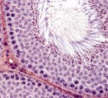
For both sample types, the team counted the number of spermatogonial cells found in a cross-section of seminiferous tubules.
“We could compare the number of spermatogonia with those found in the healthy boys as a way to estimate the effect of medical treatment or the disease itself on the future fertility of a patient,” explained study author Jan-Bernd Stukenborg, PhD, of Karolinska Institutet and University Hospital.
Impact of treatment
There was no significant difference in the mean quantity of spermatogonia per transverse tubular cross-section (S/T) between patients exposed to non-alkylating agents (1.7 ± 1.0, n=8) and biobank controls (4.1 ± 4.6, n=14).
However, samples from patients who received alkylating agents had a significantly lower mean S/T value (0.2 ± 0.3, n=6) than samples from patients treated with non-alkylating agents (P=0.003) and biobank controls (P<0.001).
“We found that the numbers of germ cells present in the cross-sections of the seminiferous tubules were significantly depleted and close to 0 in patients treated with alkylating agents,” Dr Stukenborg said.
Samples from the SCD patients also had a significantly lower mean S/T value (0.3 ± 0.6, n=6) than biobank controls (P=0.003).
Dr Stukenborg noted that the germ cell pool was totally depleted in 5 of the boys with SCD, and the pool was “very low” in the sixth SCD patient.
“This was not seen in patients who had not started treatment or were treated with non-alkylating agents or in the biobank tissues,” Dr Stukenborg said.2
He and his colleagues noted that it is possible for germ cells to recover to normal levels after treatment that is highly toxic to the testes, but high doses of alkylating agents and radiotherapy to the testicles are strongly associated with permanent or long-term infertility.
“The first group of boys who received bone marrow transplants are now reaching their thirties,” said study author Kirsi Jahnukainen, MD, PhD, of Helsinki University Central Hospital in Finland.
“Recent data suggest they may have a high chance of their sperm production recovering, even if they received high-dose alkylating therapies, so long as they had no testicular irradiation.”
Impact of disease
The researchers also found evidence to suggest that, for some boys, their disease may have affected spermatogonial cell counts before any treatment began.
Five patients with non-malignant disease who had not been exposed to chemotherapy (3 with thalassemia major, 1 with Fanconi anemia, and 1 with primary immunodeficiency) had a significantly lower mean S/T value (0.4 ± 0.5) than controls (P=0.006).
“Among patients who had not been treated previously with chemotherapy, there were several boys with a low number of germ cells for their age,” Dr Jahnukainen said.
“This suggests that some non-malignant diseases that require bone marrow transplants may affect the fertility of young boys even before exposure to therapy that is toxic for the testes.”
The researchers noted that a limitation of this study was that biobank samples had no detailed information regarding previous medical treatments and testicular volumes.
1. Testicular tissue is taken from patients under general anesthesia. The surgeon removes approximately 20% of the tissue from the testicular capsule in one of the testicles. For this study, a third of the tissue was taken to the Karolinska Institutet for analysis.
2. A recent meta-analysis showed that normal testicular tissue samples of newborns contain approximately 2.5 germ cells per tubular cross-section. This number decreases to approximately 1.2 within the first 3 years of age, followed by an increase up to 2.6 germ cells per tubular cross-section at 6 to 7 years, reaching a plateau until the age of 11. At the onset of puberty, an increase of up to 7 spermatogonia per tubular cross-section could be observed.
