User login
Doug Brunk is a San Diego-based award-winning reporter who began covering health care in 1991. Before joining the company, he wrote for the health sciences division of Columbia University and was an associate editor at Contemporary Long Term Care magazine when it won a Jesse H. Neal Award. His work has been syndicated by the Los Angeles Times and he is the author of two books related to the University of Kentucky Wildcats men's basketball program. Doug has a master’s degree in magazine journalism from the S.I. Newhouse School of Public Communications at Syracuse University. Follow him on Twitter @dougbrunk.
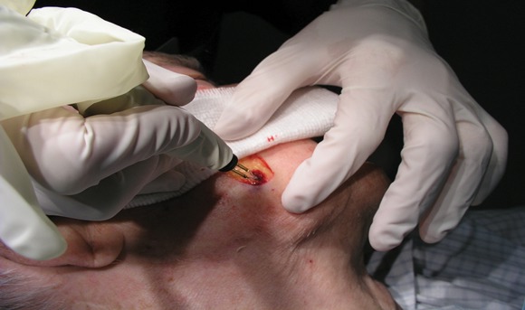
Don't Penny-Pinch on Dermatologic Surgery Instruments
SAN DIEGO – If you perform excisional surgery in your dermatology practice, don’t skimp on instrumentation, advised Dr. David E. Kent.
"Spend your money on instrumentation that’s going to get you out of trouble," Dr. Kent said at the meeting, which was sponsored by the University of California, San Diego School of Medicine and the Scripps Clinic.

His list of recommended instrumentation includes hemostats to clamp and tie off blood vessels, skin hooks to improve visualization, suction to remove excess blood, cotton tip applicators, and 4-by-4-inch gauze. "Be aware that the least expensive gauze may not have the best quality, so you want to evaluate different vendors," said Dr. Kent, a clinical instructor in the division of dermatology at the Medical College of Georgia, Augusta.
He also recommends having electrosurgical devices on hand, liquid thrombin, Gelfoam, and oxidized cellulose to place in wounds that are going to heal by second intention. Xenografts, "which can be helpful for temporary hemostasis over a wound with exposed muscle, may serve as a very nice scaffold to seal the wound and are easy to apply," he said.
Applying pressure to the wound after surgery is key, he added. "In all of our patients who are on any aspirin products, after any closure, my nurse holds pressure for 10 minutes. We’ve found that to be very helpful."
He finds the Geiger Thermal Cautery Unit useful for patients who have implantable cardiac defibrillators. "We did a study of this unit years ago and found that a setting between 6 and 7.5 is fairly ideal," Dr. Kent said. "It holds its temperature reasonably well in a wet field, compared with handheld units."
For handheld cautery, he recommends the LMA Perfect Temp device for isolated small pinpoint areas of bleeding. For solid state electrosurgical generators, "there are many manufacturers including Valleylab, Bard Medical, and Aaron Medical, to name a few," he said. "When using electrosurgical devices, it is important to avoid skin edges. This can be done by approaching the bleeding site at 90 degrees to the skin edge to avoid epidermal thermal injury. Use the lowest possible setting to control bleeding."
Another worthwhile instrument to have is a hemostatic scalpel, which provides heat energy to seal vessels and tissue. "It's excellent for skeletal muscle and large defects into muscle," Dr. Kent said. "If you're doing a lot of larger cases, it can really help you avoid excessive bleeding. But they are costly," he said. Used hemostatic scalpels can cost as much as $5,000. Blades cost $10 apiece and are not reusable.
If postoperative bleeding occurs after the patient has gone home, see the patient as soon as possible. "The next day is not soon enough," Dr. Kent said. "Have someone there to help you; make sure you have a nurse on call if you need one." On return, make sure the patient's vital signs are stable. Is the bandage soiled? Is there active bleeding? "Consider removing one or two sutures to see if there is brisk bleeding," Dr. Kent said. "Try to establish if it is a single skin edge or something more. If uncertain, you may need to take the entire closure down, inspect, and control what is bleeding."
Dr. Kent said that he had no relevant financial conflicts to disclose.
SAN DIEGO – If you perform excisional surgery in your dermatology practice, don’t skimp on instrumentation, advised Dr. David E. Kent.
"Spend your money on instrumentation that’s going to get you out of trouble," Dr. Kent said at the meeting, which was sponsored by the University of California, San Diego School of Medicine and the Scripps Clinic.
His list of recommended instrumentation includes hemostats to clamp and tie off blood vessels, skin hooks to improve visualization, suction to remove excess blood, cotton tip applicators, and 4-by-4-inch gauze. "Be aware that the least expensive gauze may not have the best quality, so you want to evaluate different vendors," said Dr. Kent, a clinical instructor in the division of dermatology at the Medical College of Georgia, Augusta.
He also recommends having electrosurgical devices on hand, liquid thrombin, Gelfoam, and oxidized cellulose to place in wounds that are going to heal by second intention. Xenografts, "which can be helpful for temporary hemostasis over a wound with exposed muscle, may serve as a very nice scaffold to seal the wound and are easy to apply," he said.
Applying pressure to the wound after surgery is key, he added. "In all of our patients who are on any aspirin products, after any closure, my nurse holds pressure for 10 minutes. We’ve found that to be very helpful."
He finds the Geiger Thermal Cautery Unit useful for patients who have implantable cardiac defibrillators. "We did a study of this unit years ago and found that a setting between 6 and 7.5 is fairly ideal," Dr. Kent said. "It holds its temperature reasonably well in a wet field, compared with handheld units."
For handheld cautery, he recommends the LMA Perfect Temp device for isolated small pinpoint areas of bleeding. For solid state electrosurgical generators, "there are many manufacturers including Valleylab, Bard Medical, and Aaron Medical, to name a few," he said. "When using electrosurgical devices, it is important to avoid skin edges. This can be done by approaching the bleeding site at 90 degrees to the skin edge to avoid epidermal thermal injury. Use the lowest possible setting to control bleeding."
Another worthwhile instrument to have is a hemostatic scalpel, which provides heat energy to seal vessels and tissue. "It's excellent for skeletal muscle and large defects into muscle," Dr. Kent said. "If you're doing a lot of larger cases, it can really help you avoid excessive bleeding. But they are costly," he said. Used hemostatic scalpels can cost as much as $5,000. Blades cost $10 apiece and are not reusable.
If postoperative bleeding occurs after the patient has gone home, see the patient as soon as possible. "The next day is not soon enough," Dr. Kent said. "Have someone there to help you; make sure you have a nurse on call if you need one." On return, make sure the patient's vital signs are stable. Is the bandage soiled? Is there active bleeding? "Consider removing one or two sutures to see if there is brisk bleeding," Dr. Kent said. "Try to establish if it is a single skin edge or something more. If uncertain, you may need to take the entire closure down, inspect, and control what is bleeding."
Dr. Kent said that he had no relevant financial conflicts to disclose.
EXPERT ANALYSIS FROM A MEETING ON SUPERFICIAL ANATOMY AND CUTANEOUS SURGERY
Early Carotid Thickening Seen in Type 1 Diabetes
SAN DIEGO – Adolescents and young adults with type 1 diabetes have thicker and stiffer carotid arteries, compared with their healthy peers, results from a multicenter study showed.
“Type 1 diabetes has an adverse effect on carotid thickness and stiffness, and we can measure this by the time patients reach young adulthood,” Dr. Elaine M. Urbina said at the meeting. “It's independent of demographics, lipids, and blood pressure, but may be influenced by adiposity. We need to control risk factors, especially obesity, in these adolescents and young adults to improve cardiovascular outcomes in type 1 diabetes.”
As part of the SEARCH CVD study, a collaboration between investigators at the University of Colorado at Denver, the Colorado School of Public Health in Aurora, and Cincinnati Children's Hospital Medical Center, Dr. Urbina and her associates set out to examine whether type 1 diabetes has a measurable effect on carotid arteries in adolescents and young adults. They studied 162 people aged 13-26 years, collecting data on demographics, anthropometrics, blood pressure, fasting lipid and hemoglobin A1c levels, and carotid ultrasound to measure the common, bulb, and internal carotid intima-media thickness (cIMT).
Of the 162 study participants, 127 (78%) had type 1 diabetes and 35 were healthy controls who attended clinics at the two locations, said Dr. Urbina, director of preventive cardiology at Cincinnati Children's Hospital. Their mean age was 20 years, 51% were male, 81% were white, and their mean duration of diabetes was 10 years.
Dr. Urbina reported that there were significantly higher proportions of males and whites among cases, compared with controls (55% vs. 34% and 90% vs. 50%, respectively), but there were no significant differences between the two groups in anthropometric or lipid values.
After adjustment for age, sex, race, mean arterial pressure by mercury sphygmomanometry, and lipids, patients with type 1 diabetes had a significantly thicker internal cIMT, compared with controls (mean, 0.56 mm vs. 0.50 mm, respectively), with a trend for a thicker common cIMT (mean, 0.63 mm vs. 0.60 mm). Bulb cIMT was the same in both groups (mean, 0.61 mm).
Patients with type 1 diabetes also had significantly stiffer carotids, compared with controls (mean PEM, 193 vs. 169 mm Hg, respectively; mean YEM, 204 vs. 182 mm Hg/mm; mean Einc, 963 vs. 862 mm Hg).
After adjustment for body mass index, there was a trend only for significantly thicker internal cIMT, although PEM remained stiffer for the patients with type 1 diabetes who were at least 20 years old.
SEARCH CVD is funded by the National Institutes of Health and is an ancillary study of the SEARCH for Diabetes in Youth study, funded by the Centers for Disease Control and Prevention and the NIH.
Dr. Urbina had no relevant disclosures.
From the Annual Scientific Sessions of the American Diabetes Association
Screening Using HbA1c Misclassifies Many
Major Finding: In patients with diabetes by an oral glucose tolerance test, HbA1c classification by International Expert Committee criteria labeled 32% correctly, 38% incorrectly as having prediabetes, and 29% incorrectly as being normal. American Diabetes Association criteria labeled 32% correctly, 50% incorrectly as having prediabetes and 18% incorrectly as being normal; and Veterans Affairs/Department of Defense criteria labeled 12% correctly, 71% incorrectly as having prediabetes, and 18% incorrectly as being normal.
Data Source: A study of 789 individuals from the Atlanta VA Medical Center that set out to examine the use of targeted screening to detect prediabetes and diabetes, and to compare HbA1c testing with the oral glucose tolerance test.
Disclosures: The study was supported by a grant from the VA's Health Services Research and Development Service. Ms. Jackson said that she had no relevant financial conflicts of interest.
SAN DIEGO – Slightly more than half of veterans targeted for screening have unrecognized diabetes or prediabetes, results from a recent analysis showed.
However, screening such patients by measuring hemoglobin A1c “would result in major misclassification – missing disease when it is present and, to a lesser extent, mislabeling normals as having disease,” Sandra L. Jackson, M.P.H., said at the meeting.
The findings are based on a study of 789 individuals from the Atlanta VA Medical Center that assessed the use of targeted screening to detect prediabetes and diabetes, and to compare HbA1c testing with the oral glucose tolerance test (OGTT), said Ms. Jackson, a graduate student in the nutrition and health sciences doctoral program at Emory University, Atlanta.
Although screening to detect unrecognized diabetes and prediabetes is recommended, the best strategy for screening in patients in primary care settings is unknown. The upside of the OGTT, Ms. Jackson said, is that it's established in clinical use, it can detect all patients with prediabetes, “and the glucose measurement itself is accurate. On the downside, it requires [fasting] and morning testing. It's burdensome for patients and health care systems, and it has poor day-to-day reproducibility.”
The upside of HbA1c, she continued, is that it does not require a fast, “and it's only a single blood draw, so it's much more convenient, there's less day-to-day variation, and there's greater preanalytic stability. On the downside, measurement may be problematic as platforms vary, point-of-care testing can be unreliable, there's a lack of agreement on cutoffs, and there may be racial and age disparities such that blacks and older persons may have higher HbA1c independent of glucose.”
The researchers defined hyperglycemia according to American Diabetes Association (ADA) criteria: prediabetes as a fasting OGTT of 100-125 mg/dL or a 2-hour OGTT of 149-199 mg/dL, and diabetes as a fasting OGTT of 126 mg/dL or greater or a 2-hour OGTT of 200 mg/dL or greater.
They categorized HbA1c results according to three sets of diagnostic criteria: the International Expert Committee (IEC) (prediabetes 6.0%-6.4%, diabetes 6.5% or greater), ADA (prediabetes 5.7%-6.4%, diabetes 6.5% or greater), and the Department of Veterans Affairs/Department of Defense (VA/DoD) (prediabetes 5.7%-6.9%, diabetes 7.0% or greater).
The mean age of the 789 study participants was 58 years, 95% were men, 74% were black, and their mean BMI was 30.5 kg/m
Screening was offered to patients meeting National Institutes of Health guidelines for screening: without known diabetes, and with age greater than 45 years or a BMI of more than 25 with another risk factor.
Fully 10% of patients met criteria for diabetes based on the OGTT, which was a higher rate compared with the HbA1c guidelines (6.7% by the IEC, 6.7% by the ADA, and 1.5% by the VA/DoD guidelines, respectively). “This would indicate that these cutoffs are insensitive compared with the OGTT for detecting diabetes,” she said.
According to the OGTT, 42% had prediabetes: 27% had isolated impaired fasting glucose, 6% had isolated impaired glucose tolerance, and 9% had both.
In patients with diabetes by OGTT criteria, HbA1c classification by IEC criteria labeled 32% correctly, 38% incorrectly as having prediabetes, and 29% incorrectly as being normal; ADA criteria labeled 32% correctly, 50% incorrectly as having prediabetes, and 18% incorrectly as being normal; and VA/DoD criteria labeled 12% correctly, 71% incorrectly as having prediabetes, and 18% incorrectly as being normal.
In patients with prediabetes by OGTT criteria, HbA1c classification by IEC criteria labeled 36% correctly, 6% incorrectly as having diabetes, and 59% incorrectly as being normal; ADA criteria labeled 61% correctly, 6% incorrectly as having diabetes, and 33% incorrectly as being normal; and VA/DoD criteria labeled 66% correctly, 1% incorrectly as having diabetes, and 33% incorrectly as being normal.
The prevalence of diabetes increased in a stepwise fashion with increasing BMI, from 1.5% among those with a normal BMI (18.5-24.9) to 15% among those who met criteria for class III obesity (BMI more than 40). “For every 1 unit increase in BMI, we observed a 10% increase in the odds of having diabetes,” she said.
Ms. Jackson also reported that with the IEC, ADA, and VA/DoD cutoffs for diabetes, screening with HbA1c was specific but insensitive, with a false-negative rate of 68% at the 6.5% cutoff and a false-negative rate of 89% at the 7.0% cutoff.
Vitals
Source Elsevier Global Medical News
From the Annual Scientific Sessions of the American Diabetes Association
Life Expectancy Improving for People With Type 1 Diabetes
SAN DIEGO – Life expectancy of patients with type 1 diabetes has improved dramatically since 1950, results from a long-term prospective study have shown.
According to the Pittsburgh Epidemiology of Diabetes Complications (EDC) Study, life expectancy at birth for those diagnosed with type 1 diabetes during 1965-1980 was 68.8 years, or about 4 years less than that of a comparable cohort of the U.S. general population, while life expectancy for those diagnosed during 1950-1964 was 53.4 years, or about 18 years less than that of a comparable cohort of the general population.
“Individuals with childhood-onset diabetes do not represent a major insurance risk and should be minimally penalized, if at all, in terms of life insurance and other mortality-based decisions,” Dr. Trevor J. Orchard, professor of epidemiology, pediatrics, and medicine at the University of Pittsburgh, said at the meeting.
The objective of the current study was to compare the life expectancy of two different cohorts of patients enrolled in the Pittsburgh EDC Study, a prospective study of childhood-onset type 1 diabetes. Of the 933 patients, 390 were diagnosed or seen within 1 year of diagnosis at Children's Hospital of Pittsburgh during 1950-1964 (cohort 1), while 543 were diagnosed or seen within a year of diagnosis during 1965-1980 (cohort 2). Half of the participants were female, and their mean age at diagnosis was 8 years. All were followed through 2009.
To ascertain mortality, the researchers used death certificates and hospital, autopsy, and coroner reports.
Dr. Orchard reported that the 30-year mortality for patients in cohort 1 was 35% compared with 12% for those in cohort 2. Similarly, the life expectancy at birth for those in cohort 1 was estimated to be 53.4 years compared with 68.8 years for those in cohort 2, a difference of about 15 years. Both differences were significant. This persisted regardless of sex or pubertal status at diagnosis of diabetes.
The life expectancy of cohort 2 is 3.6 years less than that estimated for a comparable cohort of the U.S. general population (72.4 years), Dr. Orchard said, while the life expectancy of cohort 1 is about 18 years less than that of a comparable cohort of the general population (71.5 years).
Reasons for the improvement in life expectancy between the two cohorts are multifactorial, he said, including the development of blood glucose self-monitoring and the use of the hemoglobin A1c test, which was not available in cohort 1.
Dr. Orchard is a consultant for AstraZeneca and Abbott, received research support from VeraLight, and has inherited Bristol-Myers Squibb stock.
To view an interview with Dr. Orchard, scan this using your smartphone.
'Individuals with childhood-onset diabetes do not represent a major insurance risk.'
Source DR. ORCHARD
From the Annual Scientific Sessions of the American Diabetes Association
Exenatide Delivery Device Shows Promise
SAN DIEGO – Treatment with the investigational device ITCA 650 delivering exenatide at 20 mcg/day and dose escalation to 60 mcg/day was well tolerated and led to significant reductions in hemoglobin A1c and body weight in patients with type 2 diabetes, results from a phase II, 48-week extension study showed.
ITCA 650 is manufactured by Intarcia Therapeutics of Hayward, Calif., and is a matchstick-size, osmotic mini-pump using the DUROS technology that is placed subcutaneously, providing continuous and consistent delivery of exenatide (Byetta) at specified doses. The device is inserted during a 10- to 15-minute office procedure. ITCA 650 has been shown to be effective in lowering HbA1c and having a favorable weight profile at 12 and 24 weeks, Dr. Julio Rosenstock said at the meeting, where he presented the results of the 48-week extension of the study that demonstrated sustained effects.
“Therefore, this device has the potential for greater adherence using ITCA 650 devices that can deliver 6 or 12 months of treatment with a single placement,” said Dr. Rosenstock, director of the Dallas Diabetes and Endocrine Center at Medical City and the study's principal investigator. “It also may result in enhanced efficacy and a reduced side effect profile.”
In a trial conducted at 50 sites, 155 patients treated with metformin who had baseline HbA1c levels between 7% and 10% were enrolled in a 24-week study and were randomized to receive ITCA 650 at 20, 40, 60, or 80 mcg/day following an initial 12 weeks of either ITCA 650 (20 or 40 mcg/day) or exenatide injections (10 mcg b.i.d. self-injection). At week 24, Dr. Rosenstock and his associates offered patients the option to continue treatment at their current dose for an additional 12 weeks. A total of 86 patients from 35 of the original 50 sites entered the extension study.
Dr. Rosenstock reported that continued reductions in HbA1c and weight were observed across all ITCA 650 treatment arms at week 48, compared with week 24, with the greatest reductions seen in the 60-mcg/day and 80-mcg/day arms. Between baseline and week 48 the mean HbA1c improved 1% in the 20-mcg/day arm (from 7.8% to 6.8%), 1% in the 40-mcg/day arm (from 7.8% to 6.8%), 1.5% in the 60-mcg/day arm (from 8.1% to 6.6%), and 1.4% in the 80-mcg/day arm (from 7.9% to 6.5%). Weight reductions were observed in all treatment arms (mean reductions of 6, 10.8, 7.7, and 7.9 pounds, respectively).
There were no treatment discontinuations between weeks 24 and 48 in the group initially treated with 20 mcg/day and then escalated to 60 mcg/day, Dr. Rosenstock said. The chief side effects in all of the dose groups were nausea (10.5%) and diarrhea (3.5%). Other reported side effects were related to the skin at the placement site and included irritation (7%), pain (7%), erythema (4.7%), pruritus (3.5%), and hematoma (3.5%).
According to a prepared statement from Intarcia, a phase III study planned for 2011 will evaluate treatment regimens involving initial 12-week ITCA dosing at 20 mcg/day transitioning to 60 mcg/day thereafter using ITCA 650 devices of both 6- and 12-month duration.
Dr. Rosenstock has relationships with numerous pharmaceutical and device companies, including Intarcia Therapeutics, in the form of research support, advisory board roles, and consulting honorariums.
From the Annual Scientific Sessions of the American Diabetes Association
Big Shift Seen in Prescribing Patterns for Type 2
Major Finding: The relative proportion of prescriptions for rosiglitazone, pioglitazone, and other medications was 44%, 50%, and 6% in 2005 and 2%, 18%, and 80% in 2010.
Data Source: A study of 7,846 patients with type 2 diabetes at the Joslin Diabetes Center, Boston, who had 9,178 new prescriptions for rosiglitazone, pioglitazone, and other agents from 2001 to 2010.
Disclosures: Dr. Mehta said that he had no relevant financial conflicts to disclose.
SAN DIEGO – Since the mid-2000s clinicians dramatically shifted away from prescribing rosiglitazone and pioglitazone in favor of other novel type 2 diabetes medications, results from a large, single-center study suggest.
At the meeting, Dr. Sanjeev N. Mehta presented findings from a study of electronic medical records at Joslin Diabetes Center, Boston, that evaluated 10-year prescribing patterns for rosiglitazone and pioglitazone, as well as uptake of novel drug classes (categorized as “other”) approved between 2001 and 2010. The “other” group consisted of amylin analogs, GLP-1 analogues, DPP-IV inhibitors, and bile acid sequestrants. Insulin use was not studied, nor were medications introduced prior to 2001.
The analysis aimed to measure provider response to the 2007 FDA boxed warning for rosiglitazone, which was implemented due to the drug's association with adverse cardiovascular outcomes.
“Given the availability of these new drugs, there's a need to better understand provider patterns reading the use of new and established medications, [including] responsiveness to FDA indications and safety warnings,” said Dr. Mehta, a staff physician at Joslin Diabetes Center.
Electronic health records with integrated prescribing functionality “may best describe provider behaviors, as their content is not limited to patient claims,” he added. “Further, the detailed clinical information may provide the information necessary to validate both patient conditions and health outcomes.”
Eligible patients had a diagnosis of type 2 diabetes based on an algorithm that used ICD-9 codes and a field that specified diabetes type. If a medication was used and later resumed, he and his associates used the earliest start date. For validation they conducted a manual review of 60 random electronic medical records.
Over the 10-year study period, 7,846 patients with type 2 diabetes had 9,178 new prescriptions for rosiglitazone, pioglitazone, and other agents. After 2007, the number of new prescriptions for rosiglitazone and pioglitazone declined dramatically.
By 2010, new prescriptions for rosiglitazone and pioglitazone were at 7% and 47%, respectively, of peak levels. The relative proportion of prescriptions for rosiglitazone, pioglitazone, and other medications was 44%, 50%, and 6% in 2005 and 2%, 18%, and 80% in 2010. “Prescribing patterns may be described using EHR-based clinical data,” Dr. Mehta said.
“The data suggest provider responsiveness to an FDA warning for an established medication, as well as rapid adoption of new drugs and drug classes during this period. However, the explanation for provider behaviors cannot be fully determined in the present analysis, but merit further investigation.”
To view an interview with Dr. Mehta, scan this QR code using your smartphone.
From the Annual Scientific Sessions of the American Diabetes Association
Analgesic Combination May Increase GI Bleeding
Major Finding: Combining ibuprofen and paracetamol at nonprescription doses conferred a modest improvement in pain relief after 10 days in adults with knee pain/osteoarthritis (P less than .01), but at the expense of an increase in presumed GI bleeding at 13 weeks.
Data Source: A study of 892 adults with chronic knee pain who were randomized to one of four treatment regimens, each taken three times a day.
Disclosures: The study was sponsored by Reckitt Benckiser Healthcare International Ltd. Dr. Doherty disclosed that he has received honoraria for attending two advisory boards for Reckitt Benckiser. In addition, one of the study authors is currently employed by the company and two others are former employees.
Combining ibuprofen and paracetamol at nonprescription doses conferred a modest improvement in pain relief in adults with knee pain/osteoarthritis. But this gain came at the expense of an increase in presumed gastrointestinal bleeding, results from a large randomized, controlled trial demonstrated.
The trial found that paracetamol 3 g per day may cause similar levels of blood loss as ibuprofen 1,200 mg per day, and that the combination of the two appears to be additive, or even synergistic in terms of the number of individuals with a decrease in hemoglobin greater than 2 g/dL.
“These results need to be confirmed, along with their clinical relevance and identification of the site of gastrointestinal bleeding,” wrote the researchers, who were led by Dr. Michael Doherty of the Arthritis UK Pain Center at Nottingham (England) City Hospital. “If confirmed, this observation should lead to the re-consideration of current recommendations for oral analgesic use in osteoarthritis and in chronic pain in general, and to the consideration of strategies to reduce this side effect.”
Over a period of 13 weeks, Dr. Doherty and his associates followed 892 adults with chronic knee pain who were randomized to one of four treatment regimens, each taken three times a day: ibuprofen (400 mg), paracetamol (1,000 mg), one fixed-dose combination tablet (ibuprofen 200 mg/paracetamol 500 mg), or two fixed-dose combination tablets (ibuprofen 400 mg/paracetamol 1,000 mg).
The primary short-term efficacy end point was the difference at 10 days between groups in the WOMAC (Western Ontario McMaster Universities) osteoarthritis index pain subscale, which was normalized to a 0- to 100-mm scale.
The primary long-term efficacy end point was the patient global assessment of study medication after 13 weeks. This was determined by asking patients, “Taking into account both how your medicine worked for you and any side effects you think it caused you, how would you rate your medication as a treatment for your painful knee?” Respondents used a 5-point scale for replies (1, excellent; 2, good; 3, fair; 4, poor; 5, unacceptable).
The primary safety end point was incidence of moderate and severe adverse events reported during the study period (Ann. Rheum. Dis. 2011;70:1534-41).
Criteria for inclusion were age of at least 40 years; knee pain for most of the past 3 months and on 4 of 7 preceding days; discontinuation of current analgesics; Steinbrocker functional capacity class I-III; and pain affecting the index knee (after a washout period if currently taking analgesics) of 30 mm or greater and 80 mm or less on a 100-mm visual analog scale over the previous 48 hours for one or more of the following: walking on a flat surface, going up/down stairs, at night, sitting, lying, or standing upright.
The mean age of patients was 61 years and 51% were men. More than half of the study participants (63%) had radiographic osteoarthritis, and 85% fulfilled American College of Rheumatology criteria for the condition.
After measuring the mean change in WOMAC pain scores from baseline, the researchers found that at day 10, two combination tablets provided significantly more pain relief, compared with paracetamol alone (P less than .01). At 13 weeks, a significantly greater proportion of participants taking one or two combination tablets rated their treatment as excellent/good, compared with paracetamol alone (P = .015 and .0002, respectively).
The incidence of adverse events was comparable among groups and consisted mainly of dyspepsia, diarrhea, and nausea. However, at 13 weeks a decrease in hemoglobin level by at least 1 g/dL was observed among some participants in all treatment groups. More than two-thirds of patients taking two combination tablets experienced this decrease (38%), compared with 24% taking one combination tablet, 20% taking paracetamol monotherapy, and 20% taking ibuprofen monotherapy.
At 13 weeks, a significantly greater proportion of patients in the two combination tablet group experienced a decrease in hemoglobin level by 2 g/L or greater (6.9%), compared with their counterparts in the one combination tablet (1.8%), paracetamol (0.9%), and ibuprofen (0.9%) treatment groups.
Women More Likely to Have Hand Osteoarthritis
Major Finding: The age-standardized prevalence of hand OA was modestly higher in women compared with men (44% vs. 38%, respectively), but women had a significantly higher age-standardized prevalence of erosive OA (10% vs. 3%) and symptomatic OA (16% vs. 8%).
Data Source: An analysis of 2,300 adults from the ongoing Framingham Osteoarthritis Study who had bilateral hand radiographs at baseline and at 9-year follow-up.
Disclosures: The Framingham OA Study is supported by the National Institutes of Health. Dr. Haugen disclosed that she received grants from the South-Eastern Norway Regional Health Authority and a scholarship from OARSI. Another study investigator, Dr. Martin Englund, disclosed that he received funding support from Swedish Research Council and Lund (Sweden) University.
Hand osteoarthritis becomes more prevalent with advancing age and is more common in women than men, but not in all joint areas, results from a long-term analysis demonstrated.
Over 9 years of follow-up, more men than women developed metacarpal and wrist osteoarthritis (OA), yet more women than men developed erosive and symptomatic OA, according to a report by researchers led by Dr. Ida K. Haugen of the department of rheumatology at Diakonhjemmet Hospital, Oslo.
In what they describe as the first study of its kind, the researchers analyzed data from 2,300 adults who participated in the Framingham OA Study to determine the prevalence and course of radiographic, erosive, and symptomatic hand OA in the general population.
All study participants had bilateral hand radio-graphs at baseline and at 9-year follow-up (Ann. Rheum. Dis. 2011;70:1581-6).
Dr. Ida Haugen and her associates defined radio-graphic hand OA at the joint level as a Kellgren-Lawrence scale grade 2 or greater, erosive hand OA as a Kellgren-Lawrence grade 2 or greater plus erosion, and symptomatic hand OA as a Kellgren-Lawrence grade 2 or greater plus pain/aching/stiffness.
Study participants with one or more affected joint were classified as having hand OA.
The mean age of the study participants was 59 years, 57% were women, and 96% were white.
The age-standardized prevalence of hand OA was modestly higher in women compared with men (44% vs. 38%, respectively), but women had a significantly higher age-standardized prevalence of erosive OA (10% vs. 3%) and symptomatic OA (16% vs. 8%).
Over the 9 years of follow-up, the crude incidence was similar between women and men (35% vs. 34%, respectively), whereas 96% of women and 91% of men who had hand OA at baseline showed progression during follow-up.
The researchers also reported that metacarpophalangeal and wrist OA occurred more frequently and from a younger age in men, compared with women, and that development of erosive disease occurred more frequently in women, compared with men (17% vs. 10%, respectively).
“Consistent with previous studies, we found no clear evidence of higher hand OA incidence in the right hand (usually dominant),” the researchers wrote.
“The symmetrical joint affection indicates that 'wear and tear' alone is not sufficient to explain the pattern of hand OA, and neurogenic and hormonal influences have been suggested.”
They acknowledged certain limitations of the study, including the fact that participants were from a limited geographic area, which makes it “uncertain whether the results can be generalized to larger geographical areas or [nonwhite] groups,” they wrote.
“The offspring cohort [of the Framingham OA Study] was not randomly selected from the population. However, the participants were not chosen based on joint symptoms, and previous studies have indicated that the cohort is reasonably representative of the U.S. population.
“The mean time of follow-up was 9 years, and almost all participants showed progression, making discrimination between groups difficult.
“It is possible but unproven that reading of radiographs in known time sequence may lead to overestimation of progression,” Dr. Haugen and her associates said.
Poor HbA1c Control Raises HF Risk in Type 1
Major Finding: The incidence of heart failure in type 1 diabetes patients rose as HbA1c levels did, from 1.42 to 5.20 per 1,000 patient-years in patients with HbA1c levels of below 6.5% and at least 10.5%, respectively.
Data Source: An analysis of 20,985 type 1 diabetes patients aged at least 18 with no known heart failure in the Swedish national diabetes registry, who were registered during 1998-2003 and followed through 2009.
Disclosures: The study was supported by an unrestricted grant from AstraZeneca, Novo Nordisk Scandinavia, the Swedish Heart and Lung Foundation, and the Swedish Research Council. Dr. Lind has received honoraria from or been a consultant for Bayer, Eli Lilly, Novartis, Novo Nordisk Scandinavia, Medtronic, Pfizer, and Sanofi-Aventis; and has been a member of an advisory board for Novo Nordisk Scandinavia.
SAN DIEGO – Tight control of hemoglobin A1c levels significantly reduces the risk of heart failure in patients with type 1 diabetes, results from a large, long-term study show.
In fact, patients with very poor glycemic control were four times as likely to experience heart failure, compared with their counterparts with optimal glycemic control.
“Because treatment for heart failure improves survival and quality of life, clinicians should be observant of signs of heart failure in management of patients with type 1 diabetes, starting at an early stage,” lead author Dr. Marcus Lind wrote in the study, which was presented at the meeting and simultaneously published onlinei “Echocardiography might be warranted, especially in the presence of poor glycemic control, long duration of diabetes, or an adverse risk factor profile.”
Dr. Lind of the department of medicine at Uddevalla (Sweden) Hospital and his associates used the Swedish national diabetes registry to identify 20,985 patients aged 18 years or older with type 1 disease who had no known heart failure and who were registered between January 1998 and December 2003. They followed the cohort until hospital admission for heart failure, death, or end of follow-up on Dec. 31, 2009 (Lancet 2011 June 25 [doi: 10.1016/S0140-6736(11)60471-6]).
The incidence of heart failure was determined by dividing the number of patient-years of follow-up in a particular HbA1c category, reported as events per 1,000 years of follow-up. The six HbA1c categories were less than 6.5%, from 6.5% to less than 7.5%, from 7.5% to less than 8.5%, from 8.5% to less than 9.5%, from 9.5% to less than 10.5%, and 10.5% or greater. Cox regression analysis was used to study possible associations between heart failure and patients' characteristics.
The mean age of patients was 39 years, and 45% were female; they had had diabetes for a mean of 14 years, and their mean body mass index was 25 kg/m
After adjustment for age, sex, duration of diabetes, cardiovascular disease risk factors, acute myocardial infarction, and other comorbidities, a Cox regression analysis revealed that patients with an HbA1c level of 10.5% or higher were four times more likely to develop heart failure than were those who had an HbA1c level of less than 6.5%.
Other independent predictors of heart failure included age (hazard ratio, 1.64 per 10-year increase); duration of diabetes (HR, 1.34 per 10-year increase); BMI (HR, 1.05 per 1-kg/m
“For many years there have been observations that poor glycemic control is linked to heart attack and cardiovascular mortality,” Dr. Sue Kirkman, senior vice president of medical affairs and community information for the American Diabetes Association, said in an interview at the meeting. “This may be the first time that it's been shown to be linked to heart failure in a type 1 population.”
She called the study “hypothesis generating,” and noted that a long-term randomized trial will be needed to confirm the findings. “It is interesting, because it seems that in type 1 diabetes there may be a stronger link between glucose lowering and cardiac outcomes than in type 2 diabetes,” she said.
View On The News
Tight Control 'Essential'
How tightly should glycemia be controlled in diabetes? The clear message from Dr. Lind's and his colleagues' paper is that tight control of glycemia in type 1 diabetes is essential, especially now that they have shown that such control can prevent heart failure, besides other aspects of cardiovascular disease. In the future, even established type 1 diabetes cardiomyopathy might be rescued by gene-activated prosurvival paths, as shown in a mouse model. Only in developing countries, where tight control is often not feasible, could less-strict control be acceptable for type 1 diabetes.
LIONEL H. OPIE, M.D., is director of the Hatter Cardiovascular Research Institute at the University of Cape Town (South Africa). This was adapted from an accompanying commentary published online (Lancet 2011 June 25 [doi: 10.1016/S0140-6736(11)6078703]). He reported no conflicts of interest.
CVD Risk Factors Greater in Girls With Type 1 Diabetes
SAN DIEGO – Girls with type 1 diabetes had significantly increased mean hemoglobin A1c levels, body mass index, LDL cholesterol, and C-reactive protein, compared with boys who have the disease, results from a single-center study demonstrated.
The finding suggests that adolescence “may be a critical period for CVD prevention in girls with type 1 diabetes,” Talia L. Brown said at the meeting. “Future studies should investigate factors contributing to these gender differences.”
Adults with type 1 diabetes are known to have a higher risk of cardiovascular disease compared with nondiabetic adults, said Ms. Brown, a graduate student who is a research assistant at the Barbara Davis Center for Childhood Diabetes, Aurora, Colo.
“There is a greater relative increase in women, where women with type 1 diabetes have four times the CVD risk as nondiabetic women,” Ms. Brown commented.
“Meanwhile, men with type 1 diabetes have two times greater CVD risk than nondiabetic men. It is uncertain when these gender differences begin.” she said.
To find out, she and her associates compared the CVD risk profile of 302 adolescents with type 1 diabetes with 100 nondiabetic adolescents and evaluated gender differences between the groups.
The adolescents' mean age was 15 years. Tanner stage was assessed by a physician or self-report at the visit.
Measures included fasting lipids, assays for HbA1c and C-reactive protein, diastolic and systolic blood pressure, and body mass index z score.
The researchers used questionnaires to assess physical activity and average insulin dose, and multivariate linear regression to examine each CVD risk factor.
Ms. Brown reported that physical activity was equivalent among the study participants (a mean of about 2 hours per day), and insulin dose was similar between boys and girls (a mean of 1.1 vs. 1.2 units/kg, respectively).
Compared with boys with type 1 diabetes, girls with the disease had significantly increased mean hemoglobin A1c (9.1% vs. 8.7%, respectively), BMI z score (0.72 vs. 0.49), LDL cholesterol (95 mg/dL vs. 82 mg/dL), and CRP (0.86 mg/dL vs. 0.15 mg/dL).
Boys with type 1 had higher levels of systolic blood pressure, compared with girls with the disease – 115 mm Hg vs. 111 mm Hg, respectively.
But girls with type 1 had higher levels of systolic blood pressure, compared with nondiabetic girls (111 mm Hg vs. 106 mm Hg, respectively).
“Girls with diabetes had higher LDL levels than both boys with type 1 diabetes and girls without diabetes,” Ms. Brown added.
“CRP was ninefold higher in girls with type 1 diabetes than in both girls without diabetes and boys with type 1 diabetes,” she said.
After adjustment for HbA1c and BMI z score, a significant increase in CRP and LDL in girls with type 1 diabetes remained.
The researchers also found a significant interaction between gender and diabetes, “causing type 1 diabetes to have a more detrimental effect in girls than in boys with regard to LDL cholesterol and systolic blood pressure,” according to Ms. Brown.
Increased HbA1c and body mass index “are likely to contribute to the increased blood pressure, inflammation, and cholesterol that we observed in girls with type 1 diabetes,” the researcher said.
“These findings are somewhat unexpected, because generally there can be an inverse relationship between glycemic control and weight. Increased HbA1c and obesity are likely to translate into worse CVD outcomes for females, and raises concern for the long-term effects in CVD health.”
When asked to speculate why HbA1c and body mass index were increased in girls with type 1, Ms. Brown said that girls generally “have a hard time controlling both [factors], so it's hard to know what's contributing to this.”
Prevention efforts such as maintaining a healthy diet, getting adequate physical exercise, and controlling blood pressure and cholesterol levels “may improve this problem,” she said.
Major Finding: Compared with boys who have type 1 diabetes, girls with the disease had significantly increased average hemoglobin A1c (9.1% vs. 8.7%, respectively), body mass index z score (0.72 vs. 0.49), LDL cholesterol (95 mg/dL vs. 82 mg/dL), and C-reactive protein (0.86 mg/dL vs. 0.15 mg/dL).
Data Source:
A study of gender differences and cardiovascular disease risk factors
among 302 adolescents with type 1 diabetes and 100 adolescents without
the disease.
Disclosures: The study was funded by the
Juvenile Diabetes Research Foundation, the National Institute of
Diabetes and Digestive and Kidney Diseases, and Children's Hospital
Colorado Clinical Translational Research Center. Ms. Brown said that she
had no relevant financial conflicts to disclose.