User login
Doug Brunk is a San Diego-based award-winning reporter who began covering health care in 1991. Before joining the company, he wrote for the health sciences division of Columbia University and was an associate editor at Contemporary Long Term Care magazine when it won a Jesse H. Neal Award. His work has been syndicated by the Los Angeles Times and he is the author of two books related to the University of Kentucky Wildcats men's basketball program. Doug has a master’s degree in magazine journalism from the S.I. Newhouse School of Public Communications at Syracuse University. Follow him on Twitter @dougbrunk.
Thick Carotids Found in Early Type 1 Diabetes
SAN DIEGO – Adolescents and young adults with type 1 diabetes have thicker and stiffer carotid arteries, compared with their healthy peers, results from a multicenter study showed.
“Type 1 diabetes has an adverse effect on carotid thickness and stiffness, and we can measure this by the time patients reach young adulthood,” Dr. Elaine M. Urbina said. “It's independent of demographics, lipids, and blood pressure, but may be influenced by adiposity. We need to control risk factors, especially obesity, in these adolescents and young adults to improve cardiovascular outcomes in type 1 diabetes.”
As part of the SEARCH CVD study, a collaborative effort between investigators at the University of Colorado at Denver, the Colorado School of Public Health in Aurora, and Cincinnati Children's Hospital Medical Center, Dr. Urbina and her associates set out to examine whether type 1 diabetes has a measurable effect on carotid arteries in adolescents and young adults. They studied 162 people aged 13-26 years, collecting data on demographics, anthropometrics, blood pressure, fasting lipid and hemoglobin A1c levels, and carotid ultrasound to measure the common, bulb, and internal carotid intima-media thickness (cIMT), with M-mode for calculation of carotid stiffness by Peterson's elastic modulus (PEM), Young's elastic modulus (YEM), and the incremental elastic modulus (Einc).
Of the 162 study participants, 127 (78%) had type 1 diabetes and 35 were healthy controls who attended clinics at the two locations, said Dr. Urbina, director of preventive cardiology at Cincinnati Children's Hospital Medical Center. Their mean age was 20 years, 51% were male, 81% were white, and their mean duration of diabetes was 10 years.
After adjustment for age, sex, race, mean arterial pressure, and lipids, patients with type 1 diabetes had a significantly thicker internal cIMT, compared with controls (mean, 0.56 mm vs. 0.50 mm, respectively), with a trend for a thicker common cIMT (mean, 0.63 mm vs. 0.60 mm). Bulb cIMT was the same in both groups (mean, 0.61 mm).
Patients with type 1 diabetes also had significantly stiffer carotids, compared with controls (mean PEM, 193 mm Hg vs. 169 mm Hg, respectively; mean YEM, 204 mm Hg/mm vs. 182 mm Hg/mm; mean Einc, 963 mm Hg vs. 862 mm Hg).
After adjustment for body mass index, there was a trend only for significantly thicker internal cIMT, although PEM remained stiffer for the patients with type 1 diabetes who were at least 20 years old.
SEARCH CVD is funded by the National Institutes of Health and is an ancillary study of the SEARCH for Diabetes in Youth study, a multicenter study funded by the Centers for Disease Control and Prevention and the National Institute of Diabetes and Digestive and Kidney Diseases. Dr. Urbina said she had no relevant financial disclosures.
Type 1 Diabetes Cases Often Misdiagnosed as Type 2
SAN DIEGO – More than one-third of type 1 diabetes cases from a large pediatric Medicaid population were misdiagnosed as having type 2 diabetes early in management, results from a 10-year analysis showed.
Such misclassification “may be associated with significantly increased risk of life-threatening, but potentially preventable, acute complications such as diabetic ketoacidosis,” Dr. Avnish Tripathi said at the meeting.
“These findings have implications for primary health care of diabetes and reiterate the importance of performing laboratory tests such as autoantibody titers and C-peptide levels for establishing type 1 diabetes pathology earlier in the clinical management process.”
The increasing prevalence of obesity “is changing the demographics and clinical manifestations of diabetes in children,” said Dr. Tripathi, a doctoral candidate in the Arnold School of Public Health at the University of South Carolina, Columbia.
“Then there are disease variations such as double diabetes and ketosis-prone diabetes, which have further complicated the initial pediatric presentation of diabetes in terms of clear classification between type 1 and type 2 diabetes,” he said.
Misclassification can occur both ways, he continued. Since pediatric diabetes is traditionally assumed to be type 1, “it may be diagnosed as such even if characteristics point to type 2 diabetes. Because of increased awareness of type 2 diabetes in the pediatric population, type 1 diabetes in overweight or obese patients may be diagnosed as type 2 diabetes.”
In an effort to characterize the rates of initial misclassification of type 1 diabetes as type 2 diabetes and to examine the impact of its clinical implications, Dr. Tripathi and his associates analyzed data from 4,070 subjects aged 17 years and younger enrolled in the South Carolina State Medicaid Program who had at least two initial service encounters with an ICD-9 diagnosis of type 2 diabetes between 1996 and 2006.
They also evaluated ICD-9 codes for comorbid medical complications such as obesity and dyslipidemia, and for vascular and other complications such as diabetic ketoacidosis.
Of the 4,070 children and adolescents, more than half (57%) were female, 56% were non-Hispanic black, their median age was 8 years, and they were followed for a median of 7 years.
Dr. Tripathi reported that 2,489 of the subjects (61%) maintained a diagnosis of type 2 diabetes over time while 39% were later reclassified as having type 1 (misclassification group).
Compared with their counterparts who maintained a diagnosis of type 2 diabetes over the follow-up period, a significantly higher proportion of youth in the misclassification group were treated with insulin (82% vs. 2%, respectively), and went on to develop dyslipidemia (P < .001) and hypertension (P = .0001).
After follow-up time and other variables were taken into account, older age at diagnosis increased the risk of misclassification (odds ratio 1.66), while being obese or overweight decreased the risk of being in the misclassification group (OR 0.79).
Compared with those who maintained a diagnosis of type 2 diabetes, youth in the misclassification group had a 50-fold increased risk of at least one incidence of diabetic ketoacidosis (OR 49.5), nearly a 4-fold increased risk of developing cumulative diabetic neuropathy (OR 3.75), a higher risk of cumulative renal complications (OR 1.27), and a lower risk of developing cardiac conditions (OR 0.81).
Dr. Tripathi also reported that older age was associated with increased risk of cumulative neuropathy (OR 1.79), renal complications (OR 1.17), and cardiovascular complications (OR 1.44).
He acknowledged certain limitations of the study, including ascertainment and information bias due to the use of administrative data, “but we tried to mitigate this by using more than one service encounter and use of concomitant medications to ascertain medical conditions.
“However, the direction of causality cannot be inferred from our results, and the results cannot be extrapolated to other regions and populations,” he noted
Dr. Tripathi said that he had no relevant financial disclosures.
This concept is not new, and there have been a number of publications
over the past decade regarding the difficulty in clinically separating
type 1 and type 2 diabetes, as at least one-third of type 1 patients in
our series are overweight or obese at diagnosis (Pediatr. Diabetes
2003;4:110-3; Diabetes Care 2003;26:2876-82; Diabetes Care
2003;26:2871-5), and another group has published a number of
publications showing that patients with clinical type 2 diabetes have
autoimmunity.
These findings have since been confirmed by the Today (Treatment
Options for type 2 Diabetes in Adolescents and Youth) study and the
SEARCH for Diabetes in Youth study. In the current study, I found it
difficult to evaluate what the criteria for the reclassification were.
Nonetheless, the message to pediatricians, general practitioners, and
diabetologists should be that being obese does not protect the patient
from type 1 diabetes, and thus, there needs to be other criteria to make
the diagnosis of type 2 in children.
DOROTHY BECKER, M.D., is professor of pediatrics and director of
the division of endocrinology and diabetes at Children's Hospital of
Pittsburgh and the University of Pittsburgh, who was asked to comment on
Dr. Tripathi's findings. Dr. Becker said she had no relevant financial
disclosures. Her 2003 series of diabetes studies were funded by the
National Institutes of Health.
SAN DIEGO – More than one-third of type 1 diabetes cases from a large pediatric Medicaid population were misdiagnosed as having type 2 diabetes early in management, results from a 10-year analysis showed.
Such misclassification “may be associated with significantly increased risk of life-threatening, but potentially preventable, acute complications such as diabetic ketoacidosis,” Dr. Avnish Tripathi said at the meeting.
“These findings have implications for primary health care of diabetes and reiterate the importance of performing laboratory tests such as autoantibody titers and C-peptide levels for establishing type 1 diabetes pathology earlier in the clinical management process.”
The increasing prevalence of obesity “is changing the demographics and clinical manifestations of diabetes in children,” said Dr. Tripathi, a doctoral candidate in the Arnold School of Public Health at the University of South Carolina, Columbia.
“Then there are disease variations such as double diabetes and ketosis-prone diabetes, which have further complicated the initial pediatric presentation of diabetes in terms of clear classification between type 1 and type 2 diabetes,” he said.
Misclassification can occur both ways, he continued. Since pediatric diabetes is traditionally assumed to be type 1, “it may be diagnosed as such even if characteristics point to type 2 diabetes. Because of increased awareness of type 2 diabetes in the pediatric population, type 1 diabetes in overweight or obese patients may be diagnosed as type 2 diabetes.”
In an effort to characterize the rates of initial misclassification of type 1 diabetes as type 2 diabetes and to examine the impact of its clinical implications, Dr. Tripathi and his associates analyzed data from 4,070 subjects aged 17 years and younger enrolled in the South Carolina State Medicaid Program who had at least two initial service encounters with an ICD-9 diagnosis of type 2 diabetes between 1996 and 2006.
They also evaluated ICD-9 codes for comorbid medical complications such as obesity and dyslipidemia, and for vascular and other complications such as diabetic ketoacidosis.
Of the 4,070 children and adolescents, more than half (57%) were female, 56% were non-Hispanic black, their median age was 8 years, and they were followed for a median of 7 years.
Dr. Tripathi reported that 2,489 of the subjects (61%) maintained a diagnosis of type 2 diabetes over time while 39% were later reclassified as having type 1 (misclassification group).
Compared with their counterparts who maintained a diagnosis of type 2 diabetes over the follow-up period, a significantly higher proportion of youth in the misclassification group were treated with insulin (82% vs. 2%, respectively), and went on to develop dyslipidemia (P < .001) and hypertension (P = .0001).
After follow-up time and other variables were taken into account, older age at diagnosis increased the risk of misclassification (odds ratio 1.66), while being obese or overweight decreased the risk of being in the misclassification group (OR 0.79).
Compared with those who maintained a diagnosis of type 2 diabetes, youth in the misclassification group had a 50-fold increased risk of at least one incidence of diabetic ketoacidosis (OR 49.5), nearly a 4-fold increased risk of developing cumulative diabetic neuropathy (OR 3.75), a higher risk of cumulative renal complications (OR 1.27), and a lower risk of developing cardiac conditions (OR 0.81).
Dr. Tripathi also reported that older age was associated with increased risk of cumulative neuropathy (OR 1.79), renal complications (OR 1.17), and cardiovascular complications (OR 1.44).
He acknowledged certain limitations of the study, including ascertainment and information bias due to the use of administrative data, “but we tried to mitigate this by using more than one service encounter and use of concomitant medications to ascertain medical conditions.
“However, the direction of causality cannot be inferred from our results, and the results cannot be extrapolated to other regions and populations,” he noted
Dr. Tripathi said that he had no relevant financial disclosures.
FROM THE ANNUAL SCIENTIFIC SESSIONS OF THE AMERICAN DIABETES ASSOCIATION
Current Therapies for AK and Precancerous Lesions Subpar
DANA POINT, CALIF. – Despite significant advances in dermatology in recent years, little progress has been made in reducing actinic keratoses and skin cancers, Dr. Mark G. Rubin said at the SDEF Summit in Aesthetic Medicine.
"The take-home message to me in all this is, don’t get actinic keratoses," said Dr. Rubin, who practices dermatology in Beverly Hills, Calif. "We really don’t have a great therapy, so it’s important for your patients and for yourself that you limit the amount of ultraviolet light that you get, because we don’t have a wonderful treatment option for patients at this point."

That message is especially important for immunosuppressed organ transplant patients, who face an incidence of skin cancer 64-250 times higher than that of the general population.
"Immunosuppressed patients grow four times as many squamous cells than basal cells, which is the reverse of the ratio of these cancers in immunocompetent patients," he said. "Those particular tumors are much more aggressive and have a higher incidence of metastasis. Sun protection is tremendously important in these patients."
In a randomized trial of 120 transplant patients, 60 patients applied 2 mg/cm2 of sunscreen with an SPF greater than 50 to the head, neck, forearms, and hands daily for 24 months, while 60 patients in the control group did not apply any sunscreen (Br. J. Derm. 2009;161 [Suppl. 3]:78-84). Both groups of patients had an equal number of AKs at the start of the trial, but at the end of 24 months, 82 new AKs developed in the control group compared with none in the sunscreen treatment group. In addition, eight patients in the control group developed squamous cell carcinoma, compared with none in the sunscreen group, while nine patients in the control group developed basal cell epithelioma, compared with two in the sunscreen group.
While fluorouracil in the form of Efudex 2% and 5%, Fluoroplex 1%, and Carac 0.5% has been a mainstay of AK treatment, imiquimod in the form of Aldara Cream 5% and Zyclara Cream 3.75% "has probably been the most popular recently," said Dr. Rubin, also of the University of California, San Diego. Another treatment option is diclofenac in the form of Solaraze 3%.
"None of these are fun therapies for patients," he said. "It’s hard to get patients to apply 5-FU more than once. They’ll do it once, hate the experience, and ask, ‘What do you have now doc, because I’m never facing that again.’ It’s made some of the other products like Solaraze, Carac, and Aldara more popular because they’re a little less brutal for the patient. But it’s important to realize that any of these products are going to impact your patients’ daily life. They’re all going to cause some redness, swelling, crusting, stinging, and burning that will go on for a period of weeks if not months, depending on the product that you use and the protocol that you follow."
A review of multiple trials suggests that these medical therapies show complete clearing of AKs in 36%-58% of patients within 1-4 months post treatment.
"It’s important to differentiate between reducing the overall bulk of precancer and eradication, or complete clearance," Dr. Rubin added. "You really want to look at complete clearance, because if you just improved it and the keratosis is smaller, 6 months later it will be back and look like you never touched it. Unless you’ve eradicated the lesion, you’re wasting your time."
Systemic retinoids such as Acitretin and Etretinate are another treatment option, yet they are not a long-term solution given their propensity to cause multiple side effects, including chapped lips, dry eyes, headaches, and hyperlipidemia.
"It’s almost unfair to put a patient on these because they’ll love it for awhile, and then you have to take it away, and then they’ll do horribly – unless you’re doing this with a second plan where you put the patient on it for 6 months to stabilize them before moving on to another treatment option, such as photodynamic therapy, which may be reasonable," Dr. Rubin said.
Chemical peels have some value for decreasing AKs in immunocompetent patients, but the results are no better than with topical medications, he said. The deeper the destruction, the better the result.
"Some actinic keratoses and squamous cell in situ go down the hair follicles," Dr. Rubin said. "If you’re not chasing it down the hair follicle you’re leaving the root behind in a lot of these patients, and that’s what creates a lot of these recurrences. Medical therapies are moderately effective if you look at them a couple of months later. But response rates are not wonderful, and the relapse rates are really terrible."
Dr. Rubin disclosed that he has received research support and consulting fees from Medicis. SDEF and this news organization are owned by Elsevier.
DANA POINT, CALIF. – Despite significant advances in dermatology in recent years, little progress has been made in reducing actinic keratoses and skin cancers, Dr. Mark G. Rubin said at the SDEF Summit in Aesthetic Medicine.
"The take-home message to me in all this is, don’t get actinic keratoses," said Dr. Rubin, who practices dermatology in Beverly Hills, Calif. "We really don’t have a great therapy, so it’s important for your patients and for yourself that you limit the amount of ultraviolet light that you get, because we don’t have a wonderful treatment option for patients at this point."
That message is especially important for immunosuppressed organ transplant patients, who face an incidence of skin cancer 64-250 times higher than that of the general population.
"Immunosuppressed patients grow four times as many squamous cells than basal cells, which is the reverse of the ratio of these cancers in immunocompetent patients," he said. "Those particular tumors are much more aggressive and have a higher incidence of metastasis. Sun protection is tremendously important in these patients."
In a randomized trial of 120 transplant patients, 60 patients applied 2 mg/cm2 of sunscreen with an SPF greater than 50 to the head, neck, forearms, and hands daily for 24 months, while 60 patients in the control group did not apply any sunscreen (Br. J. Derm. 2009;161 [Suppl. 3]:78-84). Both groups of patients had an equal number of AKs at the start of the trial, but at the end of 24 months, 82 new AKs developed in the control group compared with none in the sunscreen treatment group. In addition, eight patients in the control group developed squamous cell carcinoma, compared with none in the sunscreen group, while nine patients in the control group developed basal cell epithelioma, compared with two in the sunscreen group.
While fluorouracil in the form of Efudex 2% and 5%, Fluoroplex 1%, and Carac 0.5% has been a mainstay of AK treatment, imiquimod in the form of Aldara Cream 5% and Zyclara Cream 3.75% "has probably been the most popular recently," said Dr. Rubin, also of the University of California, San Diego. Another treatment option is diclofenac in the form of Solaraze 3%.
"None of these are fun therapies for patients," he said. "It’s hard to get patients to apply 5-FU more than once. They’ll do it once, hate the experience, and ask, ‘What do you have now doc, because I’m never facing that again.’ It’s made some of the other products like Solaraze, Carac, and Aldara more popular because they’re a little less brutal for the patient. But it’s important to realize that any of these products are going to impact your patients’ daily life. They’re all going to cause some redness, swelling, crusting, stinging, and burning that will go on for a period of weeks if not months, depending on the product that you use and the protocol that you follow."
A review of multiple trials suggests that these medical therapies show complete clearing of AKs in 36%-58% of patients within 1-4 months post treatment.
"It’s important to differentiate between reducing the overall bulk of precancer and eradication, or complete clearance," Dr. Rubin added. "You really want to look at complete clearance, because if you just improved it and the keratosis is smaller, 6 months later it will be back and look like you never touched it. Unless you’ve eradicated the lesion, you’re wasting your time."
Systemic retinoids such as Acitretin and Etretinate are another treatment option, yet they are not a long-term solution given their propensity to cause multiple side effects, including chapped lips, dry eyes, headaches, and hyperlipidemia.
"It’s almost unfair to put a patient on these because they’ll love it for awhile, and then you have to take it away, and then they’ll do horribly – unless you’re doing this with a second plan where you put the patient on it for 6 months to stabilize them before moving on to another treatment option, such as photodynamic therapy, which may be reasonable," Dr. Rubin said.
Chemical peels have some value for decreasing AKs in immunocompetent patients, but the results are no better than with topical medications, he said. The deeper the destruction, the better the result.
"Some actinic keratoses and squamous cell in situ go down the hair follicles," Dr. Rubin said. "If you’re not chasing it down the hair follicle you’re leaving the root behind in a lot of these patients, and that’s what creates a lot of these recurrences. Medical therapies are moderately effective if you look at them a couple of months later. But response rates are not wonderful, and the relapse rates are really terrible."
Dr. Rubin disclosed that he has received research support and consulting fees from Medicis. SDEF and this news organization are owned by Elsevier.
EXPERT ANALYSIS FROM THE SDEF SUMMIT IN AESTHETIC MEDICINE
Many Pediatric Type 1 Diabetes Cases Initially Misdiagnosed
SAN DIEGO – More than one-third of type 1 diabetes cases from a large pediatric Medicaid population were misdiagnosed as having type 2 diabetes early in management, results from a 10-year analysis showed.
Such misclassification "may be associated with significantly increased risk of life-threatening but potentially preventable acute complications such as diabetic ketoacidosis," Dr. Avnish Tripathi said at the annual scientific sessions of the American Diabetes Association. "These findings have implications for primary health care of diabetes and reiterate the importance of performing laboratory tests such as autoantibody titers and C-peptide levels for establishing type 1 diabetes pathology earlier in the clinical management process."
The increasing prevalence of obesity "is changing the demographics and clinical manifestations of diabetes in children," said Dr. Tripathi, a doctoral candidate in the Arnold School of Public Health at the University of South Carolina, Columbia. "Then there are disease variations, such as double diabetes and ketosis-prone diabetes, which have further complicated the initial pediatric presentation of diabetes in terms of clear classification between type 1 and type 2 diabetes."
Misclassification can occur both ways, he continued. Since pediatric diabetes is traditionally assumed to be type 1, "it may be diagnosed as such even if characteristics point to type 2 diabetes. Because of increased awareness of type 2 diabetes in the pediatric population, type 1 diabetes in overweight or obese patients may be diagnosed as type 2 diabetes."
In an effort to characterize the rates of initial misclassification of type 1 diabetes as type 2 diabetes and to examine the impact of its clinical implications, Dr. Tripathi and his associates analyzed data from 4,070 subjects aged 17 years and younger enrolled in the South Carolina State Medicaid Program who had at least two initial service encounters with an ICD-9 diagnosis of type 2 diabetes between 1996 and 2006. They also evaluated ICD-9 codes for comorbid medical complications such as obesity and dyslipidemia, and for vascular and other complications such as diabetic ketoacidosis.
Of the 4,070 children and adolescents, more than half (57%) were female, 56% were non-Hispanic black, their median age was 8 years, and they were followed for a median of 7 years. Dr. Tripathi reported that 2,489 of the subjects (61%) maintained a diagnosis of type 2 diabetes over time while 39% were later reclassified as having type 1 (misclassification group).
Compared with their counterparts who maintained a diagnosis of type 2 diabetes over the follow-up period, a significantly higher proportion of youth in the misclassification group were treated with insulin (82% vs. 2%, respectively) and went on to develop dyslipidemia (P < 0.001) and hypertension (P = 0.0001).
After accounting for follow-up time and other variables, older age at diagnosis increased the risk of misclassification (odds ratio [OR] 1.66), while being obese or overweight decreased the risk of being in the misclassification group (OR 0.79).
Compared with those who maintained a diagnosis of type 2 diabetes, youth in the misclassification group had a 50-fold increased risk of at least one incidence of diabetic ketoacidosis (OR 49.5), nearly a fourfold increased risk of developing cumulative diabetic neuropathy (OR 3.75), a higher risk of cumulative renal complications (OR 1.27), and a lower risk of developing cardiac conditions (OR 0.81).
Dr. Tripathi also reported that older age was associated with increased risk of cumulative neuropathy (OR 1.79), renal complications (OR 1.17), and cardiovascular complications (OR 1.44).
He acknowledged certain limitations of the study, including ascertainment and information bias due to the use of administrative data, "but we tried to mitigate this by using more than one service encounter and use of concomitant medications to ascertain medical conditions. However, the direction of causality cannot be inferred from our results, and the results cannot be extrapolated to other regions and populations."
Dr. Tripathi said that he had no relevant financial disclosures.
This concept is not new, and there have been a number of publications over the past decade regarding the difficulty in clinically separating type 1 and type 2 diabetes, as at least one-third of type 1 patients in our series are overweight or obese at diagnosis (Pediatr. Diabetes 2003;4:110-3; Diabetes Care 2003;26:2876-82; Diabetes Care 2003;26:2871-5), and another group has published a number of publications showing that patients with clinical type 2 diabetes have autoimmunity.
These findings have since been confirmed by the Today (Treatment Options for type 2 Diabetes in Adolescents and Youth) study and the SEARCH for Diabetes in Youth study. In the current study, I found it difficult to evaluate what the criteria for the reclassification were.
Nonetheless, the message to pediatricians, general practitioners, and diabetologists should be that being obese does not protect the patient from type 1 diabetes, and thus, there needs to be other criteria to make the diagnosis of type 2 in children.
Dr. Dorothy Becker is professor of pediatrics and director of the division of endocrinology and diabetes at Children’s Hospital of Pittsburgh and the University of Pittsburgh.
SAN DIEGO – More than one-third of type 1 diabetes cases from a large pediatric Medicaid population were misdiagnosed as having type 2 diabetes early in management, results from a 10-year analysis showed.
Such misclassification "may be associated with significantly increased risk of life-threatening but potentially preventable acute complications such as diabetic ketoacidosis," Dr. Avnish Tripathi said at the annual scientific sessions of the American Diabetes Association. "These findings have implications for primary health care of diabetes and reiterate the importance of performing laboratory tests such as autoantibody titers and C-peptide levels for establishing type 1 diabetes pathology earlier in the clinical management process."
The increasing prevalence of obesity "is changing the demographics and clinical manifestations of diabetes in children," said Dr. Tripathi, a doctoral candidate in the Arnold School of Public Health at the University of South Carolina, Columbia. "Then there are disease variations, such as double diabetes and ketosis-prone diabetes, which have further complicated the initial pediatric presentation of diabetes in terms of clear classification between type 1 and type 2 diabetes."
Misclassification can occur both ways, he continued. Since pediatric diabetes is traditionally assumed to be type 1, "it may be diagnosed as such even if characteristics point to type 2 diabetes. Because of increased awareness of type 2 diabetes in the pediatric population, type 1 diabetes in overweight or obese patients may be diagnosed as type 2 diabetes."
In an effort to characterize the rates of initial misclassification of type 1 diabetes as type 2 diabetes and to examine the impact of its clinical implications, Dr. Tripathi and his associates analyzed data from 4,070 subjects aged 17 years and younger enrolled in the South Carolina State Medicaid Program who had at least two initial service encounters with an ICD-9 diagnosis of type 2 diabetes between 1996 and 2006. They also evaluated ICD-9 codes for comorbid medical complications such as obesity and dyslipidemia, and for vascular and other complications such as diabetic ketoacidosis.
Of the 4,070 children and adolescents, more than half (57%) were female, 56% were non-Hispanic black, their median age was 8 years, and they were followed for a median of 7 years. Dr. Tripathi reported that 2,489 of the subjects (61%) maintained a diagnosis of type 2 diabetes over time while 39% were later reclassified as having type 1 (misclassification group).
Compared with their counterparts who maintained a diagnosis of type 2 diabetes over the follow-up period, a significantly higher proportion of youth in the misclassification group were treated with insulin (82% vs. 2%, respectively) and went on to develop dyslipidemia (P < 0.001) and hypertension (P = 0.0001).
After accounting for follow-up time and other variables, older age at diagnosis increased the risk of misclassification (odds ratio [OR] 1.66), while being obese or overweight decreased the risk of being in the misclassification group (OR 0.79).
Compared with those who maintained a diagnosis of type 2 diabetes, youth in the misclassification group had a 50-fold increased risk of at least one incidence of diabetic ketoacidosis (OR 49.5), nearly a fourfold increased risk of developing cumulative diabetic neuropathy (OR 3.75), a higher risk of cumulative renal complications (OR 1.27), and a lower risk of developing cardiac conditions (OR 0.81).
Dr. Tripathi also reported that older age was associated with increased risk of cumulative neuropathy (OR 1.79), renal complications (OR 1.17), and cardiovascular complications (OR 1.44).
He acknowledged certain limitations of the study, including ascertainment and information bias due to the use of administrative data, "but we tried to mitigate this by using more than one service encounter and use of concomitant medications to ascertain medical conditions. However, the direction of causality cannot be inferred from our results, and the results cannot be extrapolated to other regions and populations."
Dr. Tripathi said that he had no relevant financial disclosures.
FROM THE ANNUAL SCIENTIFIC SESSIONS OF THE AMERICAN DIABETES ASSOCIATION
Major Finding: Sixty-one percent of children and adolescents initially diagnosed with type 2 diabetes maintained a diagnosis of type 2 diabetes over a median of seven years while 39% were reclassified as having type 1 diabetes.
Data Source: A study of 4,070 subjects aged 17 years and younger enrolled in the South Carolina State Medicaid Program who had at least two initial service encounters with an ICD-9 diagnosis of type 2 diabetes between 1996 and 2006.
Disclosures: Dr. Tripathi said that he had no relevant financial disclosures.
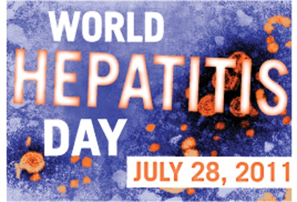
Hepatitis Prevalence Among Drug Users Varies Greatly Worldwide
Worldwide, an estimated 10 million injecting drug users have hepatitis C virus, while about 1.2 million have hepatitis B virus, based on results from an innovative and comprehensive analysis.
The researchers found wide variation in the geographic prevalence of viral hepatitis among injecting drug users (IDUs), with the highest rates seen in Eastern Europe, East Asia, and Southeast Asia.
"Investment in, and development of, comprehensive and effective strategies to prevent the transmission of viral hepatitis and reduce resultant morbidity and mortality in IDUs are urgently required," wrote Paul K. Nelson, a doctoral candidate at Australia’s National Drug and Alcohol Research Centre, University of New South Wales, Sydney, and his coauthors.
The findings – published online July 28, 2011, in The Lancet to coincide with World Hepatitis Day – suggest that the significance of viral hepatitis "needs to receive greater attention than it does at present," the study authors said (Lancet 2011 July 28 [Epub doi:10.1016/S0140-6736(11)61097-0]).
In what is believed to be the first study of its kind, the researchers reviewed 1,125 peer-reviewed and grey literature sources related to HCV and HBV in IDUs. Infection was defined as presence of HCV antibodies (anti-HCV), hepatitis B core antibodies (anti-HBC), or HBV surface antigen (HBsAg) in studies that included more than 40 participants.
Prevalence data for injecting drug use and HIV infection were obtained from a previously published systematic review by the Reference Group to the United Nations on HIV and Injecting Drug Use, with decision rules and estimates approved by all Reference Group members.
For HCV infection, the researchers located eligible reports with data on prevalence of anti-HCV in IDUs for 77 of the 152 countries or territories where injecting drug use has been reported. These 77 countries make up 82% of the world’s estimated population of IDUs.
Anti-HCV prevalence among IDUs was 60%-80% in 25 counties, including Spain (80%), Norway (76%), Germany (75%), France (74%), the United States (73%), China (67%), and Canada (64%), while the prevalence exceeded 80% in a further 12 countries, including Italy (81%), Portugal (83%), Pakistan (84%), The Netherlands (86%), Thailand (90%), and Mexico (97%).
The lowest prevalence was seen in Australia (55%), New Zealand (53%), and the United Kingdom (50%). Countries with the largest IDU populations infected with HCV were China (1.6 million), the United States (1.5 million), and Russian (1.3 million).
The data showed that worldwide, about 10 million IDUs are anti-HCV positive, which is about 3.5-fold higher than the 2.8 million IDUs who are estimated to be living with HIV.
For hepatitis B, anti-HBC positivity was measured in 43 countries that account for 65% of the world’s estimated population of IDUs. Rates varied widely among countries, from a low of 4.2% in Slovenia to a high of 85% in Mexico. The rate in the United States was 23%.
The prevalence of HBsAg among IDUs was measured in 59 countries accounting for 73% of the world’s estimated population of IDUs. The prevalence ranged from 5% to 10% in 21 counties and exceeded 10% in 10 countries, including the United States (12%). The highest rates were seen in counties that have endemic HBV in the general population, including Vietnam (20%), Estonia (19%), Saudi Arabia (18%), and Taiwan (17%).
For hepatitis B, the researchers estimated that 6.4 million IDUs worldwide are anti-HBC positive, and 1.2 million are HBsAg positive.
"Efforts to prevent, treat, and reduce harms related to liver disease in IDUs are essential – especially in situations in which HIV has successfully been prevented or managed – because the large numbers of IDUs infected with HCV and significant morbidity resulting from this infection mean that the health and economic costs of HCV transmitted by injecting drug use might be as high as (or higher than) those of HIV," the researchers wrote. "Nonetheless, HCV treatment is underused."
One reason for this neglect, they continued, "is the high cost, which will remain a substantial barrier to increasing treatment coverage in low resource settings until costs are reduced."
The researchers recommend bringing viral hepatitis treatments "into the same (lower cost) access framework as HIV antiretrovirals" as well as improving efforts to reduce the effect of other causes of progression of liver disease in people who are chronically infected with viral hepatitis, including addressing "problems of alcohol use, and provision of [hepatitis A virus] and HBV vaccination."
They acknowledged certain limitations of the study, including "the concentration of peer-reviewed data from high-income countries, the small team who undertook the analysis, and the potential for papers in languages other than English to be overlooked."
The study was funded by the National Institutes of Health and the World Health Organization’s HIV department.
Through country-by-country estimates, Nelson and colleagues provide an opportunity to examine the striking geographic disparities in rates of hepatitis B and C. Why is the prevalence of hepatitis C antibodies in injecting drug users (IDUs) in Hungary 23%, while it is about 90% in Estonia or Lithuania and 73% in Russia? Why do 85% of IDUs in Mexico have hepatitis B core antibodies compared with 20% of IDUs in Uruguay? These differences could be due to the limitations of the data; despite thousands of studies reviewed, grade A reports (multisite seroprevalence studies with several sample types for at least one hepatitis marker) were only available for 20 of the 77 countries for which any data were available, and few studies provided truly national estimates.
However, the differences may also show trends and patterns of drug use, or important differences in state policies and investment in harm reduction. The large variations between countries show that it is not inevitable for IDUs to have high rates of hepatitis B, hepatitis C, or HIV infection. Moreover, the estimates provide a powerful means for health and human rights advocates to question government officials in countries with high prevalences, and to caution governments in countries with low prevalences about the potential costs (human and economic) of failing to put in place or sustain effective, rights-based policies.
Nelson and colleagues provide us with a first step and powerful data to draw attention to the problem of viral hepatitis in people who use drugs. The next step is to challenge governments to act and hold them accountable for implementation of rights-respecting and evidence-based programs.
Joseph J. Amon, Ph.D., is director of the Health and Human Rights Division of Human Rights Watch in New York. This was adapted from a commentary published online July 28, 2011 in The Lancet (Lancet 2011 July 28 [Epub doi:10.1016/S0140-6736(11)61132-X]).
Worldwide, an estimated 10 million injecting drug users have hepatitis C virus, while about 1.2 million have hepatitis B virus, based on results from an innovative and comprehensive analysis.
The researchers found wide variation in the geographic prevalence of viral hepatitis among injecting drug users (IDUs), with the highest rates seen in Eastern Europe, East Asia, and Southeast Asia.
"Investment in, and development of, comprehensive and effective strategies to prevent the transmission of viral hepatitis and reduce resultant morbidity and mortality in IDUs are urgently required," wrote Paul K. Nelson, a doctoral candidate at Australia’s National Drug and Alcohol Research Centre, University of New South Wales, Sydney, and his coauthors.
The findings – published online July 28, 2011, in The Lancet to coincide with World Hepatitis Day – suggest that the significance of viral hepatitis "needs to receive greater attention than it does at present," the study authors said (Lancet 2011 July 28 [Epub doi:10.1016/S0140-6736(11)61097-0]).
In what is believed to be the first study of its kind, the researchers reviewed 1,125 peer-reviewed and grey literature sources related to HCV and HBV in IDUs. Infection was defined as presence of HCV antibodies (anti-HCV), hepatitis B core antibodies (anti-HBC), or HBV surface antigen (HBsAg) in studies that included more than 40 participants.
Prevalence data for injecting drug use and HIV infection were obtained from a previously published systematic review by the Reference Group to the United Nations on HIV and Injecting Drug Use, with decision rules and estimates approved by all Reference Group members.
For HCV infection, the researchers located eligible reports with data on prevalence of anti-HCV in IDUs for 77 of the 152 countries or territories where injecting drug use has been reported. These 77 countries make up 82% of the world’s estimated population of IDUs.
Anti-HCV prevalence among IDUs was 60%-80% in 25 counties, including Spain (80%), Norway (76%), Germany (75%), France (74%), the United States (73%), China (67%), and Canada (64%), while the prevalence exceeded 80% in a further 12 countries, including Italy (81%), Portugal (83%), Pakistan (84%), The Netherlands (86%), Thailand (90%), and Mexico (97%).
The lowest prevalence was seen in Australia (55%), New Zealand (53%), and the United Kingdom (50%). Countries with the largest IDU populations infected with HCV were China (1.6 million), the United States (1.5 million), and Russian (1.3 million).
The data showed that worldwide, about 10 million IDUs are anti-HCV positive, which is about 3.5-fold higher than the 2.8 million IDUs who are estimated to be living with HIV.
For hepatitis B, anti-HBC positivity was measured in 43 countries that account for 65% of the world’s estimated population of IDUs. Rates varied widely among countries, from a low of 4.2% in Slovenia to a high of 85% in Mexico. The rate in the United States was 23%.
The prevalence of HBsAg among IDUs was measured in 59 countries accounting for 73% of the world’s estimated population of IDUs. The prevalence ranged from 5% to 10% in 21 counties and exceeded 10% in 10 countries, including the United States (12%). The highest rates were seen in counties that have endemic HBV in the general population, including Vietnam (20%), Estonia (19%), Saudi Arabia (18%), and Taiwan (17%).
For hepatitis B, the researchers estimated that 6.4 million IDUs worldwide are anti-HBC positive, and 1.2 million are HBsAg positive.
"Efforts to prevent, treat, and reduce harms related to liver disease in IDUs are essential – especially in situations in which HIV has successfully been prevented or managed – because the large numbers of IDUs infected with HCV and significant morbidity resulting from this infection mean that the health and economic costs of HCV transmitted by injecting drug use might be as high as (or higher than) those of HIV," the researchers wrote. "Nonetheless, HCV treatment is underused."
One reason for this neglect, they continued, "is the high cost, which will remain a substantial barrier to increasing treatment coverage in low resource settings until costs are reduced."
The researchers recommend bringing viral hepatitis treatments "into the same (lower cost) access framework as HIV antiretrovirals" as well as improving efforts to reduce the effect of other causes of progression of liver disease in people who are chronically infected with viral hepatitis, including addressing "problems of alcohol use, and provision of [hepatitis A virus] and HBV vaccination."
They acknowledged certain limitations of the study, including "the concentration of peer-reviewed data from high-income countries, the small team who undertook the analysis, and the potential for papers in languages other than English to be overlooked."
The study was funded by the National Institutes of Health and the World Health Organization’s HIV department.
FROM THE LANCET
Major Finding: Worldwide, an estimated 10 million injecting drug users have been exposed to hepatitis C virus, while 1.2 million have hepatitis B infection.
Data Source: An analysis of 1,125 sources in the peer-reviewed and grey literature related to HCV and HBV in injecting drug users.
Disclosures: The study was funded by the National Institutes of Health and the World Health Organization.
CAD Software Does Not Improve Mammogram Accuracy
Computer-aided detection software does not help clinicians analyze and interpret film-screen screening mammograms, results from a large multicenter study demonstrated.
The findings "raise concerns that CAD, as currently implemented in clinical practice, may have little or no impact on breast cancer mortality, which may depend on earlier detection of invasive breast cancer," researchers led by Dr. Joshua J. Fenton reported online July 27 in the Journal of the National Cancer Institute.
Dr. Fenton of the department of family and community medicine at the center for health care policy and research of the University of California, Davis, and his associates analyzed records from 684,956 women who underwent more than 1.6 million film-screen screening mammograms in 1998-2006 at 90 facilities that are members of the Breast Cancer Surveillance Consortium (BCSC), a federally supported effort that links mammography data to cancer outcomes in seven states (California, Colorado, North Carolina, New Hampshire, New Mexico, Washington, and Vermont).
The researchers used random effects logistic regression to estimate the associations between CAD and specificity, sensitivity, and positive predictive value while adjusting for factors that might influence mammography findings, including patient age, breast density, and use of hormone therapy (J. Natl. Cancer Inst. 2011 July 27 [doi:10.1093/jnci/djr298]).
More than half of the women studied (61%) were aged 40-59 years. Of the 90 BCSC facilities, 25 (28%) implemented CAD and used it for an average of 28 months during the study period. All told, 793 radiologists interpreted the results, including 154 (19%) at facilities with CAD.
After adjusting for BCSC registry, patient characteristics, hormone therapy use, and year of mammography interpretation, the researchers found that CAD use was associated with a statistically significant lower specificity (odds ratio, 0.87) and positive predictive value (OR, 0.89), and an increase in sensitivity (OR, 1.06) that was not statistically significant.
When the sensitivity analysis was limited to invasive cancers only, CAD use was no longer associated with increased sensitivity (OR, 0.96). When the analysis was limited to ductal carcinoma in situ, CAD use was associated with greater sensitivity (OR, 1.55), yet this did not reach statistical significance.
There were also no statistically significant differences between CAD use and no-CAD in the odds of overall breast cancer detection (OR, 1.01), the diagnosis of stage I invasive cancer compared with later-stage invasive cancer (OR, 0.97), or the diagnosis of invasive tumors of 15 mm or less in size, compared with those greater than 15 mm (OR, 0.92).
Dr. Fenton and his associates acknowledged certain limitations of the study, including the absence of digital mammography data. "Whereas CAD algorithms perform a similar alerting function in the film-screen and digital environments, film-screen mammograms must be digitized before CAD analysis, and digitization may introduce noise and adversely affect performance," they wrote.
"However, small retrospective studies suggest that the performance impacts of CAD are similar when used in digital and film-screen environments. Because prior research suggests that facilities apply CAD on nearly all mammograms after implementation, these analyses assumed that all mammograms were interpreted with CAD after implementation – another limitation of this study."
The study was supported by grants from the National Cancer Institute and the American Cancer Society. A study coauthor disclosed holding stock options and serving as a medical consultant for Hologic Inc., a manufacturer of CAD equipment that had "no role in data collection, analysis, or interpretation."
What happens when CAD software is used with screening mammography? The second "pair of eyes" finds additional cancers, but it opens the lead-time window at most a small fraction of a doubling time. The possibility that a tumor would become metastatic precisely in any such short period of time is remote. So small is the fraction of cancers that would become metastatic in such a small interval (if there are any) that it would be difficult to impossible to detect an incremental mortality advantage of CAD even in a large, randomized trial.
|
Moreover, improving sensitivity may preferentially find less-aggressive tumors, or it might find more of those tumors that would have otherwise revealed themselves as interval tumors. Neither type of increment in sensitivity could have much impact on breast cancer mortality.
Why is CAD so popular? An obvious reason is that it is built into digital mammography equipment, which is increasingly common in the United States. Another is financial: In 2008, Medicare’s global reimbursement was $16.50 per CAD. Still another is that CAD marks are comforting to the reader, even though the comfort may be misplaced. In a related vein, relying on CAD marks (or the absence of same) in medical malpractice suits may be an effective defense, although the sword has two edges.
An argument for the use of CAD with film or digital mammograms is that it will get better over time. Fine. Researchers and device companies should work to make the software ever better. But this should happen in an experimental setting and not while exposing millions of women to a technology that may be more harmful than it is beneficial. In the meantime, economic incentives may stoke its continued proliferation.
Donald A. Berry, Ph.D., is chair and professor of biostatistics at the University of Texas M.D. Anderson Cancer Center, Houston. This was extracted from an editorial that appeared online July 27 in the Journal of the National Cancer Institute (J. Natl. Cancer Inst. 2011 July 27 [doi:10.1093/jnci/djr267]).
Computer-aided detection software does not help clinicians analyze and interpret film-screen screening mammograms, results from a large multicenter study demonstrated.
The findings "raise concerns that CAD, as currently implemented in clinical practice, may have little or no impact on breast cancer mortality, which may depend on earlier detection of invasive breast cancer," researchers led by Dr. Joshua J. Fenton reported online July 27 in the Journal of the National Cancer Institute.
Dr. Fenton of the department of family and community medicine at the center for health care policy and research of the University of California, Davis, and his associates analyzed records from 684,956 women who underwent more than 1.6 million film-screen screening mammograms in 1998-2006 at 90 facilities that are members of the Breast Cancer Surveillance Consortium (BCSC), a federally supported effort that links mammography data to cancer outcomes in seven states (California, Colorado, North Carolina, New Hampshire, New Mexico, Washington, and Vermont).
The researchers used random effects logistic regression to estimate the associations between CAD and specificity, sensitivity, and positive predictive value while adjusting for factors that might influence mammography findings, including patient age, breast density, and use of hormone therapy (J. Natl. Cancer Inst. 2011 July 27 [doi:10.1093/jnci/djr298]).
More than half of the women studied (61%) were aged 40-59 years. Of the 90 BCSC facilities, 25 (28%) implemented CAD and used it for an average of 28 months during the study period. All told, 793 radiologists interpreted the results, including 154 (19%) at facilities with CAD.
After adjusting for BCSC registry, patient characteristics, hormone therapy use, and year of mammography interpretation, the researchers found that CAD use was associated with a statistically significant lower specificity (odds ratio, 0.87) and positive predictive value (OR, 0.89), and an increase in sensitivity (OR, 1.06) that was not statistically significant.
When the sensitivity analysis was limited to invasive cancers only, CAD use was no longer associated with increased sensitivity (OR, 0.96). When the analysis was limited to ductal carcinoma in situ, CAD use was associated with greater sensitivity (OR, 1.55), yet this did not reach statistical significance.
There were also no statistically significant differences between CAD use and no-CAD in the odds of overall breast cancer detection (OR, 1.01), the diagnosis of stage I invasive cancer compared with later-stage invasive cancer (OR, 0.97), or the diagnosis of invasive tumors of 15 mm or less in size, compared with those greater than 15 mm (OR, 0.92).
Dr. Fenton and his associates acknowledged certain limitations of the study, including the absence of digital mammography data. "Whereas CAD algorithms perform a similar alerting function in the film-screen and digital environments, film-screen mammograms must be digitized before CAD analysis, and digitization may introduce noise and adversely affect performance," they wrote.
"However, small retrospective studies suggest that the performance impacts of CAD are similar when used in digital and film-screen environments. Because prior research suggests that facilities apply CAD on nearly all mammograms after implementation, these analyses assumed that all mammograms were interpreted with CAD after implementation – another limitation of this study."
The study was supported by grants from the National Cancer Institute and the American Cancer Society. A study coauthor disclosed holding stock options and serving as a medical consultant for Hologic Inc., a manufacturer of CAD equipment that had "no role in data collection, analysis, or interpretation."
FROM THE JOURNAL OF THE NATIONAL CANCER INSTITUTE
Major Finding: Using CAD to analyze and interpret mammograms was associated with statistically significant lowering of specificity (OR, 0.87) and positive predictive value (OR, 0.89), and a nonstatistically significant increase in sensitivity (OR, 1.06).
Data Source: A study of records from 684,956 women who underwent more than 1.6 million film screening mammograms in 1998-2006 at 90 BCSC facilities that are members.
Disclosures: The study was supported by grants from the National Cancer Institute and the American Cancer Society. A coauthor disclosed holding stock options and serving as a medical consultant for Hologic Inc., a manufacturer of CAD equipment that had "no role in data collection, analysis, or interpretation."
Investigational Device Promising for Type 2 Diabetes
SAN DIEGO – Treatment with the investigational device ITCA 650 delivering exenatide at 20 mcg/day and dose escalation to 60 mcg/day was well tolerated and led to significant reductions in HbA1c and body weight in patients with type 2 diabetes, results from a phase II, 48-week extension study showed.
ITCA 650 is manufactured by Intarcia Therapeutics of Hayward, Calif., and is a matchstick-size, osmotic mini-pump using the DUROS technology that is placed subcutaneously, providing continuous and consistent delivery of exenatide (Byetta) at specified doses. The device is inserted during a 10- to 15-minute office procedure. ITCA 650 has been shown to be effective in lowering HbA1c and having a favorable weight profile at 12 and 24 weeks, Dr. Julio Rosenstock said at the annual scientific sessions of the American Diabetes Association, where he presented the results of the 48-week extension of the study that demonstrated sustained effects.
"Therefore, this device has the potential for greater adherence using ITCA 650 devices that can deliver 6 or 12 months of treatment with a single placement," said Dr. Rosenstock, an endocrinologist who directs the Dallas Diabetes and Endocrine Center at Medical City and who served as the current study’s principal investigator. "It also may result in enhanced efficacy and a reduced side effect profile."
In a trial conducted at 50 sites, 155 patients treated with metformin who had baseline HbA1c levels between 7% and 10% were enrolled in a 24-week study and were randomized to receive ITCA 650 at 20, 40, 60, or 80 mcg/day following initial 12 weeks of either ITCA 650 (20 or 40 mcg/day) or exenatide injections (10 mcg b.i.d. self-injection). At week 24, Dr. Rosenstock and his associates offered patients the option to continue treatment at their current dose for an additional 12 weeks. A total of 86 patients from 35 of the original 50 sites entered the extension study.
Dr. Rosenstock reported that continued reductions in HbA1c and weight were observed across all ITCA 650 treatment arms at week 48, compared with week 24, with the greatest reductions seen in the 60 mcg/day and 80 mcg/day arms. Between baseline and week 48, the mean HbA1c improved 1% in the 20 mcg/day arm (from 7.8% to 6.8%), 1% in the 40 mcg/day arm (from 7.8% to 6.8%), 1.5% in the 60 mcg/day arm (from 8.1% to 6.6%), and 1.4% in the 80 mcg/day arm (from 7.9% to 6.5%). Weight reductions were observed in all treatment arms (mean reductions of 6, 10.8, 7.7, and 7.9 pounds, respectively).
There were no treatment discontinuations between weeks 24 and 48 in the group initially treated with 20 mcg/day and then escalated to 60 mcg/day, Dr. Rosenstock said. The chief side effects in all of the dose groups were nausea (10.5%) and diarrhea (3.5%). Other reported side effects were related to the skin at the placement site and included irritation (7%), pain (7%), erythema (4.7%), pruritus (3.5%), and hematoma (3.5%).
"These results support further evaluation of ITCA 650 using longer duration subcutaneous devices for injection-free exenatide therapy in type 2 diabetes," Dr. Rosenstock said.
According to a prepared statement from Intarcia, a phase III study planned for 2011 will evaluate treatment regimens involving initial 12-week ITCA dosing at 20 mcg/day transitioning to 60 mcg/day thereafter using ITCA 650 devices of both 6- and 12-month duration.
Dr. Rosenstock disclosed that he has relationships with numerous pharmaceutical and device companies, including Intarcia Therapeutics, in the form of research support, advisory board roles, and consulting honorariums.
FROM THE ANNUAL SCIENTIFIC SESSIONS OF THE AMERICAN DIABETES ASSOCIATION
Major Finding: Between baseline and week 48 the mean HbA1c improved 1% in the 20 mcg/day arm (from 7.8% to 6.8%), 1% in the 40 mcg/day arm (from 7.8% to 6.8%), 1.5% in the 60 mcg/day arm (from 8.1% to 6.6%), and 1.4% in the 80 mcg/day arm (from 7.9% to 6.5%). Weight reductions were observed in all treatment arms (mean reductions of 6, 10.8, 7.7, and 7.9 pounds, respectively).
Data Source: A 48-week, multicenter extension study conducted in 86 patients with type 2 diabetes who were treated with ITCA 650, a matchstick-size, osmotic mini-pump using the DUROS technology that is placed subcutaneously, providing continuous and consistent delivery of exenatide at specified doses.
Disclosures: Dr. Rosenstock disclosed that he has relationships with numerous pharmaceutical and device companies, including Intarcia Therapeutics, in the form of research support, advisory board roles, and consulting honorariums.
Second Botox Rival Joins Frown Line Market
On July 21, the Food and Drug Administration approved incobotulinumtoxinA for the temporary improvement in appearance of moderate to severe glabellar lines in adult patients.
Manufactured by Merz Aesthetics and marketed as Xeomin, incobotulinumtoxinA is the third botulinum toxin type A to enter the marketplace, following Botox and Dysport, respectively. It is expected to be available to physicians in the United States in the spring of 2012.
IncobotulinumtoxinA's approval is based on results from two clinical trials conducted at 16 sites in the United States that included 547 healthy adult patients. According to Merz, in both studies, the product significantly improved the appearance of glabellar lines 30 days after the first injection, compared with placebo.
IncobotulinumtoxinA is classified as a pregnancy category C agent and is the only botulinum toxin currently approved in the United States that does not require refrigeration prior to reconstitution.

In an interview, Dr. Joel L. Cohen, one of the trial investigators and director of AboutSkin Dermatology in Denver called incobotulinumtoxinA "more similar than it is different to other products that are on the market, particularly Botox. It’s exciting for botulinum toxin to be in the news again for something very positive in the approval of another product. I think that we should feel comfortable using Xeomin as something that has demonstrated safety and efficacy."

Dr. Christopher Zachary, professor and chair of the department of dermatology at the University of California, Irvine, predicted that Xeomin "is going to give Allergan a run for its money. The introduction of this product is likely to be more easily adopted than rival Dysport because one vial of Xeomin contains 100 units of botulinum toxin and can be diluted similarly. Dose for dose, this direct comparison with Botox is going to make staff feel quite comfortable from the get-go. Requiring staff to use different dilutions and maintain correct labeling just adds another element of confusion and might increase the potential for incorrect dosing. Having said this, Dysport remains a premier and very equivalent product and will have its devotees."
According to Merz, use of the product is contraindicated "in patients with a known hypersensitivity to the active substance botulinum toxin type A or to any of the components in the formulation and in the presence of infection at the proposed injection site(s)." The company also noted that the potency units of incobotulinumtoxinA are not interchangeable with other preparations of botulinum toxin products. "Therefore, units of biological activity of Xeomin cannot be compared to or converted into units of any other botulinum toxin products."
The most common adverse reaction was headache, which was reported by 5.4% of patients in the trials.
Xeomin is approved for the temporary improvement in the appearance of glabellar lines in 14 countries in the European Union, including Germany, the United Kingdom, France, Italy and Spain, under the brand name Bocouture.
Dr. Cohen disclosed that he has been a consultant and clinical trial investigator for Merz, Allergan, and Medicis.
Dr. Zachary disclosed that he has received support and honoraria from Merz, Allergan, and Medicis for various educational courses in recent years.
On July 21, the Food and Drug Administration approved incobotulinumtoxinA for the temporary improvement in appearance of moderate to severe glabellar lines in adult patients.
Manufactured by Merz Aesthetics and marketed as Xeomin, incobotulinumtoxinA is the third botulinum toxin type A to enter the marketplace, following Botox and Dysport, respectively. It is expected to be available to physicians in the United States in the spring of 2012.
IncobotulinumtoxinA's approval is based on results from two clinical trials conducted at 16 sites in the United States that included 547 healthy adult patients. According to Merz, in both studies, the product significantly improved the appearance of glabellar lines 30 days after the first injection, compared with placebo.
IncobotulinumtoxinA is classified as a pregnancy category C agent and is the only botulinum toxin currently approved in the United States that does not require refrigeration prior to reconstitution.
In an interview, Dr. Joel L. Cohen, one of the trial investigators and director of AboutSkin Dermatology in Denver called incobotulinumtoxinA "more similar than it is different to other products that are on the market, particularly Botox. It’s exciting for botulinum toxin to be in the news again for something very positive in the approval of another product. I think that we should feel comfortable using Xeomin as something that has demonstrated safety and efficacy."
Dr. Christopher Zachary, professor and chair of the department of dermatology at the University of California, Irvine, predicted that Xeomin "is going to give Allergan a run for its money. The introduction of this product is likely to be more easily adopted than rival Dysport because one vial of Xeomin contains 100 units of botulinum toxin and can be diluted similarly. Dose for dose, this direct comparison with Botox is going to make staff feel quite comfortable from the get-go. Requiring staff to use different dilutions and maintain correct labeling just adds another element of confusion and might increase the potential for incorrect dosing. Having said this, Dysport remains a premier and very equivalent product and will have its devotees."
According to Merz, use of the product is contraindicated "in patients with a known hypersensitivity to the active substance botulinum toxin type A or to any of the components in the formulation and in the presence of infection at the proposed injection site(s)." The company also noted that the potency units of incobotulinumtoxinA are not interchangeable with other preparations of botulinum toxin products. "Therefore, units of biological activity of Xeomin cannot be compared to or converted into units of any other botulinum toxin products."
The most common adverse reaction was headache, which was reported by 5.4% of patients in the trials.
Xeomin is approved for the temporary improvement in the appearance of glabellar lines in 14 countries in the European Union, including Germany, the United Kingdom, France, Italy and Spain, under the brand name Bocouture.
Dr. Cohen disclosed that he has been a consultant and clinical trial investigator for Merz, Allergan, and Medicis.
Dr. Zachary disclosed that he has received support and honoraria from Merz, Allergan, and Medicis for various educational courses in recent years.
Haitian Experience Reflects Success of Global Surgery Fellowship
PALM DESERT, CALIF. – Providing quality surgical care in resource-poor settings is challenging, but participants in the Paul Farmer Global Surgery Fellowship have found it to be a worthwhile endeavor.
The fellowship, founded by Dr. John Meara, plastic surgeon-in-chief at Children’s Hospital Boston, in conjunction with Dr. Paul Farmer of Partners in Health (PIH), was designed "to build surgical capacity internationally," according to Dr. Shahram Aarabi. Between June 2009 and June 2010, Dr. Aarabi served as a visiting surgeon in Haiti for 2 weeks every 2 months alongside Dr. Jason Smithers, a Paul Farmer Global Surgery Fellow with Children’s Hospital Boston.
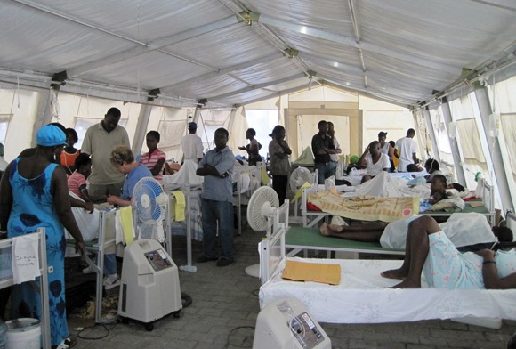
"It’s also a model for how to help meet the global surgical burden in disease. The fellowship brings individual surgeons from abroad to work in specific PIH sites over the course of one or more years in order to provide continuity of care and teaching," Dr. Aarabi said at the annual meeting of the American Pediatric Surgical Association, where he reported on the work he did in Haiti.
Dr. Aarabi, a general surgery resident at the University of Washington, Seattle, spent most of his time in Cange, Haiti, at a 104-bed full-service hospital that includes four operating rooms. This facility was the first to be founded by PIH in 1985.
"The hospital and satellite clinics in the valley provide free care to patients over a catchment area of about 1.2 million people," he said, noting that there are now more than 11 PIH health care facilities nationwide. "There are two or three full-time Haitian general surgeons who work for PIH at these facilities, and one full-time orthopedic surgeon."
Dr. Aarabi also assisted in the temporary staffing of University Hospital in Port-au-Prince after the devastating earthquake of January 2010.
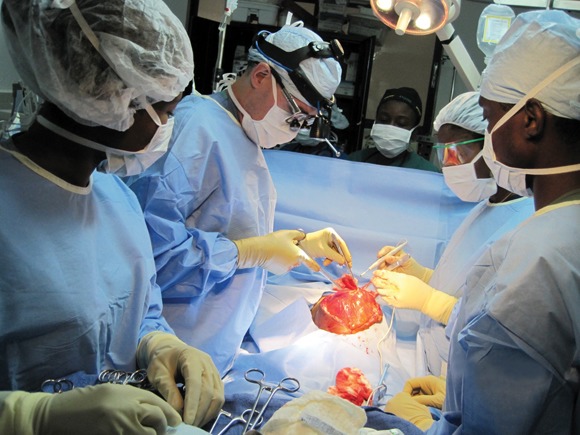
He reported on 147 operations that were performed on 131 patients at the two hospitals over a 12-week period. More than three-quarters (78%) of the procedures were elective, and 22% were emergent. Hernia repairs, imperforate anus/Hirschprung’s disease repairs, and bowel resections were the most commonly performed operations.
Nearly one-third of the operative patients (32%) were adults and the rest were children, including 15 under 1 year of age and five who were less than 2 months old. A minority of patients (29%) lived locally, while 71% came from various locations across the country, usually by foot.
"Some Haitians spent days to a week trying to get to PIH facilities for care," Dr. Aarabi said. "Our patients received regular follow-up at weekly clinics staffed either by ourselves or by Haitian general surgeons. Limits to follow-up were primarily due to social factors for our patients, not from a lack of surgical staffing."
Dr. Smithers, who is now a pediatric surgeon at Children’s Hospital Boston, said that he found it helpful to have an understanding of surgical history, because in many cases he and his associates relied on strategies used by surgeons 20 years ago, "when the equipment and capabilities that we had in the United States were similar to what Haiti currently has," he said.
For example, in the United States most Hirschprung’s disease repairs can be performed with a single surgical procedure in the neonatal period, while the same procedure in Haiti requires multiple stages.
"When it comes down to it, a lot of what surgeons need are just a scalpel and scissors, and some suture," Dr. Smithers added. "Even though we may have a variety of complex instruments at home, you can get by without those."
Dr. Aarabi said that most cases (91%) involved training of an American medical student or resident with an interest in international medicine, a Haitian resident, or a Haitian staff surgeon.
"We feel that one of the major strengths of the fellowship was that training of Haitian students and surgeons was integrated into the program. The infant cases in particular were valuable to the Haitian surgeons because there are no pediatric surgeons in Haiti," Dr. Aarabi said.
"Our nonoperative time was spent seeing patients in clinic, taking care of our inpatients, seeing new consults on the wards and in the emergency room, and working on site development and planning," he added.
The Paul Farmer Global Surgery Fellowship’s planned expansion to other sites around the world "holds promise as a way to provide a lasting collaboration of developing local surgical infrastructure and training local surgeons," he concluded.
Dr. Aarabi said that he had no relevant financial disclosures to make. The meeting was supported by a grant from Elsevier, which owns this news organization.
PALM DESERT, CALIF. – Providing quality surgical care in resource-poor settings is challenging, but participants in the Paul Farmer Global Surgery Fellowship have found it to be a worthwhile endeavor.
The fellowship, founded by Dr. John Meara, plastic surgeon-in-chief at Children’s Hospital Boston, in conjunction with Dr. Paul Farmer of Partners in Health (PIH), was designed "to build surgical capacity internationally," according to Dr. Shahram Aarabi. Between June 2009 and June 2010, Dr. Aarabi served as a visiting surgeon in Haiti for 2 weeks every 2 months alongside Dr. Jason Smithers, a Paul Farmer Global Surgery Fellow with Children’s Hospital Boston.
"It’s also a model for how to help meet the global surgical burden in disease. The fellowship brings individual surgeons from abroad to work in specific PIH sites over the course of one or more years in order to provide continuity of care and teaching," Dr. Aarabi said at the annual meeting of the American Pediatric Surgical Association, where he reported on the work he did in Haiti.
Dr. Aarabi, a general surgery resident at the University of Washington, Seattle, spent most of his time in Cange, Haiti, at a 104-bed full-service hospital that includes four operating rooms. This facility was the first to be founded by PIH in 1985.
"The hospital and satellite clinics in the valley provide free care to patients over a catchment area of about 1.2 million people," he said, noting that there are now more than 11 PIH health care facilities nationwide. "There are two or three full-time Haitian general surgeons who work for PIH at these facilities, and one full-time orthopedic surgeon."
Dr. Aarabi also assisted in the temporary staffing of University Hospital in Port-au-Prince after the devastating earthquake of January 2010.
He reported on 147 operations that were performed on 131 patients at the two hospitals over a 12-week period. More than three-quarters (78%) of the procedures were elective, and 22% were emergent. Hernia repairs, imperforate anus/Hirschprung’s disease repairs, and bowel resections were the most commonly performed operations.
Nearly one-third of the operative patients (32%) were adults and the rest were children, including 15 under 1 year of age and five who were less than 2 months old. A minority of patients (29%) lived locally, while 71% came from various locations across the country, usually by foot.
"Some Haitians spent days to a week trying to get to PIH facilities for care," Dr. Aarabi said. "Our patients received regular follow-up at weekly clinics staffed either by ourselves or by Haitian general surgeons. Limits to follow-up were primarily due to social factors for our patients, not from a lack of surgical staffing."
Dr. Smithers, who is now a pediatric surgeon at Children’s Hospital Boston, said that he found it helpful to have an understanding of surgical history, because in many cases he and his associates relied on strategies used by surgeons 20 years ago, "when the equipment and capabilities that we had in the United States were similar to what Haiti currently has," he said.
For example, in the United States most Hirschprung’s disease repairs can be performed with a single surgical procedure in the neonatal period, while the same procedure in Haiti requires multiple stages.
"When it comes down to it, a lot of what surgeons need are just a scalpel and scissors, and some suture," Dr. Smithers added. "Even though we may have a variety of complex instruments at home, you can get by without those."
Dr. Aarabi said that most cases (91%) involved training of an American medical student or resident with an interest in international medicine, a Haitian resident, or a Haitian staff surgeon.
"We feel that one of the major strengths of the fellowship was that training of Haitian students and surgeons was integrated into the program. The infant cases in particular were valuable to the Haitian surgeons because there are no pediatric surgeons in Haiti," Dr. Aarabi said.
"Our nonoperative time was spent seeing patients in clinic, taking care of our inpatients, seeing new consults on the wards and in the emergency room, and working on site development and planning," he added.
The Paul Farmer Global Surgery Fellowship’s planned expansion to other sites around the world "holds promise as a way to provide a lasting collaboration of developing local surgical infrastructure and training local surgeons," he concluded.
Dr. Aarabi said that he had no relevant financial disclosures to make. The meeting was supported by a grant from Elsevier, which owns this news organization.
FROM THE ANNUAL MEETING OF THE AMERICAN PEDIATRIC SURGICAL ASSOCIATION
HbA1c Misclassifies Patients in Diabetes Screening Program
SAN DIEGO – Slightly more than half of veterans targeted for screening have unrecognized diabetes or prediabetes, results from a recent analysis showed.
However, screening such patients by measuring hemoglobin A1c "would result in major misclassification – missing disease when it is present and, to a lesser extent, mislabeling normals as having disease," Sandra L. Jackson, M.P.H., said at the annual scientific sessions of the American Diabetes Association.

The findings are based on a study of 789 individuals from the Atlanta VA Medical Center that assessed the use of targeted screening to detect prediabetes and diabetes, and to compare HbA1c testing with the oral glucose tolerance test (OGTT), said Ms. Jackson, a graduate student in the nutrition and health sciences doctoral program at Emory University, Atlanta.
Although screening to detect unrecognized diabetes and prediabetes is recommended, the best strategy for screening in patients in primary care settings is unknown. The upside of the OGTT, Ms. Jackson said, is that it’s established in clinical use, it can detect all patients with prediabetes, and the glucose measurement itself is accurate. However, "on the downside, it requires [fasting] and morning testing. It’s burdensome for patients and health care systems, and it has poor day-to-day reproducibility."
The upside of HbA1c, she continued, is that it does not require a fast, "and it’s only a single blood draw, so it’s much more convenient, there’s less day-to-day variation, and there’s greater preanalytic stability. On the downside, measurement may be problematic as platforms vary, point-of-care testing can be unreliable, there’s a lack of agreement on cutoffs, and there may be racial and age disparities such that blacks and older persons may have higher HbA1c independent of glucose."
The researchers defined hyperglycemia according to American Diabetes Association (ADA) criteria: prediabetes as a fasting OGTT of 100-125 mg/dL or a 2-hour OGTT of 149-199 mg/dL, and diabetes as a fasting OGTT of 126 mg/dL or greater or a 2-hour OGTT of 200 mg/dL or greater.
They categorized HbA1c results according to three sets of diagnostic criteria: the International Expert Committee (IEC) (prediabetes 6.0%-6.4%, diabetes 6.5% or greater), ADA (prediabetes 5.7%-6.4%, diabetes 6.5% or greater), and Department of Veterans Affairs/Department of Defense (VA/DoD) (prediabetes 5.7%-6.9%, diabetes 7.0% or greater).
The mean age of the 789 study participants was 58 years, 95% were men, 74% were black, and their mean BMI was 30.5 kg/m2.
Screening was offered to patients meeting National Institutes of Health guidelines for screening: without known diabetes, and with age greater than 45 years or a BMI of more than 25 with another risk factor.
Fully 10% of patients met criteria for diabetes based on the OGTT, which was a higher rate compared with the HbA1c guidelines (6.7% by the IEC, 6.7% by the ADA, and 1.5% by the VA/DoD guidelines, respectively). "This would indicate that these cutoffs are insensitive compared with the OGTT for detecting diabetes," she said.
According to the OGTT, 42% had prediabetes: 27% had isolated impaired fasting glucose, 6% had isolated impaired glucose tolerance, and 9% had both.
In patients with diabetes by OGTT criteria, HbA1c classification by IEC criteria labeled 32% correctly, 38% incorrectly as having prediabetes, and 29% incorrectly as being normal; ADA criteria labeled 32% correctly, 50% incorrectly as having prediabetes, and 18% incorrectly as being normal; and VA/DoD criteria labeled 12% correctly, 71% incorrectly as having prediabetes, and 18% incorrectly as being normal.
In patients with prediabetes by OGTT criteria, HbA1c classification by IEC criteria labeled 36% correctly, 6% incorrectly as having diabetes, and 59% incorrectly as being normal; ADA criteria labeled 61% correctly, 6% incorrectly as having diabetes, and 33% incorrectly as being normal; and VA/DoD criteria labeled 66% correctly, 1% incorrectly as having diabetes, and 33% incorrectly as being normal.
The prevalence of diabetes increased in a stepwise fashion with increasing BMI, from 1.5% among those with a normal BMI (18.5-24.9) to 15% among those who met criteria for class III obesity (BMI more than 40). "For every 1 unit increase in BMI, we observed a 10% increase in the odds of having diabetes," she said.
Ms. Jackson also reported that with the IEC, ADA, and VA/DoD cutoffs for diabetes, screening with HbA1c was specific but insensitive, with a false negative rate of 68% at the 6.5% cutoff and a false negative rate of 89% at the 7.0% cutoff.
"Many veterans – and probably many Americans – targeted by national guidelines have unrecognized diabetes and prediabetes, yet screening with HbA1c would miss many," she said. "Given these findings, we should consider or develop alternative diabetes screening strategies that can be used opportunistically during outpatient visits, without fasting, but are more accurate."
The study was supported by a grant from the VA’s Health Services Research and Development Service. Ms. Jackson said that she had no relevant financial conflicts of interest.
SAN DIEGO – Slightly more than half of veterans targeted for screening have unrecognized diabetes or prediabetes, results from a recent analysis showed.
However, screening such patients by measuring hemoglobin A1c "would result in major misclassification – missing disease when it is present and, to a lesser extent, mislabeling normals as having disease," Sandra L. Jackson, M.P.H., said at the annual scientific sessions of the American Diabetes Association.
The findings are based on a study of 789 individuals from the Atlanta VA Medical Center that assessed the use of targeted screening to detect prediabetes and diabetes, and to compare HbA1c testing with the oral glucose tolerance test (OGTT), said Ms. Jackson, a graduate student in the nutrition and health sciences doctoral program at Emory University, Atlanta.
Although screening to detect unrecognized diabetes and prediabetes is recommended, the best strategy for screening in patients in primary care settings is unknown. The upside of the OGTT, Ms. Jackson said, is that it’s established in clinical use, it can detect all patients with prediabetes, and the glucose measurement itself is accurate. However, "on the downside, it requires [fasting] and morning testing. It’s burdensome for patients and health care systems, and it has poor day-to-day reproducibility."
The upside of HbA1c, she continued, is that it does not require a fast, "and it’s only a single blood draw, so it’s much more convenient, there’s less day-to-day variation, and there’s greater preanalytic stability. On the downside, measurement may be problematic as platforms vary, point-of-care testing can be unreliable, there’s a lack of agreement on cutoffs, and there may be racial and age disparities such that blacks and older persons may have higher HbA1c independent of glucose."
The researchers defined hyperglycemia according to American Diabetes Association (ADA) criteria: prediabetes as a fasting OGTT of 100-125 mg/dL or a 2-hour OGTT of 149-199 mg/dL, and diabetes as a fasting OGTT of 126 mg/dL or greater or a 2-hour OGTT of 200 mg/dL or greater.
They categorized HbA1c results according to three sets of diagnostic criteria: the International Expert Committee (IEC) (prediabetes 6.0%-6.4%, diabetes 6.5% or greater), ADA (prediabetes 5.7%-6.4%, diabetes 6.5% or greater), and Department of Veterans Affairs/Department of Defense (VA/DoD) (prediabetes 5.7%-6.9%, diabetes 7.0% or greater).
The mean age of the 789 study participants was 58 years, 95% were men, 74% were black, and their mean BMI was 30.5 kg/m2.
Screening was offered to patients meeting National Institutes of Health guidelines for screening: without known diabetes, and with age greater than 45 years or a BMI of more than 25 with another risk factor.
Fully 10% of patients met criteria for diabetes based on the OGTT, which was a higher rate compared with the HbA1c guidelines (6.7% by the IEC, 6.7% by the ADA, and 1.5% by the VA/DoD guidelines, respectively). "This would indicate that these cutoffs are insensitive compared with the OGTT for detecting diabetes," she said.
According to the OGTT, 42% had prediabetes: 27% had isolated impaired fasting glucose, 6% had isolated impaired glucose tolerance, and 9% had both.
In patients with diabetes by OGTT criteria, HbA1c classification by IEC criteria labeled 32% correctly, 38% incorrectly as having prediabetes, and 29% incorrectly as being normal; ADA criteria labeled 32% correctly, 50% incorrectly as having prediabetes, and 18% incorrectly as being normal; and VA/DoD criteria labeled 12% correctly, 71% incorrectly as having prediabetes, and 18% incorrectly as being normal.
In patients with prediabetes by OGTT criteria, HbA1c classification by IEC criteria labeled 36% correctly, 6% incorrectly as having diabetes, and 59% incorrectly as being normal; ADA criteria labeled 61% correctly, 6% incorrectly as having diabetes, and 33% incorrectly as being normal; and VA/DoD criteria labeled 66% correctly, 1% incorrectly as having diabetes, and 33% incorrectly as being normal.
The prevalence of diabetes increased in a stepwise fashion with increasing BMI, from 1.5% among those with a normal BMI (18.5-24.9) to 15% among those who met criteria for class III obesity (BMI more than 40). "For every 1 unit increase in BMI, we observed a 10% increase in the odds of having diabetes," she said.
Ms. Jackson also reported that with the IEC, ADA, and VA/DoD cutoffs for diabetes, screening with HbA1c was specific but insensitive, with a false negative rate of 68% at the 6.5% cutoff and a false negative rate of 89% at the 7.0% cutoff.
"Many veterans – and probably many Americans – targeted by national guidelines have unrecognized diabetes and prediabetes, yet screening with HbA1c would miss many," she said. "Given these findings, we should consider or develop alternative diabetes screening strategies that can be used opportunistically during outpatient visits, without fasting, but are more accurate."
The study was supported by a grant from the VA’s Health Services Research and Development Service. Ms. Jackson said that she had no relevant financial conflicts of interest.
FROM THE ANNUAL SCIENTIFIC SESSIONS OF THE AMERICAN DIABETES ASSOCIATION
Major Finding: In patients with diabetes by an oral glucose tolerance test, HbA1c classification by International Expert Committee criteria labeled 32% correctly, 38% incorrectly as having prediabetes and 29% incorrectly as being normal. American Diabetes Association criteria labeled 32% correctly, 50% incorrectly as having prediabetes and 18% incorrectly as being normal; and Veterans Affairs/Department of Defense criteria labeled 12% correctly, 71% incorrectly as having prediabetes and 18% incorrectly as being normal.
Data Source: A study of 789 individuals from the Atlanta VA Medical Center that set out to examine the use of targeted screening to detect prediabetes and diabetes, and to compare HbA1c testing with the oral glucose tolerance test.
Disclosures: The study was supported by a grant from the VA’s Health Services Research and Development Service. Ms. Jackson said that she had no relevant financial conflicts of interest.