User login
Blinatumomab bests chemo in rel/ref B-ALL

Blinatumomab proved more effective than chemotherapy in a phase 3 trial of adults with Ph-negative, relapsed/refractory B-cell precursor acute lymphoblastic leukemia (B-ALL).
Blinatumomab produced higher remission rates and nearly doubled overall survival (OS) when compared to standard care, which encompassed 4 different chemotherapy regimens (investigator’s choice).
The incidence of grade 3 or higher adverse events (AEs) was higher for patients who received chemotherapy, but the incidence of serious AEs was higher in the blinatumomab arm.
These results were published in NEJM. The trial, known as TOWER, was sponsored by Amgen, the company developing blinatumomab.
“Results from the TOWER study reinforce the potential of this single-agent, bispecific T-cell engager immunotherapy, which helped a higher percentage of patients achieve minimal residual disease response versus standard of care chemotherapy, highlighting the depth and quality of remissions achieved,” said study author Hagop M. Kantarjian, MD, of The University of Texas MD Anderson Cancer Center in Houston.
Treatment
TOWER enrolled 405 patients with Ph-negative, relapsed/refractory B-ALL, 376 of whom ultimately received treatment.
The patients received blinatumomab (n=267) or investigator’s choice of 1 of 4 protocol-defined standard chemotherapy regimens (n=109):
- FLAG (fludarabine, high-dose cytarabine arabinoside, and granulocyte-colony stimulating factor), with or without an anthracycline (n=49, 45%)
- A high-dose cytarabine arabinoside-based regimen (n=19, 17%)
- A high-dose methotrexate-based regimen (n=22, 20%)
- A clofarabine-based regimen (n=19, 17%).
Patients who received blinatumomab received it as a continuous infusion, 4 weeks on and 2 weeks off, at 9 µg/day for 7 days, then 28 µg/day on weeks 2-4. They received 2 cycles of induction, which was followed by 3 cycles of consolidation if they had ≤5% blasts.
If patients still had ≤5% blasts after consolidation, they received up to 12 months of blinatumomab maintenance. Maintenance was a continuous infusion, 4 weeks on and 8 weeks off, at 28 µg/day.
Patients in the blinatumomab arm received a median of 2 cycles of therapy (range, 1-9). And patients in the chemotherapy arm received a median of 1 cycle (range, 1 to 4).
Thirty-two percent of patients in the blinatumomab arm received consolidation, as did 3% of patients in the chemotherapy arm.
Patients
Patient characteristics were similar between the treatment arms. The mean age was 41 in both arms (range, 18-80). Nearly 60% of patients in both arms were male.
About 40% of patients in the blinatumomab arm and 50% in the chemotherapy arm had not received any prior salvage regimens.
Thirty-five percent of patients in the blinatumomab arm and 34% in the chemotherapy arm had an allogeneic hematopoietic stem cell transplant (allo-HSCT) prior to study enrollment. Seventeen percent and 20%, respectively, relapsed after HSCT.
Forty-two percent and 40%, respectively, were refractory to primary or salvage therapy. Twenty-eight percent of patients in both arms were in their first relapse, and their first remission had lasted less than 12 months. Twelve percent of patients in both arms had an untreated second or greater relapse.
Remission
Within 12 weeks of treatment initiation, complete remission (CR) rates were significantly higher in the blinatumomab arm than the chemotherapy arm. (This was in the intent-to-treat population, which included 271 patients in the blinatumomab arm and 134 patients in the chemotherapy arm.)
The rate of CR with full hematologic recovery was 34% in the blinatumomab arm and 16% in the chemotherapy arm (P<0.001). The rate of CR with full, partial, or incomplete hematologic recovery was 44% and 25%, respectively (P<0.001).
Among patients who achieved a CR with full, partial, or incomplete hematologic recovery, 76% of those in the blinatumomab arm and 48% of those in the chemotherapy arm were negative for minimal residual disease.
Survival
At a median follow-up of 11.7 months for the blinatumomab arm and 11.8 months for the chemotherapy arm, the OS was significantly longer in the blinatumomab arm.
The median OS was 7.7 months and 4.0 months, respectively (hazard ratio for death=0.71, P=0.01).
The improvement in OS with blinatumomab was consistent across subgroups, regardless of age, prior salvage therapy, or prior allo-HSCT.
The investigators also considered the effect that post-treatment allo-HSCT might have on OS. Sixty-five patients in the blinatumomab arm and 32 in the chemotherapy arm went on to receive an allo-HSCT (24% of patients in both arms).
When the investigators censored for post-treatment allo-HSCT, the median OS was 6.9 months in the blinatumomab arm and 3.9 months in the chemotherapy arm (hazard ratio=0.66, P=0.004).
Safety
Nearly all patients in both arms (99%) experienced AEs. Grade 3 or higher AEs occurred in 87% of patients in the blinatumomab arm and 92% of those in the chemotherapy arm. Serious AEs occurred in 62% and 45%, respectively.
Grade 3 or higher AEs of interest, according to the researchers, were infection (34% with blinatumomab and 52% with chemotherapy), neutropenia (38% and 58%, respectively), elevated liver enzymes (13% and 15%, respectively), neurologic events (9% and 8%, respectively), cytokine release syndrome (5% and 0%, respectively), infusion reactions (3% and 1%, respectively), and lymphopenia (2% and 4%, respectively).
Fatal AEs occurred in 19% of patients in the blinatumomab arm and 17% of those in the chemotherapy arm.
Fatal AEs that occurred in at least 1% of patients in either arm (blinatumomab and chemotherapy, respectively) were sepsis (3% and 4%), septic shock (2% and 0%), multiorgan failure (1% and 0%), respiratory failure (<1% and 2%), and bacteremia (0% and 2%).
FDA approves new unit for clot-dissolving device
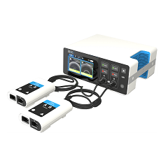
The US Food and Drug Administration (FDA) has granted 510(k) clearance for the EKOS® Control Unit 4.0, a new unit that can control the EKOS® System.
The EKOS System includes an ultrasonic device that uses acoustic pulses to enhance the effects of thrombolytic drugs and dissolve blood clots in patients with deep vein thrombosis, pulmonary embolism, and peripheral arterial occlusions.
With this system, acoustic pulses unwind and thin fibrin to expose drug receptor sites.
This allows thrombolytic drugs to reach deeper into clots, accelerating absorption and helping to dissolve clots faster and with less drug, according to BTG International LTD, the company that markets the EKOS System.
The EKOS Control Unit 4.0 replaces the EKOS System’s old control unit. The new unit can support 2 EKOS devices, allowing physicians to use a single control unit to treat both pulmonary arteries, with the goal of simplifying bilateral pulmonary embolism treatment.
The EKOS System (formerly known as the EkoSonic Endovascular System) was approved by the FDA in 2014.
In clinical studies, the system has been shown to speed time to clot dissolution, increase clot removal, and enhance clinical improvement compared to either standard catheter-directed drug therapy or thrombectomy.1,2
Research has also shown that the EKOS System requires significantly shorter treatment times and less thrombolytic compared to standard catheter-directed drug therapy3, 4,5, lowering the risk of bleeding and other complications.1,5,6 ![]()
1. Lin, P, et al, “Comparison of Percutaneous Ultrasound-Accelerated Thrombolysis versus Catheter-Directed Thrombolysis in Patients with Acute Massive Pulmonary Embolism.” Vascular, Vol. 17, Suppl. 3, 2009, S137–S147.
2. Schrijver, AM, et al, “Dutch Randomized Trial Comparing Standard Catheter-Directed Thrombolysis and Ultrasound-Accelerated Thrombolysis for Arterial Thromboembolic Infrainguinal Disease (DUET)." Journal of Endovascular Therapy 2015, Vol. 22(1):87-95.
3. Litzendorf, ME, et al, “Ultrasound-accelerated thrombolysis is superior to catheter-directed thrombolysis for the treatment of acute limb ischemia.” Journal of Vascular Surgery, Jun 2011; 53(Suppl S), p106S-107S.
4. Lin, P, et al, “Catheter-Directed Thrombectomy and Thrombolysis for Symptomatic Lower-Extremity Deep Vein Thrombosis: Review of Current Interventional Treatment Strategies.” Perspectives in Vascular Surgery and Endovascular Therapy, 2010, 22(3): 152–163.
5. Parikh, S, et al, “Ultrasound-Accelerated Thrombolysis for the Treatment of Deep Vein Thrombosis: Initial Clinical Experience.” Journal of Vascular and Interventional Radiology, Vol. 19, Issue 4, April 2008, 521–528.
6. Kucher, N, et al, “Randomized, Controlled Trial of Ultrasound-Assisted Catheter-Directed Thrombolysis for Acute Intermediate-Risk Pulmonary Embolism.” Circulation, Vol. 129, No. 4, 2014, 479–486.
CAR T-cell trial in adult ALL shut down

After 2 clinical holds in 2016 and 5 patient deaths, the Seattle biotech Juno Therapeutics is shutting down the phase 2 ROCKET trial of JCAR015.
The chimeric antigen receptor (CAR) T-cell therapy JCAR015 was being tested in adults with relapsed or refractory B-cell acute lymphoblastic leukemia (ALL).
“We have decided not to move forward . . . at this time,” CEO Hans Bishop said in a statement, “even though it generated important learnings for us and the immunotherapy field.”
He said the company remains “committed to developing better treatment for patients battling ALL.”
The first clinical hold of the ROCKET trial occurred in July after 2 patients died. The company attributed the deaths primarily to the addition of fludarabine to the regimen.
Juno removed fludarabine from the treatment protocol, the clinical hold was lifted, and the trial resumed.
Then, in November, 2 more patients died from cerebral edema, and the trial was put on hold once again.
One patient had died earlier in 2016, totaling 5 patient deaths from cerebral edema, although the earliest death was not necessarily related to treatment, the company stated.
Juno attributed the deaths to multiple factors, including the patients’ treatment history and treatment received at the beginning of the trial.
Juno plans to start a new adult ALL trial in 2018. The therapy, they say, is more similar to JCAR017, which is being tested in pediatric patients.
ROCKET is not the first trial of JCAR015 to be placed on hold.
In 2014, after 2 patients died of cytokine release syndrome, the phase 1 trial was placed on clinical hold.
Juno made changes to the enrollment criteria and dosing, and the hold was lifted. Results from this trial were presented at ASCO 2015 and ASCO 2016. ![]()
Exercise better than meds to reduce fatigue in cancer patients

Exercise and/or psychological therapy work better than medications to reduce cancer-related fatigue, according to research published in JAMA Oncology.
Researchers conducted a review and meta-analysis of more than 113 studies and found that exercise and psychological interventions, as well as a combination of both, were associated with reduced fatigue during and after cancer treatment.
However, pharmaceutical interventions were not associated with the same magnitude of improvement.
The researchers therefore concluded that exercise and psychological therapy should be recommended over medications.
“If a cancer patient is having trouble with fatigue, rather than looking for extra cups of coffee, a nap, or a pharmaceutical solution, consider a 15-minute walk,” said study author Karen Mustian, PhD, of the University of Rochester Medical Center in Rochester, New York.
“It’s a really simple concept, but it’s very hard for patients and the medical community to wrap their heads around it because these interventions have not been front-and-center in the past. Our research gives clinicians a valuable asset to alleviate cancer-related fatigue.”
Dr Mustian and her colleagues reached their conclusions after analyzing data from 113 randomized clinical trials testing various treatments for cancer-related fatigue.
There were 11,525 patients enrolled in these studies. Nearly half (46.9%) were women with breast cancer. Ten studies focused on other types of cancer and enrolled only men.
Dr Mustian and her colleagues performed a meta-analysis to establish and compare the mean weighted effect sizes (WESs) of the fatigue treatments.
The team found that exercise alone—whether aerobic or anaerobic—reduced cancer-related fatigue most significantly. The WES was 0.30 (95% CI, 0.25-0.36; P<0.001).
Psychological interventions—such as therapy designed to provide education, change personal behavior, and adapt the way a person thinks about his or her circumstances—also improved fatigue. The WES was 0.27 (95% CI, 0.21-0.330.30; P<0.001).
A combination of psychological interventions and exercise had a significant improvement on fatigue as well. The WES was 0.26 (95% CI, 0.13-0.38; P<0.001).
However, the drugs tested for treating cancer-related fatigue—paroxetine hydrochloride, modafinil, armodafinil, methylphenidate hydrochloride, dexymethylphenidate, dexamphetamine, and methylprednisolone—were not as effective as the other interventions. The WES was 0.09 (95% CI, 0.00-0.19; P=0.05).
“The literature bears out that these drugs don’t work very well, although they are continually prescribed,” Dr Mustian said. “Cancer patients already take a lot of medications, and they all come with risks and side effects. So any time you can subtract a pharmaceutical from the picture it usually benefits patients.” ![]()
Potential therapeutic strategy for BL, DLBCL

Preclinical research has revealed a potential strategy for treating Burkitt lymphoma (BL) and diffuse large B-cell lymphoma (DLBCL).
Investigators discovered that miR-28 inhibits the growth of B-cell lymphomas, but this microRNA is often lost in these lymphomas.
Re-expressing miR-28 in mouse models of BL and DLBCL inhibited tumor growth, which supports the potential of synthetic miR-28 analogs for the treatment of these lymphomas.
In fact, the investigators believe their work could lead to the development of the first miRNA analog therapy for the treatment of B-cell lymphoma and provide the basis for clinical trials.
Almudena Ramiro, PhD, of Centro Nacional de Investigaciones Cardiovasculares in Madrid, Spain, and her colleagues described the work in Blood.
The team characterized the function of miR-28 in the biology of mature B lymphocytes and in the development of lymphomas associated with this cell type.
The investigators found that miR-28 regulates the terminal differentiation of B lymphocytes, a fundamental process in the biology of these cells that generates memory B lymphocytes and highly specific plasma cells.
But the team found that miR-28 expression is lost in several germinal center-derived lymphoma subtypes, including BL, DLBCL, follicular lymphoma, and chronic lymphocytic leukemia.
In vitro experiments showed that miR-28 expression dampens B-cell receptor signaling and diminishes the proliferation and survival of primary B cells and lymphoma cells.
And in vivo experiments showed that re-establishing miR-28 expression slows tumor growth in DLBCL and BL.
The investigators re-expressed miR-28 in xenograft models of BL and DLBCL via the use of viral vectors or synthetic molecules and found that both methods blocked tumor growth. The same effect was observed in mice with established BL tumors.
Dr Ramiro and her colleagues said these results reveal the therapeutic potential of miR-28 and provide ample justification for the initiation of clinical trials of miR-28-based therapies to treat B-cell lymphomas. ![]()
Drug granted priority review for relapsed/refractory AML

The US Food and Drug Administration (FDA) has granted priority review for the new drug application (NDA) for enasidenib (AG-221), an inhibitor of mutant IDH2.
The drug is under review for the treatment of patients with relapsed or refractory acute myeloid leukemia (AML) with an IDH2 mutation.
The FDA grants priority review to applications for products that may provide significant improvements in the treatment, diagnosis, or prevention of serious conditions.
The agency’s goal is to take action on a priority review application within 6 months of receiving it, rather than the standard 10-month period.
The NDA for enasidenib has been given a Prescription Drug User Fee Act action date of August 30, 2017.
Enasidenib is being developed by Celgene Corporation and Agios Pharmaceuticals.
Phase 1/2 trial
The NDA submission for enasidenib is based on results from AG221-C-001, a single-arm, phase 1/2 study of the drug in patients with advanced hematologic malignancies with an IDH2 mutation.
Early data from the relapsed or refractory AML patients in this study were presented at the 2015 ASH Annual Meeting. (The presentation included updated data that differ from the data in the abstract.)
The trial included a dose-escalation phase and 5 expansion cohorts. The first 4 expansion cohorts had completed enrollment as of the presentation.
- Arm 1: 25 patients with IDH2-mutant-positive relapsed or refractory AML age ≥60 years, or any patient with AML regardless of age who relapsed after a bone marrow transplant (BMT)
- Arm 2: 25 patients with IDH2-mutant-positive relapsed or refractory AML age <60 years, excluding patients with AML who relapsed after a BMT
- Arm 3: 25 patients with IDH2-mutant-positive untreated AML age ≥60 years who decline standard of care chemotherapy
- Arm 4: 25 patients with IDH2-mutant-positive advanced hematologic malignancies not eligible for arms 1 to 3
- Arm 5: The phase 2 portion of the trial included 125 patients with IDH2-mutant-positive AML who were in second or later relapse, refractory to second-line induction or reinduction treatment, or relapsed after allogeneic transplant.
The data reported at ASH were from patients receiving enasidenib administered from 50-mg to 650-mg total daily doses in the dose-escalation arm and 100 mg once daily in the first 4 expansion arms, as of September 1, 2015.
The median age of these patients was 69 (range, 19-100). Patients with relapsed or refractory AML received a median of 2 prior lines of therapy (range, 1-6).
Safety data
A safety analysis was conducted for all 231 treated patients. As of the ASH presentation, a maximum tolerated dose of enasidenib had not been reached.
The majority of adverse events were mild to moderate, with the most common being nausea, diarrhea, fatigue, and febrile neutropenia.
Twenty-three percent of patients had treatment-related serious adverse events—notably, differentiation syndrome (4%), leukocytosis (4%), and nausea (2%).
Drug-related grade 5 serious adverse events include atrial flutter (n=1), cardiac tamponade (n=1), pericardial effusion (n=1), and respiratory failure (n=1).
Efficacy Data
Seventy-nine of the 209 response-evaluable patients achieved investigator-assessed objective responses, for an overall response rate of 38%.
There were 37 (18%) complete remissions (CR), 3 CRs with incomplete platelet recovery (CRp), 14 marrow CRs (mCR), 3 CRs with incomplete hematologic recovery (CRi), and 22 partial remissions (PR).
Of the 159 patients with relapsed or refractory AML, 59 (37%) achieved an objective response, including 29 (18%) CRs, 1 CRp, 9 mCRs, 3 CRis, and 17 PRs.
Of the 24 patients with AML who declined standard of care chemotherapy, 10 achieved an objective response, including 4 CRs, 1 CRp, 1 mCR, and 4 PRs.
The median duration of response was 6.9 months in patients with relapsed or refractory AML.
Responding relapsed/refractory AML patients were on study treatment for up to 18 months. The median duration of treatment was 6.8 months (range, 1.8 to 18 months). ![]()
Hospital floors pose infection risk, team says
Hospital room floors may be an overlooked source of infection, according to a study published in the American Journal of Infection Control.
Researchers surveyed 5 hospitals and found that floors in patient rooms were often contaminated with pathogens.
Certain objects, such as personal items and medical devices and supplies, were in contact with the floor, and touching these objects resulted in the transfer of pathogens to bare and gloved hands.
Abhishek Deshpande, MD, PhD, of Case Western Reserve University School of Medicine in Cleveland, Ohio, and his colleagues conducted this research.
The team cultured 318 floor sites from 159 patient rooms (2 sites per room) in 5 hospitals in the Cleveland area. The rooms included both Clostridium difficile infection (CDI) isolation rooms and non-CDI rooms.
The researchers also cultured hands (gloved and bare) as well as other “high-touch” surfaces such as clothing and medical devices/supplies.
The team found that floors in patient rooms were often contaminated with Methicillin-resistant Staphylococcus aureus (MRSA), vancomycin-resistant enterococci (VRE), and C difficile.
C difficile was recovered in 55% of CDI rooms and 47% of non-CDI rooms. MRSA was recovered in 32% of CDI rooms and 8% of non-CDI rooms. VRE was recovered in 30% of CDI rooms and 13% of non-CDI rooms.
The researchers said the frequency of contamination was similar for each of the 5 hospitals and from room and bathroom floor sites.
Of the 100 occupied rooms surveyed, 41% had one or more high-touch objects that were in contact with the floor. These included personal items (eg, clothing, canes, and cellular phone chargers), medical devices and supplies (eg, pulse oximeter, call button, heating pad, urinal, blood pressure cuff, wash basin, and heel protector), and bed linens or towels.
The findings indicate that handling such items resulted in the transfer of pathogens. All 3 pathogens were recovered from bare or gloved hand cultures—MRSA in 6 (18%), VRE in 2 (6%), and C difficile in 1 (3%).
The researchers said these results suggest hospital floors could be an underappreciated source for dissemination of pathogens and are an important area for additional research.
“Understanding gaps in infection prevention is critically important for institutions seeking to improve the quality of care offered to patients,” said Linda Greene, RN, current president of the Association for Professionals in Infection Control and Epidemiology.
“Even though most facilities believe they are taking the proper precautions, this study points out the importance of ensuring cleanliness of the hospital environment and the need for education of both staff and patients on this issue.” ![]()
Antithrombotic drugs may raise risk of subdural hematoma

Use of antithrombotic drugs is associated with a higher risk of subdural hematoma, according to a case-control study of more than 400,000 individuals in Denmark.
The highest risk of subdural hematoma was observed in patients who were taking both a vitamin K antagonist (VKA) and clopidogrel.
The study also showed that the incidence of subdural hematoma increased from 2000 to 2015, which appeared to be associated with the increased use of antithrombotic drugs over that time period.
David Gaist, MD, PhD, of Odense University Hospital and the University of Southern Denmark, and his colleagues conducted this research and reported the results in JAMA.
The researchers evaluated 10,010 patients, ages 20 to 89, with a first-ever subdural hematoma diagnosis from 2000 to 2015. The patients were matched to 400,380 individuals from the general population.
Among the patients with subdural hematoma (average age, 69), 47% were taking antithrombotic medications.
The researchers used conditional logistic regression models to estimate the association between antithrombotic drugs and subdural hematoma risk, adjusting for a range of factors. With the following data, they adjusted for age, sex, calendar period, comorbidity, education level, and income level.
The team found that current low-dose aspirin use was associated with a low risk of subdural hematoma (adjusted odds ratio [OR]=1.24).
Current clopidogrel (OR=1.80) and direct oral anticoagulant (OR=1.55) use were both associated with a moderate risk.
And current VKA use was associated with the highest risk of all the agents when given alone (OR=3.69).
Concurrent use of more than one antithrombotic drug was also associated with a high subdural hematoma risk:
- Low-dose aspirin and clopidogrel (OR=2.45)
- Low-dose aspirin and direct oral anticoagulant (OR=2.52)
- Low-dose aspirin and VKA (OR=4.00)
- Clopidogrel and VKA (OR=7.93).
There was one exception to this. Low-dose aspirin combined with the antiplatelet drug dipyridamole was associated with a risk similar to that of low-dose aspirin alone (OR=1.17).
The researchers noted that the prevalence of antithrombotic drug use increased in the general population over the time period studied, from 31.0 per 1000 individuals in 2000 to 76.9 per 1000 individuals in 2015 (P<0.001 for trend).
The overall subdural hematoma incidence rate increased as well, from 10.9 per 100,000 person-years in 2000 to 19.0 per 100,000 person-years in 2015 (P<0.001 for trend).
The largest increase in subdural hematoma incidence was among patients older than 75 years of age—from 55.1 per 100,000 person-years to 99.7 per 100,000 person-years (P<0.001 for trend). ![]()
Use of antithrombotic drugs is associated with a higher risk of subdural hematoma, according to a case-control study of more than 400,000 individuals in Denmark.
The highest risk of subdural hematoma was observed in patients who were taking both a vitamin K antagonist (VKA) and clopidogrel.
The study also showed that the incidence of subdural hematoma increased from 2000 to 2015, which appeared to be associated with the increased use of antithrombotic drugs over that time period.
David Gaist, MD, PhD, of Odense University Hospital and the University of Southern Denmark, and his colleagues conducted this research and reported the results in JAMA.
The researchers evaluated 10,010 patients, ages 20 to 89, with a first-ever subdural hematoma diagnosis from 2000 to 2015. The patients were matched to 400,380 individuals from the general population.
Among the patients with subdural hematoma (average age, 69), 47% were taking antithrombotic medications.
The researchers used conditional logistic regression models to estimate the association between antithrombotic drugs and subdural hematoma risk, adjusting for a range of factors. With the following data, they adjusted for age, sex, calendar period, comorbidity, education level, and income level.
The team found that current low-dose aspirin use was associated with a low risk of subdural hematoma (adjusted odds ratio [OR]=1.24).
Current clopidogrel (OR=1.80) and direct oral anticoagulant (OR=1.55) use were both associated with a moderate risk.
And current VKA use was associated with the highest risk of all the agents when given alone (OR=3.69).
Concurrent use of more than one antithrombotic drug was also associated with a high subdural hematoma risk:
- Low-dose aspirin and clopidogrel (OR=2.45)
- Low-dose aspirin and direct oral anticoagulant (OR=2.52)
- Low-dose aspirin and VKA (OR=4.00)
- Clopidogrel and VKA (OR=7.93).
There was one exception to this. Low-dose aspirin combined with the antiplatelet drug dipyridamole was associated with a risk similar to that of low-dose aspirin alone (OR=1.17).
The researchers noted that the prevalence of antithrombotic drug use increased in the general population over the time period studied, from 31.0 per 1000 individuals in 2000 to 76.9 per 1000 individuals in 2015 (P<0.001 for trend).
The overall subdural hematoma incidence rate increased as well, from 10.9 per 100,000 person-years in 2000 to 19.0 per 100,000 person-years in 2015 (P<0.001 for trend).
The largest increase in subdural hematoma incidence was among patients older than 75 years of age—from 55.1 per 100,000 person-years to 99.7 per 100,000 person-years (P<0.001 for trend).
Hemophilia repository open to US scientists

A new resource has been made available for US-based scientists studying hemophilia A and B.
The resource—known as the My Life, Our Future Research Repository—is a collection of genetic data and blood samples that are linked to phenotypic data from more than 5000 hemophilia patients in the US.
Academic and industry scientists within the US can now access the repository but must apply to do so.
Scientists outside of the US should have access to the repository in 2018.
My Life, Our Future is a program founded by the American Thrombosis and Hemostasis Network, Bloodworks Northwest, the National Hemophilia Foundation, and Bioverativ.
My Life, Our Future was launched in 2012 to increase genotyping among individuals with hemophilia A and B, as well as potential and confirmed carriers of the disorder. The program will be available at participating hemophilia treatment centers through the end of 2017.
My Life, Our Future provides hemophilia patients (and potential/confirmed carriers) with free genotyping, and patients can then consent to add their de-identified genetic data and blood sample to the My Life, Our Future Research Repository.
Eighty-three percent of genotyped patients have contributed their data and blood samples. This is more than 5000 individuals representing more than 25% of the US hemophilia population.
The information provided by these individuals has led to the discovery of more than 600 new genetic variants that cause hemophilia.
“Hemophilia is a complex disorder, and, prior to today, there was no genetic and phenotypic database of this scale aiding scientific innovation,” said Barbara Konkle, MD, associate chief scientific officer of Bloodworks Northwest and principal investigator for My Life, Our Future.
“With this new resource, researchers now have one source for the genetic data, repository samples, and clinical data needed to investigate the many unanswered questions about hemophilia.”
Accessing the repository
To access the samples and data in the repository, scientists must first submit a letter of intent to the American Thrombosis and Hemostasis Network.
Scientists may then be asked to submit a full research proposal, which will be evaluated by a research review committee—a group of experts including a molecular pathologist, genetic counselor, research scientist, molecular biologist, genetic epidemiologist, molecular epidemiologist, geneticist, patient representative, and hematologists.
Approved scientists will be selected based on the scientific merit of their proposals and level of benefit to those with bleeding disorders.
For details on how to apply, visit ATHN.org/MLOF.
CCSs’ subsequent cancer risk decreased from ’70s to ’90s

Childhood cancer survivors (CCSs) who were diagnosed in the 1990s have a lower risk of subsequent malignancies than CCSs diagnosed in the 1970s, according to research published in JAMA.
The data suggest this outcome is associated with a reduction in the overall use and median dose of therapeutic radiation over time.
Past research has shown an association between therapeutic radiation and the development of subsequent neoplasms in CCSs. Studies have also linked specific chemotherapeutic agents to subsequent neoplasms.
This information has been used to modify childhood cancer treatment over time, with the hope of reducing the risk of subsequent neoplasms while maintaining or improving 5-year survival.
To assess the effects of these treatment modifications, Gregory Armstrong, MD, of St. Jude Children’s Research Hospital in Memphis, Tennessee, and his colleagues conducted a study of CCSs.
The researchers evaluated 23,603 five-year CCSs (mean age at diagnosis, 7.7 years) treated at pediatric hospitals in the US and Canada from 1970 through 1999, with follow-up through December 2015.
The most common initial diagnoses were acute lymphoblastic leukemia (35.1%), Hodgkin lymphoma (11.1%), and astrocytoma (9.6%).
Subsequent neoplasms, malignancies
At a mean follow-up of 20.5 years, 1639 CCSs had experienced 3115 subsequent neoplasms, including 1026 malignancies, 233 benign meningiomas, and 1856 non-melanoma skin cancers. The most common neoplasms were breast and thyroid cancers.
The 15-year cumulative incidence of subsequent neoplasms decreased by decade of diagnosis. The incidence was 2.9% for patients diagnosed in the 1970s, 2.4% for those diagnosed in the ’80s, and 1.5% for those diagnosed in the ’90s. For the 1970s vs 1980s, the P value was 0.02. For the 1970s vs 1990s and for the 1980s vs 1990s, the P value was <0.001.
The 15-year cumulative incidence of subsequent malignancies also decreased by decade of diagnosis—2.1% for the ’70s, 1.7% for the ’80s, and 1.3% for the ’90s. The P value was <0.001 for the ’70s vs the ’90s and the ’80s vs the ’90s.
Risk factors
In multivariable analyses, female CCSs had a higher rate of subsequent neoplasms (including malignancies) than males.
In addition, high doses of alkylating agents and platinum agents were associated with increased rates of subsequent malignancies.
The researchers noted that, although there was a decrease in the median cumulative dose of alkylating agents over time, the proportion of CCSs receiving these agents increased. And both the median cumulative dose of platinum agents and the proportion of CCSs receiving these agents increased from the ’70s to the ’90s.
Finally, therapeutic radiation was associated with increased rates of subsequent malignant neoplasms, meningiomas, and non-melanoma skin cancers.
This corresponded with the researchers’ findings that the proportion of individuals receiving radiation and the median dose of radiation both decreased over time.
The proportion of individuals receiving any radiation therapy was 77.7% in the ’70s, 56.7% in the ’80s, and 36.8% in the ’90s. The median dose of radiation was 30.0 Gy in the ’70s, 24.0 Gy in the ’80s, and 26.0 Gy in the ’90s.
“The most ominous late effect of pediatric cancer treatment is a second malignancy,” Dr Armstrong said. “This study shows efforts to reduce the late effects of treatment are paying off. The risk of second cancers for survivors increases with age, so it is good to see the reduction emerging early in survivorship while survivors are still young.”