User login
Company withdraws MAA for biosimilar pegfilgrastim

Image by Volker Brinkmann
Gedeon Richter Plc. has withdrawn its marketing authorization application (MAA) for the biosimilar pegfilgrastim product Cavoley from the European Medicines Agency (EMA).
Richter was seeking approval of Cavoley for the same indications as the reference product, Neulasta, a pegylated recombinant granulocyte-colony stimulating factor used to reduce the duration of neutropenia and the occurrence of febrile neutropenia in adults receiving cytotoxic chemotherapy to treat malignancies (except chronic myeloid leukemia and myelodysplastic syndromes).
The MAA filing for Cavoley was based on data from Richter’s completed biosimilar development program.
The company presented to the EMA results of studies in healthy volunteers designed to show that Cavoley is highly similar to Neulasta in terms of chemical structure, purity, the way it works, and how the body handles the drug.
Richter also presented results of a study comparing the safety and effectiveness of Cavoley and Neulasta in breast cancer patients receiving cytotoxic chemotherapy (EudraCT 2013-003166-14).
Richter withdrew the MAA for Cavoley after the EMA’s Committee for Medicinal Products for Human Use (CHMP) evaluated the documentation provided by the company and formulated lists of questions.
After the CHMP had assessed the company’s responses to the last round of questions, there were still some unresolved issues.
Based on the review of the data and Richter’s response to the CHMP’s list of questions, at the time of the withdrawal, the CHMP had some concerns and was of the provisional opinion that Cavoley could not have been approved.
The CHMP said the company had not demonstrated that Cavoley is highly similar to Neulasta.
In its letter notifying the EMA of the MAA withdrawal, Richter said it would continue developing Cavoley and follow the CHMP’s advice to eliminate the remaining uncertainty that Cavoley is highly similar to Neulasta. ![]()
Drug can improve upon chelation therapy in thalassemia major

An anti-hypertensive drug can improve the effects of chelation therapy in patients with thalassemia major and cardiac iron overload, according to research published in Blood.
Researchers found that administering the drug, amlodipine, in conjunction with conventional chelation therapy significantly reduced myocardial iron concentration (MIC) in patients with cardiac iron overload at baseline, when compared to chelation therapy alone.
In addition, amlodipine did not cause any serious adverse events.
“The drug has been used clinically for decades and is considered safe for adults and children,” said study author Juliano de Lara Fernandes, MD, PhD, of José Michel Kalaf Research Institute in Campinas, São Paulo State, Brazil.
“As an adjunct to standard treatment, it can be greatly beneficial to patients and has few side effects.”
Dr Fernandes said he and his colleagues decided to test amlodipine in patients with thalassemia major because although chelation therapy works well in peripheral organs, it’s difficult to remove iron from the heart.
“Myocardial dysfunctions are currently the main cause of death among patients with thalassemia and can emerge in children from the age of 10,” Dr Fernandes said.
The most serious problem of all, he added, is caused by an accumulation of non-transferrin bound iron (NTBI) in myocardial cells. NTBI enters and leaves the liver without causing much damage to the organ, but it enters the heart via a channel whose main role is to carry calcium into cells.
“It occurred to us that drugs capable of blocking the calcium channel could also prevent NTBI from entering the heart and therefore increase the efficacy of chelation therapy,” Dr Fernandes said.
He and his colleagues tested this hypothesis in 62 patients with thalassemia major. The patients were divided into 2 treatment groups. One group received conventional chelation therapy along with amlodipine (5 mg/day), and the other received chelation therapy plus oral placebo.
Patients were further divided according to cardiac iron levels at baseline. Cardiac iron overload was defined as T2* < 35 ms. Fifty percent of patients in the amlodipine arm and 52% of those in the placebo arm had cardiac iron overload at baseline.
Results
The study’s main outcome was change in MIC, as determined by magnetic resonance imaging at 12 months.
At that point, among patients with cardiac iron overload at baseline, there was a significant decrease in MIC in the amlodipine arm but not the placebo arm. The median change in MIC was -0.26 mg/g and 0.01 mg/g, respectively (P=0.02).
The median MIC in the amlodipine arm went from 1.31 mg/g at baseline to 1.05 mg/g at 12 months (P=0.02). The median MIC in the placebo arm went from 0.77 mg/g at baseline to 0.75 at 12 months (P=0.76).
“Myocardial iron concentration fell 21% in patients with initial iron overload who were treated with chelation plus amlodipine, whereas it increased by 2% in those with initial overload who were treated with chelation plus placebo,” Dr Fernandes said.
On the other hand, there were no significant differences in MIC from baseline to 12 months among patients who received amlodipine but did not have cardiac iron overload at baseline.
For patients without cardiac iron overload at baseline, the median change in MIC was +0.04 mg/g in the amlodipine arm and -0.01 in the placebo arm (P=0.07).
The median MIC in the amlodipine arm went from 0.54 mg/g at baseline to 0.55 mg/g at 12 months. The median MIC in the placebo arm went from 0.55 mg/g at baseline to 0.52 mg/g at 12 months.
“Perhaps we would have needed to monitor these patients for a longer period to see the benefits of preventive therapy with amlodipine for people who don’t have excess iron in their organs,” Dr Fernandes said.
“For those who do, however, the results show it’s worth using amlodipine. There’s no need to change the existing therapy. It’s enough to administer the anti-hypertensive orally every day.”
There were 4 mild adverse events in the amlodipine arm but none in the placebo arm (13% vs 0%, P=0.11).
Three patients (10%) in the amlodipine arm had their initial dose of 5 mg/day reduced to 2.5 mg/day because of mild ankle edema (n=2) and dizziness (n=1). One patient had a mild cutaneous allergic reaction and stopped receiving amlodipine after 15 days but continued on study.
There were no deaths or hospital admissions due to cardiovascular complications during the trial. ![]()
FDA places AML trials on full, partial clinical hold

The US Food and Drug Administration (FDA) has placed holds on 3 early stage trials of vadastuximab talirine (SGN-CD33A) in acute myeloid leukemia (AML).
A phase 1/2 trial of vadastuximab talirine monotherapy in pre- and post-allogeneic transplant patients has been placed on full clinical hold.
This means no new subjects can be enrolled on the trial, and there can be no further dosing of subjects who are already enrolled.
Two phase 1 trials of vadastuximab talirine have been placed on partial clinical hold. This means no new subjects can be enrolled, but existing patients may continue treatment with re-consent.
In one of the trials on partial hold, researchers are investigating vadastuximab talirine alone and in combination with hypomethylating agents in AML patients who either relapsed after induction/consolidation or declined treatment with high-dose induction/consolidation.
In the other trial on partial hold, researchers are testing vadastuximab talirine in combination with 7+3 chemotherapy in newly diagnosed AML patients. Results from this trial were presented at the 2016 ASH Annual Meeting.
All 3 clinical holds were initiated to evaluate the potential risk of hepatotoxicity in patients who were treated with vadastuximab talirine and received allogeneic stem cell transplant either before or after treatment.
There have been 6 patients with hepatotoxicity, including several cases of veno-occlusive disease, with 4 fatal events.
Seattle Genetics, Inc., the company developing vadastuximab talirine, said it is working with the FDA to determine whether there is any association between hepatotoxicity and treatment with vadastuximab talirine to identify appropriate protocol amendments for patient safety and to enable continuation of these trials.
No new studies of vadastuximab talirine will be initiated until the clinical holds are lifted.
Seattle Genetics’ other ongoing trials of vadastuximab talirine, including the phase 3 CASCADE trial in older AML patients and phase 1/2 trial in patients with myelodysplastic syndrome (MDS), are proceeding with enrollment.
Overall, more than 300 patients have been treated with vadastuximab talirine in clinical trials across multiple treatment settings.
Vadastuximab talirine is an investigational antibody-drug conjugate (ADC) targeted to CD33, which is expressed on most AML and MDS blast cells. The CD33 engineered cysteine antibody is stably linked to a DNA binding agent called a pyrrolobenzodiazepine (PBD) dimer via site-specific conjugation technology (EC-mAb).
PBD dimers are said to be significantly more potent than systemic chemotherapeutic drugs, and the EC-mAb technology allows uniform drug-loading onto an ADC. The ADC is designed to be stable in the bloodstream and to release its PBD agent upon internalization into CD33-expressing cells. ![]()
FDA expands approval for hemophilia A drug
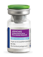
Photo courtesy of Baxalta
The US Food and Drug Administration (FDA) has expanded the approved use of Adynovate, a recombinant pegylated factor VIII (FVIII) product, in patients with hemophilia A.
Adynovate was previously approved as routine prophylaxis and for on-demand treatment of bleeding episodes in patients age 12 and older.
Now, Adynovate is approved for the same indications in patients younger than 12 and for perioperative management in patients of all ages.
Adynovate is built on the full-length Advate molecule, which was approved by the FDA in 2003. Adynovate leverages proprietary pegylation technology designed to extend the amount of FVIII available for use in the body.
The technology was selected because it maintains the integrity of the parent molecule (Advate) while reducing the time at which the body clears Adynovate, resulting in an extended circulating half-life.
Adynovate and Advate are registered trademarks of Baxalta Incorporated, a wholly owned, indirect subsidiary of Shire plc.
For more details on Adynovate, see the full prescribing information.
Trials of Adynovate
The FDA’s approval of Adynovate in children is based on data from a phase 3 trial, which were presented at the World Federation of Hemophilia 2016 World Congress.
The study enrolled previously treated children younger than 12 with no history of FVIII inhibitors. The patients received twice-weekly prophylaxis with Adynovate (50 ± 10 IU/kg) for at least 6 months or 50 exposure days, whichever occurred last.
Sixty-six patients were evaluable. None of them developed inhibitory antibodies, and there were no adverse events related to Adynovate.
The median annualized bleeding rate was 2.0, and 38% of patients did not have any bleeding episodes.
The FDA’s approval of Adynovate for perioperative management was based on interim results of an ongoing phase 3 study in 15 patients with severe hemophilia A undergoing surgical procedures. The interim results were presented at the 2015 ASH Annual Meeting.
The patients underwent 11 major surgical procedures (3 knee replacements, 2 arthroscopic synovectomies, 1 cyst extirpation, 1 port placement, 1 gastric band placement, and 3 multiple tooth extractions including 1 radicular cyst removal) and 4 minor surgeries (1 synoviorthesis, 1 radiosynovectomy, 1 tooth extraction, and 1 dermatological surgery).
Perioperative hemostatic efficacy was rated as “excellent” for most procedures. Excellent hemostatic efficacy was defined as blood loss less than or equal to that expected for the same type of procedure performed in a non-hemophilic patient and requiring blood components for transfusions less than or similar to that expected in the non-hemophilic population.
The median observed intraoperative blood loss (n=10) was 10.0 mL, compared to the predicted average blood loss (n=11) of 50.0 mL for major surgeries. ![]()
Why kids with cancer have a higher risk of treatment-related toxicity
Preclinical research appears to explain why certain tissues in very young children are more sensitive to collateral damage from cancer treatment than tissues in older individuals.
Researchers found evidence to suggest that, early in life, cells in the brain, heart, and kidney are primed for apoptosis.
On the other hand, cells in the healthy adult brain, heart, and kidneys are apoptosis-refractory.
Kristopher A. Sarosiek, PhD, of the Harvard T.H. Chan School of Public Health in Boston, Massachusetts, and his colleagues reported these findings in Cancer Cell.
The researchers used BH3 profiling to measure the relative dominance of pro-survival or pro-death signals inside cells.
A cancer cell in which apoptotic signals are dominant is said to be “highly primed” for self-destruction and therefore easily killed by therapy, while a cell with low priming is more resistant to death or damage.
Dr Sarosiek and his colleagues measured the priming of cells in tissues from adult mice and young mice.
In the adult mice, cells of the hematopoietic lineage from the periphery, thymus, spleen, and bone marrow were the most primed for apoptosis. Cells from the large intestine, small intestine, lungs, and liver were relatively unprimed. And cells in brain, heart, and kidney tissues were far less primed.
However, in embryonic and very young mice, cells in the brain, heart, and kidney were extremely primed for apoptosis.
The researchers found that, in the adult brains, hearts, and kidneys, the molecular machinery needed to perform apoptosis was nearly completely absent.
In contrast, this machinery was abundant in the brains, hearts, and kidneys of young mice. As a result, brain, heart, and kidney cells were much more vulnerable to cell death when exposed to chemotherapy or radiation.
After determining in mouse models that certain cells grew more resistant to treatment toxicity with age, the researchers tested human cells. The team obtained fresh samples of tissue that had been removed from brains of children and adults to prevent intractable epileptic seizures.
As in the mice, the youngest human brain cells were more highly primed with apoptotic machinery and vulnerable to chemotherapy and radiation damage.
The researchers said there was a period of higher heterogeneity in apoptotic priming among patients between 2 and 6 years of age. After that, the brain transitions to full apoptotic resistance.
The team also found that, in young tissues, expression of the apoptotic protein machinery is driven by c-Myc. This transcription factor drives an apoptotically primed state by directly activating transcription of the pro-apoptotic genes Bax, Bim, and Bid.
“[This research] has uncovered some opportunities to selectively block apoptosis in our healthy tissues and prevent toxicity from radiation or chemotherapy while still maintaining sensitivity within cancer cells,” Dr Sarosiek said. “We are actively pursuing the identification of new medicines that can be used exactly for this purpose.” ![]()
Anticoagulant receives priority review

Image by Andre E.X. Brown
The US Food and Drug Administration (FDA) has granted priority review to the new drug application (NDA) for betrixaban, an oral factor Xa inhibitor, for extended-duration prophylaxis of venous thromboembolism (VTE) in acute medically ill patients with risk factors for VTE.
A priority review shortens the FDA review timeline to 6 months from the standard review period of 10 months.
The application for betrixaban has been given a Prescription Drug User Fee Act action date of June 24, 2017. (Betrixaban also has fast track designation from the FDA.)
Meanwhile, the European Medicines Agency (EMA) is reviewing a marketing authorization application (MAA) for betrixaban for extended-duration prophylaxis of VTE in adults with acute medical illness and risk factors for VTE.
However, the EMA’s Committee for Medicinal Products for Human Use is reviewing the application under a standard 210-day review period.
“With the filing of the betrixaban NDA and the MAA validation, we now look forward to working with the FDA and EMA to bring this drug to market,” said Bill Lis, chief executive officer of Portola Pharmaceuticals, Inc., the company developing betrixaban.
The NDA and MAA for betrixaban are supported by data from Portola’s phase 3 APEX study, which enrolled 7513 patients at more than 450 clinical sites worldwide.
In this study, researchers compared extended-duration anticoagulation with oral betrixaban for 35-42 days to standard-duration enoxaparin for 10±4 days in preventing VTE in high-risk acute medically ill patients.
Patients who received betrixaban had a significantly lower incidence of VTE than those who received enoxaparin, and there was no significant difference in major bleeding between the treatment groups.
Full results from this study were presented at the 62nd Annual International Society on Thrombosis and Haemostasis Scientific and Standardization Committee Meeting and published in NEJM in May 2016. ![]()
Combo produces high response rate in CLL trial

Results of a phase 2 trial suggest a 2-drug combination may be effective in patients with chronic lymphocytic leukemia (CLL), particularly those with high-risk disease.
The combination consists of ublituximab (TG-1101), a glycoengineered anti-CD20 monoclonal antibody, and the oral BTK inhibitor ibrutinib.
Six months after starting treatment, the overall response rate was 88% among all evaluable patients and 95% among those with high-risk CLL.
Researchers said the long-term clinical benefit of the combination will be defined by an ongoing phase 3 trial.
The team reported results from the phase 2 trial in the British Journal of Haematology. The study was sponsored by TG Therapeutics, Inc., the company developing ublituximab.
The trial included 45 patients. Their median age was 71 (range, 39-86), about half were female, and the median ECOG performance score was 1.
Nearly half of patients (47%, n=21) had high-risk CLL. Twelve patients had del 17p, 12 had del 11q, 5 patients had both, and 2 had a TP53 mutation.
The patients had a median of 2 (range, 1-7) prior treatments, including purine analogues (n=22), bendamustine (n=21), idelalisib (n=2), a spleen-tyrosine kinase inhibitor (n=2), and the BTK inhibitor CC-292 (n=1).
Treatment
For this study, patients received ibrutinib at 420 mg once daily and 2 different doses of ublituximab. The study had a dose-confirmation safety run-in period that was followed by an open enrollment into phase 2.
The dose-confirmation safety assessment enrolled 6 patients in each of 2 cohorts. Patients in cohort 1 received ublituximab at 600 mg on days 1, 8, and 15 of cycle 1. If there was ≤1 dose-limiting toxicity (DLT) in this cohort, the dose escalation would proceed to cohort 2.
In cohort 2, patients’ ublituximab dose increased to 900 mg on days 1, 8, and 15 of cycle 1. If ≤ 1 DLT was reported in this cohort, the dose was considered safe for phase 2.
There were no DLTs observed in either cohort. So subsequent patients were enrolled into the open phase 2 part of the study, in which they received ublituximab at 900 mg on days 1, 8, and 15 of cycle 1, as well as on day 1 of cycles 2 to 6.
Patients had response assessments at cycles 3 and 6. After that, they continued on ibrutinib monotherapy off study.
Safety
All 45 patients were evaluable for safety. The most common adverse events (AEs) were infusion-related reactions (IRRs, 53%), diarrhea (40%), fatigue (33%), cough (27%), rash (27%), and nausea (24%).
Grade 3/4 AEs included anemia (11%), neutropenia (11%), IRRs (7%), thrombocytopenia (7%), diarrhea (4%), and arthralgia (2%).
All rash and grade 3/4 diarrhea events were attributed to ibrutinib, and all IRRs were related to ublituximab. Twenty-one patients (47%) had dose interruptions due to IRRs, and 1 patient had a dose reduction to 600 mg.
Four patients had ublituximab-related dose interruptions—2 due to neutropenia and 2 because of elevated aspartate aminotransferase.
Two patients had ibrutinib-related dose reductions (for diarrhea and dizziness). Ten patients had ibrutinib-related dose interruptions—3 due to rash, 2 due to neutropenia, and 1 each because of anemia, thrombocytopenia, nausea, hypercalcemia, and dehydration.
Efficacy
Forty-one patients were evaluable for efficacy. Two patients were lost to follow-up, and 2 discontinued due to AEs. One of the AEs, diarrhea, was considered related to ibrutinib. The other patient discontinued due to pneumonia and pleural effusion, which were not attributed to study treatment.
At 6 months, the overall response rate was 88% among evaluable patients and 95% among high-risk patients. The median time to response was 8 weeks.
Two patients had a complete response, 34 had a partial response, and 3 had stable disease.
Both complete responders and 1 of the partial responders achieved minimal residual disease negativity. All 3 of these patients had high-risk disease.
“[T]he addition of ublituximab to ibrutinib not only produced high response rates but also allowed patients to achieve deeper responses, with complete responses and minimal residual disease negativity seen, which is rare with ibrutinib alone,” said study author Jeff Sharman, MD, of Willamette Valley Cancer Institute in Eugene, Oregon.
“We look forward to exploring how the increased depth of response may affect the sequence of treatments given to patients.” ![]()
Results of a phase 2 trial suggest a 2-drug combination may be effective in patients with chronic lymphocytic leukemia (CLL), particularly those with high-risk disease.
The combination consists of ublituximab (TG-1101), a glycoengineered anti-CD20 monoclonal antibody, and the oral BTK inhibitor ibrutinib.
Six months after starting treatment, the overall response rate was 88% among all evaluable patients and 95% among those with high-risk CLL.
Researchers said the long-term clinical benefit of the combination will be defined by an ongoing phase 3 trial.
The team reported results from the phase 2 trial in the British Journal of Haematology. The study was sponsored by TG Therapeutics, Inc., the company developing ublituximab.
The trial included 45 patients. Their median age was 71 (range, 39-86), about half were female, and the median ECOG performance score was 1.
Nearly half of patients (47%, n=21) had high-risk CLL. Twelve patients had del 17p, 12 had del 11q, 5 patients had both, and 2 had a TP53 mutation.
The patients had a median of 2 (range, 1-7) prior treatments, including purine analogues (n=22), bendamustine (n=21), idelalisib (n=2), a spleen-tyrosine kinase inhibitor (n=2), and the BTK inhibitor CC-292 (n=1).
Treatment
For this study, patients received ibrutinib at 420 mg once daily and 2 different doses of ublituximab. The study had a dose-confirmation safety run-in period that was followed by an open enrollment into phase 2.
The dose-confirmation safety assessment enrolled 6 patients in each of 2 cohorts. Patients in cohort 1 received ublituximab at 600 mg on days 1, 8, and 15 of cycle 1. If there was ≤1 dose-limiting toxicity (DLT) in this cohort, the dose escalation would proceed to cohort 2.
In cohort 2, patients’ ublituximab dose increased to 900 mg on days 1, 8, and 15 of cycle 1. If ≤ 1 DLT was reported in this cohort, the dose was considered safe for phase 2.
There were no DLTs observed in either cohort. So subsequent patients were enrolled into the open phase 2 part of the study, in which they received ublituximab at 900 mg on days 1, 8, and 15 of cycle 1, as well as on day 1 of cycles 2 to 6.
Patients had response assessments at cycles 3 and 6. After that, they continued on ibrutinib monotherapy off study.
Safety
All 45 patients were evaluable for safety. The most common adverse events (AEs) were infusion-related reactions (IRRs, 53%), diarrhea (40%), fatigue (33%), cough (27%), rash (27%), and nausea (24%).
Grade 3/4 AEs included anemia (11%), neutropenia (11%), IRRs (7%), thrombocytopenia (7%), diarrhea (4%), and arthralgia (2%).
All rash and grade 3/4 diarrhea events were attributed to ibrutinib, and all IRRs were related to ublituximab. Twenty-one patients (47%) had dose interruptions due to IRRs, and 1 patient had a dose reduction to 600 mg.
Four patients had ublituximab-related dose interruptions—2 due to neutropenia and 2 because of elevated aspartate aminotransferase.
Two patients had ibrutinib-related dose reductions (for diarrhea and dizziness). Ten patients had ibrutinib-related dose interruptions—3 due to rash, 2 due to neutropenia, and 1 each because of anemia, thrombocytopenia, nausea, hypercalcemia, and dehydration.
Efficacy
Forty-one patients were evaluable for efficacy. Two patients were lost to follow-up, and 2 discontinued due to AEs. One of the AEs, diarrhea, was considered related to ibrutinib. The other patient discontinued due to pneumonia and pleural effusion, which were not attributed to study treatment.
At 6 months, the overall response rate was 88% among evaluable patients and 95% among high-risk patients. The median time to response was 8 weeks.
Two patients had a complete response, 34 had a partial response, and 3 had stable disease.
Both complete responders and 1 of the partial responders achieved minimal residual disease negativity. All 3 of these patients had high-risk disease.
“[T]he addition of ublituximab to ibrutinib not only produced high response rates but also allowed patients to achieve deeper responses, with complete responses and minimal residual disease negativity seen, which is rare with ibrutinib alone,” said study author Jeff Sharman, MD, of Willamette Valley Cancer Institute in Eugene, Oregon.
“We look forward to exploring how the increased depth of response may affect the sequence of treatments given to patients.”
Explaining lack of response to malaria vaccines

Image by Peter H. Seeberger
Researchers say they have uncovered one potential reason why it has been difficult to generate protective immunity against the early liver stage of malaria infection in regions where the incidence of malaria is high.
Their research, conducted in mice and published in Cell Reports, suggests that exposure to the blood stage of malaria infection inhibits the formation of the protective immune cells (and their antibodies) that can prevent liver-stage infection.
“The blood stage of malaria infection has a very profound impact on the liver stage immune response, and that impact had never been dissected and visualized at this level,” said study author Marion Pepper, PhD, of the University of Washington School of Medicine in Seattle.
“These studies really suggest that you need a vaccine that is protective against both stages of infection to effectively prevent malaria.”
To track how the blood stage of malaria infection overpowers the liver-stage immune response, Dr Pepper and her colleagues infected 2 groups of mice with different forms of malaria parasites.
One group of mice was infected with Plasmodium yoelii wild-type sporozoites, which complete the pre-erythrocytic stage of infection and establish a blood-stage infection.
The other group was infected with a genetically attenuated Plasmodium yoelii parasite that arrests late in liver stage development and does not cause blood-stage infection.
Six days after infection, the researchers found the levels of antibodies were significantly lower in the mice with the blood stage infection than in mice that only had the parasite targeted to the liver.
To understand this discrepancy, the team tracked the differentiation of Plasmodium liver stage-specific B cells. B cells can differentiate into antibody-secreting early effector cells or long-lived memory cells, both of which contribute to protection against malaria.
The researchers discovered that, 14 days after infection, the B cells in the blood-stage-infected mice never went through the necessary changes to make rapidly responsive memory cells.
However, in the mice that received the liver-stage attenuated version of the parasite, the B cells were still able to differentiate and create the necessary antibodies and memory cells for an effective immune response.
“This work really highlights the importance of looking at antigen-specific B cells,” Dr Pepper said. “These data also suggest that if you’re getting a vaccine while you have an ongoing blood-stage infection, there is a chance that the vaccine will not generate good memory cells because the blood stage disrupts all the processes that are involved in making that immunological memory.”
Dr Pepper and her colleagues are now looking into the possibility of treatment to solve this problem.
The team found that when they treated the second stage of the infection with the anti-malarial drug atovaquone, the B cells were able to create the optimally responsive memory cells.
For now, the researchers hope their work can be used to answer immediate questions about the efficacy of malaria vaccines in regions that are most significantly affected by the disease.
“Malaria has evolved with us throughout human existence and therefore has some potent immune evasion strategies,” Dr Pepper said. “We really tried to tease apart some of the factors that could be driving the loss of protective immunity during natural infection and with current vaccine strategies in areas of high malaria transmission.”
“Our next step is to compare malaria-specific B cells after vaccination or natural infection in humans so we can translate these findings and start to determine how to solve this problem.”
Bendamustine approved for new indication in Japan

Bendamustine hydrochloride (TREAKISYM®) has been approved in Japan as first-line treatment for patients with low-grade B-cell non-Hodgkin lymphoma (NHL) and mantle cell lymphoma (MCL).
The drug will now be available for adjunctive use with rituximab in these patients.
Bendamustine hydrochloride is already approved in Japan as monotherapy for relapsed or refractory low-grade B-cell NHL and MCL, as well as chronic lymphocytic leukemia.
Bendamustine hydrochloride is the subject of a licensing agreement concluded between Eisai Co., Ltd and SymBio Pharmaceuticals Limited. Under the licensing agreement, Eisai has been marketing the product since December 2010.
Bendamustine hydrochloride is available at doses of 25 mg and 100 mg for intravenous infusion. The recommended dosage and administration is as follows:
- For low-grade B-cell NHL and MCL
- As first-line treatment
When used adjunctively with rituximab, the usual adult dose of bendamustine hydrochloride is 90 mg/m2 body surface area infused intravenously over 60 minutes on days 1 and 2 of repeated 28-day cycles.
- For relapsed or refractory disease
The usual adult dose of bendamustine hydrochloride is 120 mg/m2 body surface area infused intravenously over 60 minutes on days 1 and 2 of repeated 21-day cycles.
- As first-line treatment
- For chronic lymphocytic leukemia
- The usual adult dose of bendamustine hydrochloride is 100 mg/m2 body surface area infused intravenously over 60 minutes on days 1 and 2 of repeated 28-day cycles.
All of the aforementioned doses may be reduced appropriately according to the patient’s condition.
Assay could make blood supply safer, groups say

Photo by Alex Luster
with The Storyhive
Two groups of researchers have reported that an assay can accurately diagnose patients with variant Creutzfeldt-Jakob disease (vCJD), and this could allow for effective detection of prion contamination in donated blood.
The groups both said they were able to detect vCJD with 100% sensitivity and specificity.
One group even detected abnormal prion proteins in the blood of 2 subjects before the individuals exhibited any signs of vCJD.
The researchers said this work paves the way to a noninvasive, early diagnostic screen for vCJD and possibly other conditions involving protein misfolding.
Both studies were published in Science Translational Medicine.
“Our findings, which need to be confirmed in further studies, suggest that our method of detection could be useful for the noninvasive diagnosis of this disease in pre-symptomatic individuals,” said Claudio Soto, MD, author of one of the studies and a professor at the University of Texas Medical School in Houston.
“Early diagnosis would allow any potential therapy to be given before substantial brain damage has occurred. In the case of the blood supply, availability of a procedure to efficiently detect small quantities of the infectious agent would allow removal of blood units contaminated with prions so that new cases can be minimized substantially.”
For their study, Dr Soto and his colleagues used a protein misfolding cyclic amplification assay (PMCA) they developed, which mimics the prion replication process in vitro that occurs in prion disease.
The team used the assay to screen for abnormal prion proteins in blood from 14 individuals with vCJD and 153 control subjects.
In another study, Daisy Bougard, PhD, of Etablissement Français du Sang, INSERM, Université de Montpellier in France, and her colleagues tested a similar technique on blood samples from 18 individuals with vCJD and 238 without vCJD.
Dr Bougard’s group used the same PMCA as Dr Soto’s group. But Dr Bougard and her colleagues first captured prions from blood using plasminogen-coated beads.
In both studies, the PMCA diagnosed vCJD with 100% sensitivity and 100% specificity.
Dr Bougard and her colleagues were able to detect small amounts of prions in 2 blood donors more than a year before the onset of symptoms.
The researchers stressed that these results will need to be confirmed in a larger number of blood samples.