User login
Blood disorders prove costly for European economy

chemotherapy
Photo by Rhoda Baer
Malignant and non-malignant blood disorders cost 31 European countries a total of €23 billion in 2012, according to a pair of papers published in The Lancet Haematology.
Healthcare costs accounted for €16 billion of the total costs, with €7 billion for hospital inpatient care and €4 billion for medications.
Informal care (from friends and relatives) cost €1.6 billion, productivity losses due to mortality cost €2.5 billion, and morbidity cost €3 billion.
Researchers determined these figures by analyzing data from international health organizations (WHO and EUROSTAT), as well as national ministries of health and statistical institutes.
The team estimated the economic burden of malignant and non-malignant blood disorders in 2012 for all 28 countries in the European Union (EU), as well as Iceland, Norway, and Switzerland.
The costs considered were healthcare costs (primary care, accident and emergency care, hospital inpatient and outpatient care, and drugs), informal care costs (from friends and relatives), and productivity losses (due to premature death and people being unable to work due to illness).
Malignant blood disorders
In one paper, the researchers noted that the total economic cost of blood cancers to the 31 countries studied was €12 billion in 2012. Healthcare costs measured €7.3 billion (62% of total costs), productivity losses cost €3.6 billion (30%), and informal care cost €1 billion (8%).
In the 28 EU countries, blood cancers represented 8% of the total cancer costs (€143 billion), meaning that blood cancers are the fourth most expensive type of cancer after lung (15%), breast (12%), and colorectal (10%) cancers.
When considering healthcare costs alone, blood cancers were second only to breast cancers (12% vs 13% of healthcare costs for all cancers).
In 2012, blood cancers cost, on average, €14,674 per patient in the EU (€15,126 in all 31 countries), which is almost 2 times higher than the average cost per patient across all cancers (€7929 in the EU).
The researchers said this difference may be due to the longer length of hospital stay observed for patients with blood cancers (14 days, on average, compared to 8 days across all cancers).
Another potential reason is that blood cancers are increasingly treated with complex, long-term treatments (including stem cell transplants, multi-agent chemotherapy, and radiotherapy) and diagnosed via extensive procedures.
The costs of blood cancers varied widely between the countries studied, but the reasons for this were unclear. For instance, the average healthcare costs in Finland were nearly twice as high as in Belgium (€18,014 vs €9596), despite both countries having similar national income per capita.
Non-malignant blood disorders
In the other paper, the researchers said the total economic cost of non-malignant blood disorders to the 31 countries studied was €11 billion in 2012. Healthcare costs accounted for €8 billion (75% of total costs), productivity losses for €2 billion (19%), and informal care for €618 million (6%).
Averaged across the population studied, non-malignant blood disorders represented an annual healthcare cost of €159 per 10 citizens.
“Non-malignant blood disorders cost the European economy nearly as much as all blood cancers combined,” said Jose Leal, DPhil, of the University of Oxford in the UK.
“We found wide differences in the cost of treating blood disorders in different countries, likely linked to the significant differences in the access and delivery of care for patients with blood disorders. Our findings suggest there is a need to harmonize care of blood disorders across Europe in a cost-effective way.” ![]()
EC grants immunotherapy orphan designation
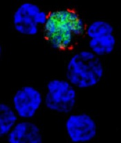
among uninfected cells (blue)
Image courtesy of
Benjamin Chaigne-Delalande
The European Commission (EC) has granted orphan drug designation for CMD-003 (baltaleucel-T) as a treatment for nasal type extranodal NK/T-cell lymphoma and post-transplant lymphoproliferative disorder.
CMD-003 consists of patient-derived T cells that have been activated to kill malignant cells expressing antigens associated with Epstein-Barr virus (EBV).
The T cells specifically target 4 EBV epitopes—LMP1, LMP2, EBNA, and BARF1.
CMD-003 is being developed by Cell Medica and the Baylor College of Medicine, with funding provided, in part, by the Cancer Prevention and Research Institute of Texas.
About orphan designation
Orphan designation from the EC provides regulatory and financial incentives for companies to develop and market therapies that treat a life-threatening or chronically debilitating condition affecting no more than 5 in 10,000 people in the European Union, and where no satisfactory treatment is available.
Orphan designation provides a 10-year period of marketing exclusivity in the European Union if the drug receives regulatory approval. The designation also provides incentives for companies seeking protocol assistance from the European Medicines Agency during the product development phase and direct access to the centralized authorization procedure.
CMD-003 also has orphan designation from the US Food and Drug Administration to treat all EBV-associated non-Hodgkin lymphomas.
CMD-003-related research
CMD-003 is currently under investigation in a phase 2 trial, CITADEL, for the treatment of advanced NK/T-cell lymphoma.
Researchers have not published results from any trials of CMD-003, but they have published results with EBV-specific T-cell products related to CMD-003.
In one study, published in the Journal of Clinical Oncology, researchers administered cytotoxic T lymphocytes (CTLs) in 50 patients with EBV-associated Hodgkin or non-Hodgkin lymphoma.
Twenty-nine of the patients were in remission when they received CTL infusions, but they were at a high risk of relapse. The remaining 21 patients had relapsed or refractory disease at the time of CTL infusion.
Twenty-seven of the patients who received CTLs as an adjuvant treatment remained in remission at 3.1 years after treatment.
Their 2-year event-free survival rate was 82%. None of the patients died of lymphoma, but 9 died from complications associated with the chemotherapy and radiation they had received.
Of the 21 patients with relapsed or refractory disease, 13 responded to CTL infusions, and 11 patients achieved a complete response. In this group, the 2-year event-free survival rate was about 50%.
The researchers said there were no toxicities that were definitively related to CTL infusion.
One patient had central nervous system deterioration 2 weeks after infusion. This was attributed to disease progression but could possibly have been treatment-related.
Another patient developed respiratory complications about 4 weeks after a second CTL infusion that may have been treatment-related. However, the researchers attributed it to an intercurrent infection, and the patient made a complete recovery. ![]()
Early VTE prophylaxis better for sTBI, team says

Results of an observational study support early initiation of venous thromboembolism (VTE) prophylaxis in patients with severe traumatic brain injury (sTBI).
The research suggests that starting anticoagulant therapy within 72 hours of hospital arrival significantly lowers the risk of deep vein thrombosis (DVT) and pulmonary embolism (PE) in patients with sTBI, without increasing the risk of bleeding complications or death.
These results were published in the Journal of the American College of Surgeons.
“Physicians have traditionally been hesitant to initiate pharmacological blood clot prophylaxis early in patients with severe brain injuries because, while thinning the blood might prevent PE and DVT, it might also increase the risk of complications related to worsening intracranial hemorrhage,” said study author James P. Byrne, MD, of the University of Toronto in Ontario, Canada.
“We performed this study because there wasn’t clear evidence that starting prophylaxis early actually prevented blood clots, or whether this benefit would outweigh the risk of complications from intracranial hemorrhage. Current evidence-based guidelines don’t address the optimal timing for starting prophylaxis in patients with severe TBI.”
The researchers looked at data on 3634 adult patients with sTBI who were treated at 186 trauma centers participating in the American College of Surgeons Trauma Quality Improvement Program. The patients received VTE prophylaxis with either low-molecular-weight heparin or unfractionated heparin between 2012 and 2014.
The researchers divided patients into 2 groups: early prophylaxis (started within 72 hours of hospital arrival) or late prophylaxis (started after 72 hours). The primary outcomes were PE or DVT. The secondary outcomes were late neurosurgical interventions (performed after 72 hours) and in-hospital death.
The team used propensity-score matching to emulate the design of a randomized, controlled trial and minimize selection bias. This method took into account a large set of patient baseline and injury factors, and yielded a cohort of 2468 patients. Outcomes were then compared between early and late prophylaxis groups.
Results
The researchers found that early VTE prophylaxis was associated with significantly lower rates of PE (adjusted odds ratio [aOR]=0.48; 95% CI 0.25–0.91) and DVT (aOR=0.51; 95% CI 0.36–0.72) than late prophylaxis.
“No previous study has shown that patients who receive early prophylaxis have lower rates of pulmonary embolism, which is important because this complication is a potentially fatal one,” Dr Byrne noted.
“We also found that trauma centers that most frequently used early prophylaxis in their patients had significantly lower rates of deep vein thrombosis, compared with counterparts where fewer patients received early prophylaxis, with no difference in rates of late neurosurgical intervention or mortality.”
Specifically, there was no significant difference between early prophylaxis and late prophylaxis groups with respect to rates of in-hospital mortality (aOR=1.10; 95% CI 0.84–1.45) or late neurosurgical interventions, including craniotomy/craniectomy (aOR=0.86; 95% CI 0.53–1.40) and intracranial monitor (aOR=0.76; 95% CI 0.37–1.58).
The researchers said this is the largest study to date comparing the effectiveness and safety of early versus late VTE prophylaxis in patients with sTBI.
A limitation of this study was the fact that statistical methods could not account for confounding factors that were not measured in the study dataset. One such factor was changes in patterns of intracranial hemorrhage on head CT scan, which would influence physician decision-making.
“The takeaway message is that early prophylaxis really does matter in patients with severe traumatic brain injury, in terms of reducing a patient’s risk of pulmonary embolism or deep vein thrombosis,” Dr Byrne said.
“Our findings suggest that this is possible without increasing the risk of the most feared complications, such as the need to take a patient to the operating room to evacuate intracranial hemorrhage or death. In other words, it’s possible to prevent PE or DVT with early prophylaxis, without putting patients at risk of bad outcomes, and we should be striving to achieve this.” ![]()
Gene therapy granted breakthrough designation for hemophilia B

The US Food and Drug Administration (FDA) has granted breakthrough therapy designation to SPK-9001 for the treatment of hemophilia B.
SPK-9001 is a bio-engineered adeno-associated virus capsid expressing a codon-optimized, high-activity human factor IX variant enabling endogenous production of factor IX.
SPK-9001 is intended to control and prevent bleeding episodes in patients with hemophilia B, without the need for regular infusions.
SPK-9001 is under investigation in an ongoing phase 1/2 trial. The therapy is being developed by Spark Therapeutics and Pfizer Inc.
“We are extremely pleased to have been granted breakthrough therapy designation for SPK-9001, which has shown early promise in achieving our goal of eliminating the need for regular infusions to control and prevent bleeding episodes in patients with hemophilia B through a potentially one-time, intravenous administration of a highly optimized gene therapy,” said Jeffrey D. Marrazzo, chief executive officer of Spark Therapeutics.
The FDA’s breakthrough therapy designation is intended to expedite the development and review of new therapies for serious or life-threatening conditions.
To earn the designation, a treatment must show encouraging early clinical results demonstrating substantial improvement over available therapies with regard to a clinically significant endpoint, or it must fulfill an unmet need. ![]()
Study may explain how LSCs evade treatment
Image by Robert Paulson
New research suggests leukemia stem cells (LSCs) can “hide” in gonadal adipose tissue (GAT) and transform the tissue so they can survive treatment.
Experiments in a mouse model of chronic myeloid leukemia (CML) showed that LSCs are enriched in GAT.
While there, the LSCs create a microenvironment that supports leukemic growth and resistance to treatment, and expression of the fatty acid transporter CD36 makes LSCs particularly resistant.
Craig Jordan, PhD, of University of Colorado in Aurora, and his colleagues conducted this research and detailed their findings in Cell Stem Cell.
The researchers began by examining cancer cells found in GAT from mice with blast crisis CML. Rather than containing the expected mix of regular leukemia cells and LSCs, the tissue was enriched for LSCs.
And these GAT-resident LSCs used a different energy source than LSCs in the bone marrow microenvironment. The GAT-resident LSCs powered their survival and growth with fatty acids, manufacturing energy by the process of fatty acid oxidization.
In fact, the GAT-resident LSCs actively signaled fat to undergo lipolysis, which released fatty acids into the microenvironment.
“The basic biology was fascinating,” Dr Jordan said. “The tumor adapted the local environment to suit itself.”
Dr Jordan and his colleagues also found that CD36 played a role. CD36+ LSCs were enriched in GAT, were more likely to migrate to GAT than to bone marrow, and were protected from treatment by GAT.
The researchers tested the effects of several drugs (cytarabine, doxorubicin, etoposide, SN-38, irinotecan, and dasatinib) on CD36+ LSCs, CD36- LSCs, and bulk leukemia cells ex vivo.
Both CD36+ and CD36- LSCs were more resistant to treatment than bulk leukemia cells, but CD36+ LSCs were preferentially drug-resistant.
The researchers observed similar results in leukemic mice and found evidence to suggest that CD36 plays a similar role in patients with blast crisis CML and those with acute myeloid leukemia. ![]()
Ibrutinib approved for first-line treatment of CLL

Photo courtesy of Janssen
Health Canada has approved the Bruton’s tyrosine kinase inhibitor ibrutinib (Imbruvica®) as a first-line treatment for patients with active chronic lymphocytic leukemia (CLL).
This is the fourth approval for ibrutinib in Canada. The drug is now approved for use in all CLL patients, patients with Waldenström’s macroglobulinemia, and patients with relapsed or refractory mantle cell lymphoma (conditional approval).
Ibrutinib is jointly developed and commercialized by Pharmacyclics LLC, an AbbVie company, and Janssen Biotech, Inc.
The latest approval of ibrutinib is based on results from the phase 3 RESONATE-2 trial
(PCYC-1115), which were presented at the 2015 ASH Annual Meeting and
simultaneously published in NEJM.
RESONATE-2 enrolled 269 treatment-naïve patients with CLL or small lymphocytic lymphoma who were 65 or older.
Patients were randomized to receive ibrutinib (n=136) at 420 mg once a day until progression or unacceptable toxicity, or chlorambucil (n=133) on days 1 and 15 of each 28-day cycle for up to 12 cycles. The starting dose for chlorambucil in cycle 1 was 0.5 mg/kg and was increased based on tolerability in cycle 2 by increments of 0.1 mg/kg to a maximum of 0.8 mg/kg.
The primary endpoint of the study was progression-free survival (PFS), as assessed by an independent review committee (IRC) according to the International Workshop on Chronic Lymphocytic Leukemia (iWCLL) 2008 criteria, with modification for treatment-related lymphocytosis.
Key secondary endpoints included overall response rate (based on the same iWCLL criteria), overall survival (OS), and safety.
Ibrutinib significantly prolonged PFS, as determined by the IRC, reducing the risk of progression or death by 84% compared to chlorambucil. The hazard ratio was 0.16 (P<0.001). The median PFS was not reached in the ibrutinib arm but was 18.9 months for the chlorambucil arm.
Ibrutinib significantly prolonged OS as well, although the median OS was not reached in either treatment arm. The OS rate at 24 months was 98% with ibrutinib and 85% with chlorambucil. The relative risk of death with ibrutinib was 84% lower than that with chlorambucil. The hazard ratio was 0.16 (P=0.001).
Ibrutinib was associated with a significantly higher IRC-assessed overall response rate compared to chlorambucil—82% and 35%, respectively (P<0.0001). Five
patients (4%) in the ibrutinib arm achieved a complete response, as did 2 patients (2%) in the chlorambucil arm.
The median duration of treatment was 17.4 months in the ibrutinib arm and 7.1 months in the chlorambucil arm.
The most common adverse events of any grade—in the ibrutinib and chlorambucil arms, respectively—were diarrhea (42% and 17%), fatigue (30% and 38%), cough (22% and 15%), nausea (22% and 39%), peripheral edema (19% and 9%), dry eye (17% and 5%), arthralgia (16% and 7%), neutropenia (16% and 23%), and vomiting (13% and 20%).
Adverse events of grade 3 or higher—in the ibrutinib and chlorambucil arms, respectively—were neutropenia (10% and 18%), anemia (6% and 8%), hypertension (4% and 0%), pneumonia (4% and 2%), diarrhea (4% and 0%), maculopapular rash (3% and 2%), decreased platelet count (3% and 1%), abdominal pain (3% and 1%), hyponatremia (3% and 0%), thrombocytopenia (2% and 6%), febrile neutropenia (2% and 2%), upper respiratory tract infection (2% and 2%), pleural effusion (2% and 1%), cellulitis (2% and 0%), fatigue (1% and 5%), syncope (1% and 2%), and hemolytic anemia (0% and 2%). ![]()
Radiologists no longer have higher risk of cancer-related death

Photo by Rhoda Baer
Radiologists who graduated from medical school after 1940 do not have an increased risk of dying from radiation-related causes such as cancers, according to a study published in Radiology.
However, the study suggested that male radiologists who graduated before 1940 had a higher risk of death from certain cancers, including acute myeloid leukemia and non-Hodgkin lymphoma.
Researchers said these findings point to the success of efforts to reduce occupational radiation doses over the past several decades.
The team noted that female radiologists did not have an increased risk of all-cause mortality or cancer-related mortality, regardless of when they graduated from medical school.
However, the small number of women in this study prevented the researchers from studying the subjects’ mortality rates in detail. And very few female radiologists worked during the early period of the study, when radiation exposures were likely highest.
To conduct this study, the researchers analyzed records from the American Medical Association Physician Masterfile, a database established in 1906 that has grown to include current and historical data for more than 1.4 million physicians, residents, and medical students in the US.
The team compared cancer incidence and mortality rates between 43,763 radiologists and 64,990 psychiatrists who graduated from medical school between 1916 and 2006. Psychiatrists were chosen as a comparison group because they are unlikely to have had occupational radiation exposure.
“Our most important finding is that radiologists have lower death rates from all causes of death combined, compared to psychiatrists, and had similar risks of cancer deaths overall,” said study author Martha Linet, MD, of the National Cancer Institute in Bethesda, Maryland.
Results in males
The researchers found that, among male subjects who graduated after 1940, the risk of all-cause mortality was lower for the radiologists than the psychiatrists (relative risk [RR]=0.94; 95% CI: 0.90, 0.97), and the risk of death from cancer was similar (RR=1.00; 95% CI: 0.93, 1.07).
In contrast, male radiologists who graduated before 1940 had higher mortality rates from certain cancers.
They had a higher risk of skin cancer mortality (RR=6.38; 95% CI: 1.75, 23.20) that was driven by an excess of melanoma (RR=8.75; 95% CI: 1.89, 40.53).
They had an increased risk of death from all myeloid leukemias (RR=1.43; 95% CI: 1.00, 2.05) that was driven by acute myeloid leukemia and/or myelodysplastic syndromes (RR=4.68; 95% CI: 0.91, 24.18).
And they had an increased risk of death from lymphomas (RR=2.24; 95% CI: 1.31, 3.86) that was driven by non-Hodgkin lymphoma (RR=2.69; 95% CI: 1.33, 5.45).
The researchers also found an increased risk of cerebrovascular deaths in the male radiologists who graduated before 1940 (RR=1.49; 95% CI: 1.11, 2.01).
The team said the reduced health risks for more recent radiology graduates are likely due to developments and improvements in radiation protection and monitoring, along with improvements in equipment safety.
“Most of the findings of increased risk were in the earlier radiologists,” Dr Linet noted. “We do feel there is evidence that decreases in dose in the United States and other countries seem to have paid off, reducing risks in recent graduates.”
Results in females
The researchers said there were no clear increases in mortality in the female radiologists compared with the female psychiatrists.
The risk of all-cause mortality was lower in the radiologists, as was the risk of death from circulatory diseases, but the risk of cancer-related mortality was similar between the radiologists and the psychiatrists.
However, the researchers said the relatively small number of female deaths in this study prevented detailed investigation. Only 2% of female radiologists (208/8851) and 3% of female psychiatrists (524/17,493) died, compared to 12% of male radiologists (4260/43,763) and 16% of male psychiatrists (7815/47,443). ![]()
Photo by Rhoda Baer
Radiologists who graduated from medical school after 1940 do not have an increased risk of dying from radiation-related causes such as cancers, according to a study published in Radiology.
However, the study suggested that male radiologists who graduated before 1940 had a higher risk of death from certain cancers, including acute myeloid leukemia and non-Hodgkin lymphoma.
Researchers said these findings point to the success of efforts to reduce occupational radiation doses over the past several decades.
The team noted that female radiologists did not have an increased risk of all-cause mortality or cancer-related mortality, regardless of when they graduated from medical school.
However, the small number of women in this study prevented the researchers from studying the subjects’ mortality rates in detail. And very few female radiologists worked during the early period of the study, when radiation exposures were likely highest.
To conduct this study, the researchers analyzed records from the American Medical Association Physician Masterfile, a database established in 1906 that has grown to include current and historical data for more than 1.4 million physicians, residents, and medical students in the US.
The team compared cancer incidence and mortality rates between 43,763 radiologists and 64,990 psychiatrists who graduated from medical school between 1916 and 2006. Psychiatrists were chosen as a comparison group because they are unlikely to have had occupational radiation exposure.
“Our most important finding is that radiologists have lower death rates from all causes of death combined, compared to psychiatrists, and had similar risks of cancer deaths overall,” said study author Martha Linet, MD, of the National Cancer Institute in Bethesda, Maryland.
Results in males
The researchers found that, among male subjects who graduated after 1940, the risk of all-cause mortality was lower for the radiologists than the psychiatrists (relative risk [RR]=0.94; 95% CI: 0.90, 0.97), and the risk of death from cancer was similar (RR=1.00; 95% CI: 0.93, 1.07).
In contrast, male radiologists who graduated before 1940 had higher mortality rates from certain cancers.
They had a higher risk of skin cancer mortality (RR=6.38; 95% CI: 1.75, 23.20) that was driven by an excess of melanoma (RR=8.75; 95% CI: 1.89, 40.53).
They had an increased risk of death from all myeloid leukemias (RR=1.43; 95% CI: 1.00, 2.05) that was driven by acute myeloid leukemia and/or myelodysplastic syndromes (RR=4.68; 95% CI: 0.91, 24.18).
And they had an increased risk of death from lymphomas (RR=2.24; 95% CI: 1.31, 3.86) that was driven by non-Hodgkin lymphoma (RR=2.69; 95% CI: 1.33, 5.45).
The researchers also found an increased risk of cerebrovascular deaths in the male radiologists who graduated before 1940 (RR=1.49; 95% CI: 1.11, 2.01).
The team said the reduced health risks for more recent radiology graduates are likely due to developments and improvements in radiation protection and monitoring, along with improvements in equipment safety.
“Most of the findings of increased risk were in the earlier radiologists,” Dr Linet noted. “We do feel there is evidence that decreases in dose in the United States and other countries seem to have paid off, reducing risks in recent graduates.”
Results in females
The researchers said there were no clear increases in mortality in the female radiologists compared with the female psychiatrists.
The risk of all-cause mortality was lower in the radiologists, as was the risk of death from circulatory diseases, but the risk of cancer-related mortality was similar between the radiologists and the psychiatrists.
However, the researchers said the relatively small number of female deaths in this study prevented detailed investigation. Only 2% of female radiologists (208/8851) and 3% of female psychiatrists (524/17,493) died, compared to 12% of male radiologists (4260/43,763) and 16% of male psychiatrists (7815/47,443).
Drug can prevent nausea, vomiting caused by chemo

Photo by Bill Branson
Results of a phase 3 study suggest the antipsychotic agent olanzapine can also be used to reduce nausea and vomiting caused by chemotherapy.
In this study, cancer patients receiving highly emetogenic chemotherapy also received combination anti-emetic therapy including olanzapine or placebo.
Those patients who received olanzapine were significantly less likely to experience nausea and vomiting in the 120 hours after starting chemotherapy.
These results were published in NEJM.
“We’ve long known the nausea and vomiting that come along with chemotherapy are a major problem and affect the quality of life of our patients,” said study author Steven Powell, MD, of Sanford Cancer Center in Sioux Falls, South Dakota.
“The findings of this study, fortunately, provide physicians with a tool to better address the needs of those they are treating for cancer.”
Dr Powell and his colleagues evaluated cancer patients who had received no previous chemotherapy but were receiving cisplatin or cyclophosphamide and doxorubicin during the study period.
To prevent nausea and vomiting, all of the patients received a 5-HT3–receptor antagonist, dexamethasone, and an NK1-receptor antagonist. Roughly half also received olanzapine, and the other half received placebo.
Overall, 380 patients were evaluable—192 assigned to olanzapine and 188 to placebo.
In the first 24 hours after starting chemotherapy, the proportion of patients who did not have chemotherapy-induced nausea was significantly greater in the olanzapine arm than the placebo arm—74% and 45%, respectively (P=0.002).
The same was true at 25 hours to 120 hours after the start of chemotherapy—42% and 25%, respectively (P=0.002)—and for the overall 120-hour period—37% and 22%, respectively (P=0.002).
The complete response rate—defined as no vomiting and no rescue therapy—was significantly higher in the olanzapine arm than the placebo arm in the first 24 hours—86% and 65% (P<0.001)—at 25 hours to 120 hours—67% and 52%, respectively (P=0.007)—and overall—64% and 41%, respectively (P<0.001).
There were two grade 3 adverse events and three grade 4 adverse events in the olanzapine arm, but none of these were attributed to olanzapine.
Patients in the olanzapine arm had significantly increased sedation on day 2 compared with baseline, but this resolved on days 3, 4, and 5, although the patients were still receiving olanzapine on days 3 and 4.
BTK inhibitor may treat ibrutinib-resistant cancers

Photo by Aaron Logan
KOLOA, HAWAII—Preclinical research suggests that ARQ 531, a reversible Bruton’s tyrosine kinase (BTK) inhibitor, might prove effective against ibrutinib-resistant hematologic malignancies.
The study showed that ARQ 531 inhibits wild-type BTK and the ibrutinib-resistant BTK-C481S mutant with similar potency.
The compound also suppressed proliferation of hematologic cancer cells in vitro and inhibited tumor growth in a mouse model of B-cell lymphoma.
Researchers disclosed these results in a poster presentation at the 2016 Pan Pacific Lymphoma Conference. The research was supported by ArQule Inc., the company developing ARQ 531.
The researchers first demonstrated that ARQ 531 enacts biochemical inhibition of both wild-type and C481S-mutant BTK at sub-nanomolar levels and cellular inhibition in C481S-mutant BTK cells that are resistant to ibrutinib.
The team then tested ARQ 531 in a range of cell lines encompassing a variety of leukemias and lymphomas, as well as multiple myeloma.
They found that ARQ 531 can inhibit proliferation in many types of hematologic cancer cells, but it “potently inhibits” cell lines that are addicted to BCR, PI3K/AKT, and Notch signaling pathways.
The researchers also tested ARQ 531 in the BTK-driven TMD8 xenograft mouse model (B-cell lymphoma). They said the compound demonstrated strong target and pathway inhibition, with sustained tumor growth inhibition.
The team noted that ARQ 531 exhibits a distinct kinase selectivity profile, with strong inhibitory activity against several key oncogenic drivers from TEC, Trk, and Src family kinases. And the compound inhibits the RAF/MEK/ERK and PI3K/AKT/mTOR pathways.
The researchers said these results support further investigation of ARQ 531, particularly in the setting of ibrutinib resistance.
It is currently estimated that about 10% of patients treated with ibrutinib develop resistance, and more than 80% of these patients present with the C481S mutation.
“We are beginning to see increasing resistance to ibrutinib, which is creating the need for a BTK inhibitor, like ARQ 531, that targets the C481S mutation,” said Brian Schwartz, head of research and development and chief medical officer at ArQule.
“The preclinical profile of ARQ 531 as a potent and reversible inhibitor of wild-type and mutant BTK presents the potential for a first-in-class and best-in-class molecule. We are working toward completing GLP [good laboratory practice] toxicology studies and filing an IND [investigational new drug application] in early 2017.”
FDA rejects pegfilgrastim biosimilar
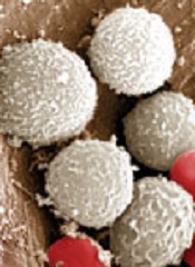
The US Food and Drug Administration (FDA) has decided not to approve Novartis’s application for a biosimilar of Amgen’s Neulasta, also known by the generic name pegfilgrastim.
The FDA issued a complete response letter for the pegfilgrastim biosimilar last month.
Novartis has not provided details about the agency’s decision or the contents of the letter, but the company said it is working with the FDA to answer its questions about the drug.
Novartis was seeking approval for its pegfilgrastim biosimilar for the same indication as Amgen’s Neulasta.
Neulasta is a leukocyte growth factor that is FDA-approved for the following indications:
- To decrease the incidence of infection, as manifested by febrile neutropenia, in patients with non-myeloid malignancies receiving myelosuppressive anticancer drugs associated with a clinically significant incidence of febrile neutropenia.
- To increase survival in patients acutely exposed to myelosuppressive doses of radiation.
Neulasta is not FDA-approved for the mobilization of peripheral blood progenitor cells for hematopoietic stem cell transplantation.