User login
Phototherapy may increase risk of pediatric cancers
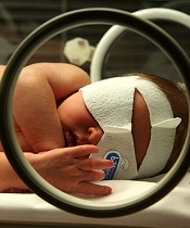
phototherapy to treat
neonatal jaundice
Photo by Martin Pot
Two new studies raise questions about a possible link between childhood cancer and phototherapy for newborn jaundice.
One study showed a significant positive association between phototherapy and 2 cancer types—myeloid leukemia and kidney cancer—but the other study did not.
Although these results are inconclusive, researchers say clinicians should exercise caution in prescribing phototherapy for infants whose jaundice is likely to resolve on its own.
However, the findings should not discourage the use of phototherapy in infants who otherwise would be at risk of brain damage or hearing loss.
The studies were published in Pediatrics alongside a related editorial.
“Phototherapy has been perceived by most as causing minimal risk to the infant,” said editorial author A. Lindsay Frazier, MD, of the Dana-Farber Cancer Institute in Boston, Massachusetts.
“Although these studies are inconclusive and do not prove a relationship between phototherapy and cancer, they should give us pause. That being said, however, the brain damage and hearing loss from high bilirubin levels are real and well-documented, and the suggested risk of cancer from these new studies is both unclear and very small.”
Kaiser Permanente study
Thomas B. Newman, MD, of University of California, San Francisco, and his colleagues conducted a study of children born in Kaiser Permanente Northern California hospitals from 1995 to 2011. The researchers analyzed data on 499,621 children born at 35 weeks’ gestation or later.
There were 60 cases of cancer among the 39,403 children exposed to phototherapy and 651 cases of cancer among the 460,218 children who were not exposed to phototherapy. That translates to 25 per 100,000 person-years and 18 per 100,000 person-years, respectively, for an incidence rate ratio (IRR) of 1.4 (P=0.01).
In an unadjusted analysis, phototherapy was associated with increased rates of any leukemia (IRR=2.1, P=0.0007), nonlymphocytic leukemia (IRR=4.0, P=0.0004), and liver cancer (IRR=5.2, P=0.04).
However, when the researchers adjusted for bilirubin levels, chromosomal disorders, congenital anomalies, and other covariates, these associations were no longer statistically significant.
Study of California hospitals
Andrea C. Wickremasinghe, MD, of Kaiser Permanente Northern California in Santa Clara, and her colleagues conducted a study of children born in California hospitals from 1998 to 2007.
The team analyzed data from the California Office of Statewide Health Planning and Development, which included 5,144,849 infants born at 35 weeks’ gestation or later.
There were 58 cases of cancer among the 178,017 infants exposed to phototherapy and 1042 cancer cases among the 4,966,832 infants who were not exposed to phototherapy. That translated to 32.6 per 100,000 and 21.0 per 100,000, respectively, for a relative risk of 1.6 (95% confidence interval [CI], 1.2–2.0; P=0.002).
In propensity-adjusted analyses, there were significant positive associations between phototherapy and overall cancer (adjusted odds ratio [aOR]=1.4; 95% CI, 1.1–1.9; P=0.007), myeloid leukemia (aOR=2.6; 95% CI, 1.3–5.0; P=0.005), and kidney cancer (aOR=2.5; 95% CI, 1.2–5.1; P=0.02).
Dr Frazier noted that these studies come at a time when the number of infants receiving phototherapy is increasing, perhaps because of the availability of light therapy units that can be used in the home. In the Kaiser Permanente study, 15.9% of infants received phototherapy in 2011, compared to 2.7% in 1995.
“What is concerning is the fact that, at least in the Kaiser Permanente Northern California healthcare system, the number of children receiving phototherapy has dramatically increased,” Dr Frazier said.
“The risks associated with such a prevalent exposure require close scrutiny. If I were the one prescribing phototherapy today, I would want to be sure it was indicated.” ![]()
Health Canada approves pathogen inactivation system for plasma

Photo by Cristina Granados
Health Canada has approved the INTERCEPT Blood System for plasma, a pathogen inactivation system used to reduce the risk of transfusion-transmitted infections.
The system can be used to reduce pathogens in plasma derived from whole blood or obtained by apheresis.
The system inactivates pathogens through a photochemical process involving controlled exposure to ultraviolet light and the chemical amotosalen.
The plasma is then purified to remove the chemical and its byproducts.
Plasma, platelets, and red blood cells do not require functional DNA or RNA for therapeutic efficacy, but pathogens and white blood cells do require these nucleic acids in order to replicate.
The INTERCEPT Blood System targets this basic biological difference. Ultraviolet light is used to activate amotosalen, which binds to DNA and RNA, thereby preventing nucleic acid replication and rendering pathogens inactive.
In studies, the INTERCEPT Blood System for plasma has proven effective in reducing a broad spectrum of viruses, bacteria, spirochetes, and parasites.
However, no pathogen inactivation process has been shown to eliminate all pathogens. Certain viruses (eg, human parvovirus B19) and spores formed by certain bacteria are known to be resistant to the INTERCEPT process.
The INTERCEPT Blood System for plasma, which is marketed by Cerus Corporation, has been approved for use in Europe since 2002 and in the US since 2014.
The safety and efficacy of plasma prepared with the INTERCEPT Blood System has been evaluated in 8 clinical studies including more than 700 patients.
For more information on these studies and the system itself, see the package insert, which is available at http://intercept-canada.com. ![]()
EC grants drug conditional approval to treat MM

Photo courtesy of Janssen
The European Commission (EC) has granted conditional marketing authorization for daratumumab (Darzalex), a monoclonal antibody targeting CD38.
The conditional approval is for daratumumab as monotherapy in adults with relapsed and refractory multiple myeloma (MM) who progressed on their last therapy and have received treatment with a proteasome inhibitor and an immunomodulatory agent.
Daratumumab is the first human CD38 monoclonal antibody approved for use in Europe.
About conditional marketing authorization
A product may receive conditional marketing authorization if the EC finds that, although comprehensive clinical data on the safety and efficacy of the product are not available, all of the following requirements are met:
- The risk-benefit balance of the product is positive
- The company developing the product will likely be in a position to provide comprehensive clinical data in the future
- Unmet medical needs will be fulfilled
- The benefit to public health of the immediate availability of the product outweighs the risk inherent in the fact that additional data are still required.
Conditional marketing authorizations are valid for 1 year, on a renewable basis. The holder will be required to complete ongoing studies or to conduct new studies with a view to confirming that the benefit-risk balance of a product is positive. In addition, specific obligations may be imposed in relation to the collection of pharmacovigilance data.
About daratumumab
Daratumumab works by binding to CD38, a signaling molecule highly expressed on the surface of MM cells. In binding to CD38, daratumumab triggers the patient’s own immune system to attack the cancer cells, resulting in tumor cell death through multiple, immune-mediated mechanisms of action and through immunomodulatory effects, in addition to direct tumor cell death via apoptosis.
The EC’s decision to grant conditional marketing authorization for daratumumab was based on a review of data from the phase 2 SIRIUS study, the phase 1/2 GEN501 study, and 3 additional supportive studies.
The GEN501 study enrolled 102 patients with relapsed MM or relapsed MM that was refractory to 2 or more prior lines of therapy. The patients received daratumumab at a range of doses and on a number of different schedules.
The results suggested daratumumab is most effective at a dose of 16 mg/kg. At this dose, the overall response rate was 36%. Most adverse events in this study were grade 1 or 2, although serious events did occur.
The SIRIUS study enrolled 124 MM patients who had received 3 or more prior lines of therapy. They received daratumumab at different doses and on different schedules, but 106 patients received the drug at 16 mg/kg.
Twenty-nine percent of the 106 patients responded to treatment, and the median duration of response was 7 months. Thirty percent of patients experienced serious adverse events.
Findings from a combined efficacy analysis of the GEN501 and SIRIUS trials demonstrated that, after a mean follow-up of 14.8 months, the estimated median overall survival for patients who received single-agent daratumumab at 16 mg/kg was 20 months.
Five phase 3 clinical studies of daratumumab in MM patients—in relapsed and frontline settings—are ongoing. Additional studies are ongoing or planned to assess the drug’s potential in other malignant and pre-malignant diseases in which CD38 is expressed.
Janssen Biotech, Inc., has exclusive worldwide rights to the development, manufacturing, and commercialization of daratumumab for all potential indications. Janssen licensed daratumumab from Genmab A/S in August 2012. ![]()
Study reveals how BET inhibitors kill cancer cells

Image courtesy of PNAS
Researchers say they have determined how BET inhibitors fight hematologic malignancies.
Previous studies showed that BET inhibitors are effective at halting tumor growth, but it wasn’t clear whether the drugs kill cancer cells outright or merely pause their growth.
The new study provides an answer and reveals potential ways in which cancer cells may develop resistance to BET inhibitors.
The findings have been published in Leukaemia.
Researchers tested the BET inhibitors JQ1 and IBET151 in a range of hematopoietic cancer cell lines (leukemias, lymphomas, and multiple myeloma) and in mice (with and without malignancy).
The team found that JQ1’s ability to kill cancer cells principally relies on the activation of BAX/BAK-dependent mitochondrial apoptosis. They said this is largely triggered by upregulation of the protein BIM when BET inhibitors suppress miR-17-92, a post-transcriptional repressor of BIM expression.
“We found that when apoptosis was impaired—for instance, by loss of BIM—the BET inhibitors were no longer effective,” said study author Zhen Xu, PhD, of Walter and Eliza Hall Institute of Medical Research in Melbourne, Victoria, Australia.
“This suggests that cancer cells that acquire mutations in genes that drive apoptosis will lose sensitivity to BET inhibitors and thus will be able to survive treatment, leading to disease relapse.”
The researchers also found that BET inhibitors could induce apoptosis in normal hematopoietic cells, particularly those of lymphoid origin. The team said this suggests the cells’ susceptibility to BET inhibitors did not arise from oncogenic transformation.
These findings could help researchers improve strategies for using BET inhibitors to treat cancers, according to study author Stefan Glaser, PhD, of the Walter and Eliza Hall Institute of Medical Research.
“Understanding how the drugs work gives us the opportunity to investigate new treatments—for example, by using combination therapies or altering the dosage and timing of treatment to prevent drug resistance from emerging,” Dr Glaser said. ![]()
Fertility concerns of female cancer survivors

Photo by Vera Kratochvil
A new study indicates that many young adult female cancer survivors do not receive adequate information about their fertility as part of their survivorship care, despite having concerns about their ability to bear children in the future.
The research, published in Cancer, suggests a need for better resources to support cancer survivors in making informed decisions about their reproductive options after they complete treatment.
To conduct this study, Catherine Benedict, PhD, of North Shore-Long Island Jewish Medical Center in Manhasset, New York, and her colleagues asked female cancer survivors to complete a web-based, anonymous survey.
There were 346 participants. They had an average age of 29.9 and had completed treatment an average of 4.9 years earlier.
The investigators focused on a subgroup of 179 women with uncertain fertility status who had not previously undergone or attempted fertility preservation, either before or after their cancer treatment, and who either wanted future children or were unsure.
Many of these women said they did not have enough information concerning their risk of infertility (58%), risk of early menopause (60%), options to assess their fertility (62%), options to preserve their fertility (51%), or options for alternative family building (43%).
The women’s greatest reproductive concerns were potential fertility problems and the health of a future child. Sixty-four percent of the women said they were concerned about not being able to have children (or more children), and 59% were worried about passing the risk of cancer on to their future children.
Only 13% of women said they were well informed about options for preserving fertility, and 74% were unclear about their personal values regarding fertility preservation.
Seventy percent of the women said they hadn’t received enough advice on fertility preservation, and 35% said they didn’t have enough support to make a decision about fertility preservation.
The investigators found a significant association between greater unmet information needs and higher levels of decisional conflict about fertility preservation (P<0.001).
On the other hand, having undergone a fertility evaluation after treatment was associated with lower decisional conflict (P=0.02).
The investigators said these findings establish the need for support services to help young female cancer survivors make decisions about fertility preservation and family-building as part of survivorship care.
The literature has largely focused on the clinical and support needs of women making fertility decisions before their treatment begins, but most patients do not preserve their fertility before treatment for a number of reasons, despite wanting children in the future.
“The potential loss of fertility has been described in the literature as being almost as painful, if not more so, than the cancer diagnosis itself,” Dr Benedict said.
“Failure to provide information and address concerns with respect to fertility-related decisions may have lasting consequences for young women who hope to move on from their cancer experience to achieve important life goals such as having children. For women at risk for early menopause, delaying fertility-related decisions may cause them to miss their narrowed window of opportunity to preserve their fertility, if desired.” ![]()
Cognitive impairment in ALL survivors

Photo by Bill Branson
New research indicates that survivors of pediatric acute lymphoblastic leukemia (ALL) suffer from brain injury even if they have no history of central nervous system disease or cranial radiation.
The study suggests the neurotoxic effects of chemotherapeutic drugs on the developing brains of young ALL patients may impair their cognitive functioning by disrupting the formation of neural networks that connect brain regions and transfer information.
Shelli Kesler, PhD, of the University of Texas MD Anderson Cancer Center in Houston, and her colleagues reported these findings in Brain Connectivity.
The researchers used diffusion tensor imaging to analyze and compare the gray matter connectome of 31 pediatric ALL survivors and 39 matched control subjects.
The team found significantly greater cognitive impairment among the ALL survivors (P=0.027), as well as significantly lower connectivity, based on small-worldness (P=0.007) and network clustering coefficient (P=0.019).
The researchers noted that clustered connectivity was altered in the parietal, frontal, hippocampal, amygdalar, thalamic, and occipital regions in the ALL survivors.
The team also described a model that can be used to predict cognitive impairment in ALL survivors. The model’s classification accuracy was 89.39% (P<0.0001), its sensitivity was 95.83%, and specificity was 85.71%.
“As survival rates for cancer patients increase, issues related to survivorship, such as chemotherapy-induced cognitive impairment, become more important to the cancer research community,” said Christopher Pawela, PhD, co-editor-in-chief of Brain Connectivity and an assistant professor at the Medical College of Wisconsin in Milwaukee.
“Dr Kesler and colleagues are developing new MRI-based biomarkers to measure brain changes associated with the neurotoxic effects of chemotherapy in the brain. These biomarkers may find utility in providing insight into the mechanisms of brain damage caused by chemotherapeutic drugs and could be used to develop neuroprotective therapies to mitigate the harmful effects of these drugs on the brain.” ![]()
Drug granted breakthrough designation for AML

Image by Lance Liotta
The US Food and Drug Administration (FDA) has granted breakthrough therapy designation for CPX-351 (Vyxeos), a fixed-ratio combination of cytarabine and daunorubicin inside a lipid vesicle, to treat adults with therapy-related acute myeloid leukemia (AML) or AML with myelodysplasia-related changes.
The FDA’s breakthrough therapy designation is intended to expedite the development and review of new therapies for serious or life-threatening conditions.
To earn the designation, a treatment must show encouraging early clinical results demonstrating substantial improvement over available therapies with regard to a clinically significant endpoint, or it must fulfill an unmet need.
Phase 3 trial
The breakthrough designation for CPX-351 is primarily based on positive results from a phase 3 trial in older patients with previously untreated, high-risk, secondary AML.
The trial was sponsored by Celator Pharmaceuticals, Inc., the company developing CPX-351.
The company has released some results from the trial, and additional data are scheduled to be presented at the 2016 ASCO Annual Meeting (abstract 7000).
The trial enrolled 309 patients, ages 60 to 75, with one of the following:
- Untreated AML and a history of prior cytotoxic treatment
- Antecedent myelodysplastic syndrome (MDS) or chronic myelomonocytic leukemia, with or without prior hypomethylating therapy
- AML with WHO-defined, MDS-related cytogenetic abnormalities.
One hundred and fifty-three patients were randomized to receive CPX-351, and 156 were randomized to 7+3 (cytarabine continuously for 7 days and a single dose of daunorubicin for the first 3 days).
The treatment arms were well-balanced for sex, race, age, performance status, AML subtype, MDS-related cytogenetics, and prior hypomethylating therapy.
The trial met its primary endpoint, demonstrating a significant improvement in overall survival with CPX-351. The median overall survival was 9.56 months in the CPX-351 arm and 5.95 months in the 7+3 arm. The hazard ratio was 0.69 (P=0.005).
According to researchers, there was no substantial difference between the treatment arms with regard to grade 3-5 adverse events.
About CPX-351
CPX-351 is a liposomal formulation of a 5:1 molar ratio of cytarabine and daunorubicin.
In one phase 2 trial, CPX-351 conferred a significant improvement in survival—over investigator’s choice of therapy—when used as salvage therapy in poor-risk patients with AML.
In another phase 2 trial, CPX-351 conferred a significant survival benefit—over 7+3—in patients with secondary AML.
The FDA previously granted CPX-351 fast track designation to treat elderly patients with secondary AML. The agency established the fast track designation process to expedite the review of drugs that are intended to treat serious or life-threatening conditions and that demonstrate the potential to address unmet medical needs.
The designation allows a drug’s developer to submit sections of a new drug application (NDA) on a rolling basis, so the FDA can review portions of the NDA as they are received instead of waiting for the entire NDA submission. A fast-track-designated product could be eligible for priority review if supported by clinical data at the time of NDA submission. ![]()
Image by Lance Liotta
The US Food and Drug Administration (FDA) has granted breakthrough therapy designation for CPX-351 (Vyxeos), a fixed-ratio combination of cytarabine and daunorubicin inside a lipid vesicle, to treat adults with therapy-related acute myeloid leukemia (AML) or AML with myelodysplasia-related changes.
The FDA’s breakthrough therapy designation is intended to expedite the development and review of new therapies for serious or life-threatening conditions.
To earn the designation, a treatment must show encouraging early clinical results demonstrating substantial improvement over available therapies with regard to a clinically significant endpoint, or it must fulfill an unmet need.
Phase 3 trial
The breakthrough designation for CPX-351 is primarily based on positive results from a phase 3 trial in older patients with previously untreated, high-risk, secondary AML.
The trial was sponsored by Celator Pharmaceuticals, Inc., the company developing CPX-351.
The company has released some results from the trial, and additional data are scheduled to be presented at the 2016 ASCO Annual Meeting (abstract 7000).
The trial enrolled 309 patients, ages 60 to 75, with one of the following:
- Untreated AML and a history of prior cytotoxic treatment
- Antecedent myelodysplastic syndrome (MDS) or chronic myelomonocytic leukemia, with or without prior hypomethylating therapy
- AML with WHO-defined, MDS-related cytogenetic abnormalities.
One hundred and fifty-three patients were randomized to receive CPX-351, and 156 were randomized to 7+3 (cytarabine continuously for 7 days and a single dose of daunorubicin for the first 3 days).
The treatment arms were well-balanced for sex, race, age, performance status, AML subtype, MDS-related cytogenetics, and prior hypomethylating therapy.
The trial met its primary endpoint, demonstrating a significant improvement in overall survival with CPX-351. The median overall survival was 9.56 months in the CPX-351 arm and 5.95 months in the 7+3 arm. The hazard ratio was 0.69 (P=0.005).
According to researchers, there was no substantial difference between the treatment arms with regard to grade 3-5 adverse events.
About CPX-351
CPX-351 is a liposomal formulation of a 5:1 molar ratio of cytarabine and daunorubicin.
In one phase 2 trial, CPX-351 conferred a significant improvement in survival—over investigator’s choice of therapy—when used as salvage therapy in poor-risk patients with AML.
In another phase 2 trial, CPX-351 conferred a significant survival benefit—over 7+3—in patients with secondary AML.
The FDA previously granted CPX-351 fast track designation to treat elderly patients with secondary AML. The agency established the fast track designation process to expedite the review of drugs that are intended to treat serious or life-threatening conditions and that demonstrate the potential to address unmet medical needs.
The designation allows a drug’s developer to submit sections of a new drug application (NDA) on a rolling basis, so the FDA can review portions of the NDA as they are received instead of waiting for the entire NDA submission. A fast-track-designated product could be eligible for priority review if supported by clinical data at the time of NDA submission.
Broad-spectrum antibiotics may worsen GVHD

Broad-spectrum antibiotics may increase the severity of graft-versus-host disease (GVHD), according to research published in Science Translational Medicine.
Researchers evaluated the relationship between antibiotics and GVHD using data from more than 850 transplant patients and by conducting experiments in mice.
Their results suggested that selecting antibiotics that spare “good” bacteria may help protect patients from GVHD.
Results in patients
Yusuke Shono, MD, PhD, of Memorial Sloan Kettering Cancer Center in New York, New York, and his colleagues conducted this study, first mining the clinical records of 857 patients who underwent allogeneic hematopoietic stem cell transplant
(HSCT).
The team found that patients who received imipenem-cilastatin and piperacillin-tazobactam antibiotics to treat neutropenic fever had a higher incidence of GVHD-related mortality at 5 years than patients who did not receive these drugs.
The incidence of GVHD-related death was 21.5% in patients who received imipenem-cilastatin and 13.1% in those who did not (P=0.025). The incidence was 19.8% in patients who received piperacillin-tazobactam and 11.9% in those who did not (P=0.007).
Two other antibiotics used to treat neutropenic fever, aztreonam and cefepime, were not associated with an increased risk of GVHD-related mortality.
The incidence of GVHD-related death was 13.8% in patients who received cefepime and 14.6% in those who did not (P=0.98). The incidence was 17.5% in patients who received aztreonam and 14.2% in those who did not (P=0.78).
The researchers also found that piperacillin-tazobactam and imipenem-cilastatin were both associated with an increased incidence of grade 2-4 GVHD (P=0.0167 and P=0.0165, respectively), upper gastrointestinal GVHD (P=0.002 and P=0.045, respectively), and lower gastrointestinal GVHD (P=0.019 and P=0.036, respectively).
When the team analyzed patients’ stool samples, they found that piperacillin-tazobactam perturbed the gut microbiome, killing off protective bacteria. The therapy was associated with a greater loss of Bacteroidetes and Lactobacillus, when compared to treatment with aztreonam or cefepime.
The change in abundance of Enterococcus, Akkermansia, and Erysipelotrichia was similar with the 3 therapies. But there was a trend toward a decrease in Clostridia and Actinobacteria with piperacillin-tazobactam.
The researchers said they could not assess the effects of imipenem-cilastatin on bacterial populations because, at their center, imipenem-cilastatin is almost always given to HSCT patients as second-line therapy for neutropenic fever.
Results in mice
In mice treated with various antibiotic regimens following HSCT, those given broad-spectrum antibiotics developed more severe GVHD.
Specifically, the researchers observed aggravated GVHD mortality with imipenem-cilastatin or piperacillin-tazobactam compared to aztreonam (P<0.01 and P<0.05, respectively).
They also found evidence of increased GVHD pathology localized in the colon of mice that received imipenem-cilastatin (P<0.05). Mice with GVHD that received imipenem-cilastatin experienced loss of the protective mucus lining of the colon (P<0.01) and compromised intestinal barrier function (P<0.05).
When the researchers sequenced stool samples from mice with GVHD that received imipenem-cilastatin, they saw an increase in Akkermansia muciniphila (P<0.001), a commensal bacterium with mucus-degrading capabilities. They said this raises the possibility that mucus degradation may contribute to murine GVHD.
The researchers noted that these findings must be confirmed in clinical trials, but they caution against the use of broad-spectrum antibiotics in HSCT patients at risk of GVHD.
Method could make injectable drugs safer

Photo by Bill Branson
A new drug-making technique could reduce the risk of hemolysis, thrombosis, and other serious side effects that can occur with injectable drugs, according to researchers.
The team used the technique to remove potentially harmful additives—surfactants—from 12 common injectable drugs.
Jonathan F. Lovell, PhD, of University at Buffalo in New York, and his colleagues described the technique in Nature Communications.
Pharmaceutical companies use surfactants to dissolve a medicine into a liquid solution, making it suitable for injection. Unfortunately, solutions loaded with surfactants and other nonessential ingredients may increase the risk of thrombosis, hemolysis, anaphylactic shock, and other side effects.
Researchers have tried to address this problem in two ways, each with varying degrees of success.
Some have taken the “top-down” approach, in which they shrink drug particles to nanoscale sizes to eliminate excess additives. While promising, the method doesn’t work well with injectable medicine because the drug particles are still too large to safely inject.
Other researchers have worked from the “bottom up,” using nanotechnology to build new drugs from scratch. This can yield the desired results, but developing new drug formulations takes years, and drugs are coupled with new additives that can produce new side effects.
The technique described in Nature Communications differs from both of these approaches.
Dr Lovell and his colleagues dissolved 12 drugs—vitamin K1, cyclosporine, cabazitaxel, and others—one at a time into a surfactant called Pluronic. Then, by lowering the solution’s temperature to 4° C, they were able to remove the excess Pluronic via a membrane.
The end result was drugs that contain 100 to 1000 times less excess additives.
“For the drugs we looked at, this is as close as anyone has gotten to introducing pure, injectable medicine into the body,” Dr Lovell said. “Essentially, it’s a new way to package drugs.”
The findings are significant, he said, because they show that many injectable drug formulations may be improved through an easy-to-adopt process. He and his colleagues are now working to refine the method further.
Mutation in mice may have affected research results

Researchers say they have discovered a mutation in a subline of C57BL/6 mice that could compromise results from previous studies.
“We found an unexpected mutation with potentially important consequences in strains of mice that had been separately engineered in labs in California and Japan,” said Shiv Pillai, MD, PhD, of The Ragon Institute of MGH, MIT and Harvard in Cambridge, Massachusetts.
“[W]e traced the problem to a subline of B6 mice from one specific company that have been sold in Asia, North America, Europe, and Israel. We have notified this company—Harlan Laboratories, which is now part of Envigo—of our findings, and they have been very responsive and will use approaches we have provided to check all of their colonies.”
Dr Pillai and his colleagues described their discovery of the mutation in Cell Reports.
The team noted that, in immunological studies, mutant mice are backcrossed for many generations into B6 mice so that all genes other than the mutant gene are derived from the B6 strain. This allows the comparison of data from laboratories in different parts of the world and simplifies creating mice with mutations in several genes.
A 2009 study out of Dr Pillai’s lab showed that 2 strains of mice engineered to lack the Siae or Cmah genes—both of which code for enzymes involved with sialic acid proteins—also had significant defects in the development of B cells, which were assumed to be the result of the knockout genes.
However, when Siae-deficient mice were further backcrossed with a different group of C57BL/6 mice, the result was a strain of Siae-knockout mice that did not have the B-cell development defects. A newly engineered strain of mice with a different Siae mutation also had normal B-cell development.
Detailed genetic sequencing of the first knockout line revealed a previously unsuspected mutation in a gene called Dock2, located on a chromosome 11, instead of chromosome 9 where Siae is located.
The same mutation was previously reported in 2 colonies of a different knockout mouse developed in Japan.
Dr Pillai’s team also found the Dock2 mutation in a completely different group of mice from the University of California, San Diego—where their Siae-mutant strain had been developed—and realized that 3 different engineered strains with the same unwanted mutation had probably acquired it from a common source. The team eventually traced it back to a subline of C57BL/6 mice from Harlan/Envigo.
Since most research papers using C57BL/6 mice or other such “background” strains do not indicate the specific subline, the researchers said they have no way of knowing how many studies might be affected by their findings.
But they hope the publication of these results will alert other research teams to the potential need to review their results.
“While embryonic stem cells from C57BL/6 mice have recently become available, which allows the generation of knockout strains with less backcrossing, B6 mice are used for many different kinds of experiments, including as controls,” Dr Pillai said.
“Researchers who have used them need to re-genotype the mice to look for the Dock2 mutation and, if they find it, check to see whether their results are preserved if the Dock2 mutation is bred out.”