User login
Intracranial bleeding in older adults on warfarin

Results of a large study suggest warfarin may pose a higher risk of traumatic intracranial bleeding than previously reported, at least among older patients with atrial fibrillation (AF).
The study included more than 30,000 US veterans with AF who were 75 years or older when starting warfarin.
These patients had a higher rate of traumatic intracranial bleeding than reported in previous trials.
John A. Dodson, MD, of the New York University School of Medicine in New York, New York, and his colleagues conducted this research and reported the results in JAMA Cardiology.
The researchers studied 31,951 subjects with AF who were 75 years or older and were new referrals to Veterans Affairs anticoagulation clinics for warfarin therapy between January 1, 2002, and December 31, 2012.
The patients had a mean age of 81.1, and 98.1% were male. Comorbidities included hypertension (82.5%), coronary artery disease (42.6%), and diabetes mellitus (33.8%).
The researchers found the incidence rate of hospitalization for any intracranial bleeding among these patients was 14.58 per 1000 person-years.
And the incidence rate of hospitalization for traumatic intracranial bleeding was 4.80 per 1000 person-years.
The researchers said this was “considerably higher” than reported in two previous trials, one published in The Lancet in 1996 and one published in JAMA Internal Medicine in 1998.
Dr Dodson and his colleagues also looked at factors associated with traumatic intracranial bleeding in their patient population.
In unadjusted analyses, the following factors were significant predictors of traumatic intracranial bleeding: dementia, fall within the past year, anemia, depression, abnormal renal or liver function, anticonvulsant use, labile international normalized ratio, and antihypertensive use.
However, when the researchers adjusted their analyses for potential confounders, fewer factors remained significant predictors. These were dementia (hazard ratio [HR]=1.76), anemia (HR=1.23), depression (HR=1.30), anticonvulsant use (HR=1.35), and labile international normalized ratio (HR=1.33).
The researchers noted that risk factors for traumatic intracranial bleeding were different from risk factors for ischemic stroke.
They also said the high overall rate of intracranial bleeding in this study suggests a need to more systematically evaluate the benefits and harms of warfarin in older adults. ![]()
FDA lifts partial clinical hold on pidilizumab

The US Food and Drug Administration (FDA) has lifted the partial clinical hold on the investigational new drug (IND) application for pidilizumab (MDV9300) in hematologic malignancies.
This means the phase 2 trial of pidilizumab in patients with relapsed or refractory diffuse large B-cell lymphoma (DLBCL), as well as other studies that cross-reference the IND for the drug, may now proceed.
The partial clinical hold on pidilizumab was not related to any safety concerns.
The FDA placed the hold because the company developing pidilizumab, Medivation Inc., determined that the drug is not an inhibitor of PD-1, as researchers previously thought.
The phase 2 trial of pidilizumab in DLBCL was launched in late 2015 but had not enrolled any patients before the FDA placed the partial clinical hold.
Patients who were receiving pidilizumab through investigator-sponsored trials have continued to receive treatment despite the hold, and those investigators have been told to update their protocols and informed consent documents to reflect that pidilizumab is not an anti-PD-1 antibody.
Medivation has likewise revised the investigator brochure, protocols, and informed consent documents related to the phase 2 trial of DLBCL patients.
The company said it is still trying to determine pidilizumab’s mechanism of action.
“We are delighted that the FDA has lifted the partial clinical hold and that we may proceed with our potentially pivotal trial in this area of high unmet medical need,” said David Hung, MD, founder, president, and chief executive officer of Medivation.
“As we move forward, we also are working to determine the compound’s exact binding mechanism which, we believe, modulates the body’s innate immune response and differentiates it from the heavily crowded immuno-oncology space that targets the adaptive side of immunity.”
Medivation said it intends to submit an amendment to the Chemistry, Manufacturing, and Controls section of the IND for pidilizumab to provide for larger manufacturing lot sizes to better support the current and planned clinical activities for the drug.
The company also said it plans to resume the phase 2 trial of pidilizumab in DLBLCL in the second half of this year.
The trial is expected to enroll approximately 180 patients who had an incomplete response to salvage therapy or autologous stem cell transplant for relapsed or refractory CD20+ DLBCL, transformed indolent lymphoma, or primary mediastinal B-cell lymphoma.
The patients will be assessed in 2 parallel cohorts of approximately 90 patients each. One cohort will enroll patients who have received an autologous stem cell transplant, and the other will enroll patients who have received salvage chemotherapy but are ineligible for transplant.
Pidilizumab will be given at a dose of 200 mg by intravenous infusion. The primary endpoint of the trial is best overall response rate. ![]()
Team finds precursors to HSPCs in mice
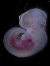
Image by Matthias Zepper
Researchers have reportedly identified precursor cells that can be matured into transplantable hematopoietic stem/progenitor cells (HSPCs) in the lab.
The investigators discovered the precursor cells in the placentas and embryos of mice, but the team believes their findings could aid the development of patient-specific HSPCs and more differentiated blood products for cell-replacement therapy in humans.
“To cure disease in the long-term, we need to be able to transplant something that can keep producing new blood cells and won’t be rejected by the patient’s body,” said study author Kateri Moore, DVM, of the Icahn School of Medicine at Mount Sinai in New York, New York.
“We are excited by the results of our study. The precursor cells can be matured in the lab to transplantable HSPCs. Our reprogramming process can inform developmental hematopoiesis and vice-versa.”
Dr Moore and her colleagues described this work in Developmental Cell.
With previous work, the researchers reprogrammed mouse fibroblasts to become HSPCs. They showed that this process occurred through hemogenic precursors that are Prom1+Sca1+CD34+CD45− (PS34CD45−).
So for the current study, the investigators examined mouse placentas and embryos, looking for cells with the same phenotype. They were able to find and analyze PS34CD45− cells.
Investigation revealed that PS34CD45− cells express endothelial and hematopoietic markers. And the cells originate in embryonic tissue and localize to the vascular labyrinth.
Furthermore, the researchers said global gene expression profiles of PS34CD45− cells correlate with reprogrammed precursors and establish a hemogenic precursor cell molecular signature.
In culture, PS34CD45− cells gave rise to multi-lineage hematopoietic colonies. PS34CD45− cells generated B and T lymphocytes and engrafted in primary and secondary immunodeficient mice.
The investigators said the next step is to test these findings in humans.
“Our ultimate goal is to grow blood-forming cells in the lab and improve efficiencies to generate patient-specific blood cells,” Dr Moore said. “This study brings us a step closer to reaching this goal.” ![]()
Tool provides insight into T cells’ behavior

Image courtesy of NIAID
A new tool can help scientists determine how different types of T cells detect, destroy, and remember antigens, according to research published in Nature Methods.
The tool is TraCeR, a computational method that allows researchers to reconstruct full-length, paired T-cell receptor sequences from single-cell RNA sequence data.
TraCeR reveals clonal relationships between T cells as well as their transcriptional profiles.
In the current study, TraCeR helped scientists detect T-cell clonotypes in mice infected with Salmonella.
“This new tool for single-cell sequencing gives us a new approach to the study of T cells and opens up new opportunities to explore immune responses in disease, vaccination, cancer, and autoimmunity,” said study author Sarah Teichmann, PhD, of the Wellcome Trust Sanger Institute in Cambridge, UK.
She and her colleagues noted that T-cell receptors are extremely variable, and a combination of paired sequences determines which protein a receptor will detect. So to understand what is happening at the molecular level, it is imperative to find both sequences in each cell.
TraCeR is designed to allow scientists to look at the DNA and RNA profiles of these highly variable T-cell receptors at the same time.
Dr Teichmann and her colleagues found that receptor sequences are unique, unless the T cells have the same parent cell. The presence of “sibling” cells proves that an infection has triggered the division of a particular T cell, which indicates it is multiplying to fight an antigen.
Using TraCeR, the researchers were able to accurately identify sibling T cells and explore their different responses to Salmonella infection in mice.
“This technique helps us see whether all the ‘children’ of a particular T cell do the same thing at the same time, which is an open question in biology,” said study author Tapio Lönnberg, PhD, of the European Molecular Biology Laboratory-European Bioinformatics Institute in Cambridge, UK.
“We can start to see whether the antigen itself plays a role in how a T cell will respond, and even whether it’s possible to determine what the invader is, just based on the sequence of a T-cell receptor.”
The researchers said their next step is to apply similar methods to the study of B cells to better understand the adaptive immune system as a whole. ![]()
VTE linked to survival after bladder cancer surgery

Image by Andre E.X. Brown
Patients who undergo surgery for bladder cancer may require long-term prophylaxis for venous thromboembolism (VTE), according to research published in BJU International.
The study showed an increase in VTE incidence over time, with more than half of VTEs occurring after patients were discharged from the hospital.
In addition, VTE was associated with an increased risk of death—from the cancer or any cause.
The study included 3879 patients from the Ontario Cancer Registry who had radical cystectomy to treat bladder cancer between 1994 and 2008.
Within 1 month of their surgical admission date, 3.6% of patients had been diagnosed with VTE. The VTE incidence increased to 4.7% by 2 months and 5.4% by 3 months.
In all, 55% of VTE events occurred after hospital discharge, which is generally when patients are not receiving VTE prophylaxis.
“Although the findings in this population-based study confirmed some previous understanding of the frequency of VTE for this complex surgery, we were surprised at the number that were diagnosed after hospital discharge,” said study author D. Robert Siemens, MD, of Queen’s University in Kingston, Ontario, Canada.
“Furthermore, we were unable to identify strong predictive factors associated with VTE, suggesting to us that most all patients should receive prolonged VTE prophylaxis well beyond their hospital discharge.”
More specifically, the researchers conducted a multivariate analysis and found that higher surgeon volume and increased length of hospital stay were the only factors significantly associated with VTE (P=0.004 and P<0.001, respectively).
The team also discovered that patients with VTE tended to die earlier. VTE was associated with inferior cancer-specific survival and overall survival. The hazard ratios were 1.35 and 1.27, respectively.
Dr Siemens said these results suggest VTE could represent a marker of more aggressive disease. ![]()
Compound could treat T-ALL subtype

Photo courtesy of
The Ottawa Hospital
An experimental compound is “highly promising” as a potential treatment for a subtype of T-cell acute lymphoblastic leukemia (T-ALL), according to researchers.
The team found the histone demethylase UTX is important for the maintenance of TAL1-positive T-ALL.
Inhibiting UTX with a compound known as GSK-J4 proved toxic to TAL1-positive T-ALL cells in vitro and in vivo, but normal cells were spared.
The researchers reported these findings in Genes & Development.
“It’s very exciting because this is the first time anyone has found a potential personalized treatment for this aggressive disease,” said study author Marjorie Brand, PhD, of The Ottawa Hospital Research Institute in Ontario, Canada.
“Unlike current therapies, ours targets the offending gene without harming the rest of the body.”
Dr Brand and her colleagues decided the best way to find a better treatment for TAL1-positive T-ALL was to investigate exactly how it works at a molecular level. So they analyzed the TAL1 gene, which, in certain circumstances, can transform T-cell precursors into leukemic cells.
They found that TAL1 needs UTX to trigger leukemia production. This was surprising because UTX was previously described as a tumor suppressor in T-ALL.
The team said their work suggests UTX is actually a pro-oncogenic cofactor that is essential for leukemia maintenance in TAL1-positive—but not TAL1-negative—T-ALL.
The researchers found that targeting UTX with the H3K27 demethylase inhibitor GSK-J4 could completely halt the growth of TAL1-positive leukemia cells and induce apoptosis in these cells in vitro. But the compound did not have the same effect in TAL1-negative T-ALL.
The team also tested GSK-J4 in mouse models of T-ALL. After 3 weeks of treatment, there was a “dramatic decrease” in the percentage of leukemic blasts in the bone marrow of mice with TAL1-positive T-ALL. These mice also had a reduction in the infiltration of leukemic cells in the spleen.
However, mice with TAL1-negative T-ALL did not experience the same benefits.
The researchers said GSK-J4 appeared to be well-tolerated. None of the treated mice experienced weight loss or adverse effects in the intestine, spleen, liver, kidney, or hematopoietic system.
“While our study is a proof-of-concept, these promising results might one day lead to a similar targeted treatment for humans,” said study author Carmen Palii, PhD, also of The Ottawa Hospital Research Institute.
In the meantime, the researchers are conducting additional studies in mice, testing higher doses of GSK-J4 and evaluating long-term side effects of the compound. ![]()
EMA initiative aims to accelerate drug development

Photo courtesy of the FDA
The European Medicines Agency (EMA) has launched PRIME (PRIority MEdicines), an initiative intended to accelerate the development of drugs that target unmet medical needs.
These “priority medicines” may offer a major therapeutic advantage over existing treatments or benefit patients who currently have no treatment options in the European Union (EU).
Through PRIME, the EMA will offer early support to drug developers to optimize the generation of robust data on a treatment’s benefits and risks and enable accelerated assessment of an application for marketing authorization.
By engaging with developers early, the EMA aims to strengthen the design of clinical trials to facilitate the generation of high quality data to support an application for marketing authorization.
PRIME builds on the existing regulatory framework and available tools such as scientific advice and accelerated assessment.
To be accepted for PRIME, a treatment has to show the potential to benefit patients with unmet medical needs based on early clinical data.
Once a candidate medicine has been selected for PRIME, the EMA:
- Appoints a rapporteur from the Committee for Medicinal Products for Human Use (CHMP), or from the Committee on Advanced Therapies (CAT) in the case of an advanced therapy, to provide continuous support and help to build knowledge before a company submits an application for marketing authorization
- Organizes a kick-off meeting with the CHMP/CAT rapporteur and a multidisciplinary group of experts from relevant EMA scientific committees and working parties, and provides guidance on the overall development plan and regulatory strategy
- Assigns a dedicated EMA contact point
- Provides scientific advice at key development milestones, involving additional stakeholders such as health technology assessment bodies to facilitate quicker access to the new drug
- Confirms potential for accelerated assessment when an application for marketing authorization is submitted.
PRIME is open to all drug companies on the basis of preliminary clinical evidence. However, micro-, small- and medium-sized enterprises and applicants from the academic sector can apply earlier on the basis of compelling non-clinical data and tolerability data from initial clinical trials. They may also request fee waivers for scientific advice.
The EMA has released guidance documents on PRIME as well as a comprehensive overview of the EU early access regulatory tools—ie, accelerated assessment, conditional marketing authorization, and compassionate use.
The agency has also published revised guidelines on the implementation of accelerated assessment and conditional marketing authorization. These tools are reserved for treatments addressing major public health needs, and the revised guidelines provide more detailed information based on past experience.
Although PRIME is specifically designed to promote accelerated assessment, the EMA said the initiative will also help drug developers make the best use of other EU early access tools and initiatives, which can be combined whenever a drug fulfills the respective criteria.
PRIME was developed in consultation with the EMA’s scientific committees, the European Commission and its expert group on Safe and Timely Access to Medicines for Patients, as well as the European medicines regulatory network.
The main principles of PRIME were released for a 2-month public consultation in 2015, and the comments received were taken into account in the final version. ![]()
Photo courtesy of the FDA
The European Medicines Agency (EMA) has launched PRIME (PRIority MEdicines), an initiative intended to accelerate the development of drugs that target unmet medical needs.
These “priority medicines” may offer a major therapeutic advantage over existing treatments or benefit patients who currently have no treatment options in the European Union (EU).
Through PRIME, the EMA will offer early support to drug developers to optimize the generation of robust data on a treatment’s benefits and risks and enable accelerated assessment of an application for marketing authorization.
By engaging with developers early, the EMA aims to strengthen the design of clinical trials to facilitate the generation of high quality data to support an application for marketing authorization.
PRIME builds on the existing regulatory framework and available tools such as scientific advice and accelerated assessment.
To be accepted for PRIME, a treatment has to show the potential to benefit patients with unmet medical needs based on early clinical data.
Once a candidate medicine has been selected for PRIME, the EMA:
- Appoints a rapporteur from the Committee for Medicinal Products for Human Use (CHMP), or from the Committee on Advanced Therapies (CAT) in the case of an advanced therapy, to provide continuous support and help to build knowledge before a company submits an application for marketing authorization
- Organizes a kick-off meeting with the CHMP/CAT rapporteur and a multidisciplinary group of experts from relevant EMA scientific committees and working parties, and provides guidance on the overall development plan and regulatory strategy
- Assigns a dedicated EMA contact point
- Provides scientific advice at key development milestones, involving additional stakeholders such as health technology assessment bodies to facilitate quicker access to the new drug
- Confirms potential for accelerated assessment when an application for marketing authorization is submitted.
PRIME is open to all drug companies on the basis of preliminary clinical evidence. However, micro-, small- and medium-sized enterprises and applicants from the academic sector can apply earlier on the basis of compelling non-clinical data and tolerability data from initial clinical trials. They may also request fee waivers for scientific advice.
The EMA has released guidance documents on PRIME as well as a comprehensive overview of the EU early access regulatory tools—ie, accelerated assessment, conditional marketing authorization, and compassionate use.
The agency has also published revised guidelines on the implementation of accelerated assessment and conditional marketing authorization. These tools are reserved for treatments addressing major public health needs, and the revised guidelines provide more detailed information based on past experience.
Although PRIME is specifically designed to promote accelerated assessment, the EMA said the initiative will also help drug developers make the best use of other EU early access tools and initiatives, which can be combined whenever a drug fulfills the respective criteria.
PRIME was developed in consultation with the EMA’s scientific committees, the European Commission and its expert group on Safe and Timely Access to Medicines for Patients, as well as the European medicines regulatory network.
The main principles of PRIME were released for a 2-month public consultation in 2015, and the comments received were taken into account in the final version.
CDC quantifies threat of resistant HAIs in US

in the intensive care unit
New data from the US Centers for Disease Control and Prevention (CDC) suggest the incidence of certain healthcare-associated infections (HAIs) has fallen in recent years, but antibiotic-resistant bacteria remain a threat.
Therefore, the CDC is advising healthcare workers to use a combination of infection control recommendations to better protect patients from these infections.
“New data show that far too many patients are getting infected with dangerous, drug-resistant bacteria in healthcare settings,” said CDC Director Tom Frieden, MD.
The facts and figures are available in the CDC’s latest Vital Signs report and the agency’s annual progress report on HAI prevention.
The data indicate that 1 in 7 catheter- and surgery-related HAIs in acute care hospitals can be caused by 6 types of antibiotic-resistant bacteria. That number increases to 1 in 4 infections in long-term acute care hospitals.
The 6 antibiotic-resistant threats are carbapenem-resistant Enterobacteriaceae (CRE), methicillin-resistant Staphylococcus aureus (MRSA), ESBL-producing Enterobacteriaceae (extended-spectrum β-lactamases), vancomycin-resistant Enterococcus (VRE), multidrug-resistant Pseudomonas aeruginosa, and multidrug-resistant Acinetobacter.
Prevention and resistance
According to the CDC’s data, acute care hospitals saw a 50% decrease in central line-associated bloodstream infections between 2008 and 2014. But 1 in 6 remaining central line-associated bloodstream infections is caused by urgent or serious antibiotic-resistant bacteria.
Between 2008 and 2014, acute care hospitals saw a 17% decrease in surgical site infections related to 10 procedures that were tracked in previous HAI progress reports. One in 7 remaining surgical site infections is caused by urgent or serious antibiotic-resistant bacteria.
Acute care hospitals saw no change in the incidence of catheter-associated urinary tract infections (CAUTIs) between 2009 and 2014. However, there was a reduction in CAUTIs between 2013 and 2014. One in 10 CAUTIs is caused by urgent or serious antibiotic-resistant bacteria.
The Vital Signs report also examines Clostridium difficile, which caused almost half a million infections in the US in 2011 alone. The CDC’s annual progress report shows that hospital-onset C difficile infections decreased by 8% between 2011 and 2014.
In addition to the reports, the CDC has released a web app with interactive data on HAIs caused by antibiotic-resistant bacteria.
The tool, known as the Antibiotic Resistance Patient Safety Atlas, provides national, regional, and state map views of superbug/drug combinations showing percent resistance over time. The Atlas uses data reported to the CDC’s National Healthcare Safety Network from 2011 to 2014 from more than 4000 healthcare facilities.
Inhibitor exhibits activity against myeloma

A dual kinase inhibitor has shown early promise as a potential treatment for multiple myeloma (MM), according to investigators.
The inhibitor, ON123300, targets both AMPK-related protein kinase 5 (ARK5) and cyclin-dependent kinase 4 (CDK4), proteins responsible for maintaining energy balance within the cell.
Investigators found that ON123300 can induce apoptosis in MM cells in vitro and halt tumor growth in mouse models of MM.
The team reported these findings in Cancer Research.
“ARK5 is critical for myeloma survival, and this study suggests a novel function for ARK5 in bridging the mTOR and MYC pathways,” said study author Deepak Perumal, PhD, of the Icahn School of Medicine at Mount Sinai in New York, New York.
“Given that MYC is critically overexpressed in myeloma, we sought to determine whether selective inhibition of ARK5 and CDK4 could be an effective way to target MYC-driven proliferation in myeloma.”
The team at Mount Sinai developed ON123300 in collaboration with Onconova Therapeutics, Inc. And they tested the drug in MM cell lines and primary samples from patients with recurring MM, as well as human HS-5 bone marrow stromal cells.
ON123300 induced rapid cell-cycle arrest and apoptosis in MM cells but was not toxic to normal peripheral blood cells.
In mouse models of MM, ON123300 decreased tumor growth without causing significant toxicity.
The investigators also discovered that MM cells that are sensitive to ON123300 have a unique genomic signature, which could guide the clinical development of the drug.
“Our study results show that ON123300 induces cell death and negatively regulates key oncogenic pathways in multiple myeloma cells,” said Samir Parekh, MD, also of the Icahn School of Medicine at Mount Sinai.
“This is the first report showing potent cytotoxicity of CDK4/ARK5 inhibition in MM and provides the foundation for further clinical trials using CDK4/ARK5 inhibitors to improve outcomes for MM patients.”
System could aid assessment of sickle cell disease

and a normal one
Image by Betty Pace
A microfluidic system can measure the deformability and adhesion of red blood cells (RBCs) in samples from patients with sickle cell disease (SCD), according to research published in Technology.
The researchers noted that RBC deformability has been associated with vaso-occlusion in SCD, but we have limited knowledge on deformation characteristics of RBCs adhered to endothelium-associated proteins in microphysiological fluid flow conditions.
In the past, various approaches have been used to measure RBC deformability, including optical tweezers, micropipette aspiration, and atomic force microscopy. These methods have enabled sensitive and controlled measurement of RBC mechanical properties, but they are typically performed in open environments without fluid flow.
“Microfluidic techniques allow incorporation of physiological flow conditions, as well as biologically relevant adhesion surfaces in a closed setting, which better mimic the natural physiological environment of the RBCs in blood flow,” said study author Umut Gurkan, PhD, of the Case Western Reserve University in Cleveland, Ohio.
For their system, Dr Gurkan and his colleagues integrated a microfluidic approach with a cell-dimensioning algorithm.
They introduced a new parameter to assess the deformability of RBCs. It is known as the dynamic deformability index (DDI), which they defined as the time-dependent change of the cell’s aspect ratio in response to fluid flow shear stress.
The researchers assessed the deformability and adhesion of RBCs containing healthy hemoglobin A (HbA) and homozygous sickle hemoglobin (HbS). And they found the DDI of HbS-containing RBCs was significantly lower than the DDI of HbA-containing RBCs.
The team also found they could divide HbS-containing RBCs into 2 groups—deformable and non-deformable RBCs.
“We report, for the first time, on the subpopulations of RBCs in terms of dynamic deformation characteristics in SCD: deformable and non-deformable RBCs,” said Yunus Alapan, a PhD candidate at Case Western Reserve University.
“Furthermore, we analyzed adhesion of non-deformable RBCs, in comparison to deformable RBCs, quantitatively at physiological and above physiological flow shear stresses in blood samples obtained from SCD patients.”
“We observed significantly greater numbers of adhered non-deformable sickle RBCs than deformable sickle RBCs at flow shear stresses well above the physiological range, suggesting an interplay between dynamic deformability and increased adhesion of RBCs in vaso-occlusive events.”
Now, the researchers are working to further characterize deformability and adhesion of RBCs in a greater number of SCD patients to analyze their associations with clinical phenotypes and complications.
The team said their system may provide important biophysical insights into disease pathophysiology when widely applied in SCD.
They also believe the microfluidic platform has the potential to be used as an in vitro assay for monitoring disease activity at baseline, during clinical flux after treatment, during painful episodes, and in association with long-term complications.