User login
Fertility preservation in young cancer patients

Image courtesy of NHS
Young patients with cancer, particularly females, may be uninformed about their options for preserving fertility, according to a study published in Cancer.
The research showed that males were both more likely to have discussed fertility preservation with their physicians and more likely to have taken steps to preserve fertility.
Other factors such as education and insurance status also appeared to have an impact on fertility preservation.
Margarett Shnorhavorian, MD, of the University of Washington in Seattle, and her colleagues conducted this research.
The team enlisted 459 adolescents and young adults who were diagnosed with cancer in 2007 or 2008, asking them to complete questionnaires on fertility preservation.
Eighty percent of males and 74% of females said they had been told that cancer therapy might affect their fertility. For females, multivariable analysis revealed no significant factors associated with this discussion.
However, multivariable analysis showed that males with an unknown treatment fertility risk were more likely to be uninformed of the potential risk (odds ratio [OR]= 2.73; 95% CI, 1.09-6.86), as were males who did not consult a medical oncologist (OR=2.28; 95% CI, 1.03-5.00).
Twenty-nine percent of males and 56.3% of females said they did not discuss fertility preservation with their doctors before they began cancer treatment. Males raising children younger than 18 were more likely than males without children to miss out on the discussion (OR=2.45; 95% CI, 1.24-4.85).
Males were also more likely to miss the discussion if they had a treatment fertility risk classified as “none/low” rather than “intermediate/high” (OR=3.39; 95% CI, 1.60-7.16) and if they had no insurance or government insurance rather than private insurance (OR=2.91; 95% CI, 1.41-5.97).
Males diagnosed in 2008 were less likely than those diagnosed in 2007 to miss out on the discussion (OR=0.43; 95% CI, 0.20-0.80).
Females raising children under 18 were more likely than females without children to say they did not discuss fertility preservation with their doctors (OR=3.38; 95% CI, 1.43-8.02). Females without private insurance were more likely to miss the discussion as well (OR=5.46; 95% CI, 1.59-18.72).
Females diagnosed in 2008 were less likely to miss the discussion than those diagnosed in 2007 (OR=0.36; 95% CI, 0.15-0.85).
Sixty-nine percent of males and 93.2% of females said they did not make fertility preservation arrangements. Men were more likely to lack arrangements if they were raising children younger than 18 years (OR=3.53; 95% CI, 1.63-7.65) or had less than a college degree (OR, 1.98; 95% CI, 1.00-3.97).
The researchers did not conduct a multivariable analysis for women because so few women made arrangements for fertility preservation.
“The access and health-related reasons for not making arrangements for fertility preservation reported by participants in this study further highlight the need for decreased cost, improved insurance coverage, and partnerships between cancer healthcare providers and fertility experts to develop strategies that increase awareness of fertility preservation options and decrease delays in cancer therapy as fertility preservation for adolescent and young adult cancer patients improves,” Dr Shnorhavorian concluded. ![]()
New hope for treating fatal subtype of ALL

Photo by Aaron Logan
Preclinical research has revealed potential therapeutic options for TCF3-HLF-positive acute lymphoblastic leukemia (ALL).
Investigators discovered a range of mutations in this subtype of ALL and identified features that appear to contribute to treatment resistance.
However, the team also found that TCF3-HLF-positive ALL is sensitive to treatment with glucocorticoids, anthracyclines, and certain agents in clinical development.
The BCL2-specific inhibitor venetoclax (ABT-199) proved particularly active against the disease.
Jean-Pierre Bourquin, MD, PhD, of the University Children’s Hospital Zurich in Switzerland, and his colleagues reported these findings in Nature Genetics.
The investigators sequenced samples from patients with TCF3-HLF-positive ALL and found a range of mutations. Most samples (67%) had deletions in PAX5, and most of the samples without PAX5 deletions had deletions in VPREB1.
The team also found recurrent mutations of TCF3, a new fusion gene (KHDRBS1-LCK), and activating mutations in RAS signaling pathway genes (NRAS, KRAS, and PTPN11), among other mutations.
After additional investigation, Dr Bourquin and his colleagues hypothesized that the initiating TCF3-HLF fusion in this disease occurs in a B-cell progenitor, and the specific lineage context is constrained further in a restricted developmental stage by additional mutations.
The investigators also tested various treatments in mouse models of TCF3-HLF-positive ALL. They observed resistance to dasatinib, nucleotide analogs, mitotic spindle inhibitors, and polo-like and aurora kinase inhibitors.
On the other hand, the disease was sensitive to glucocorticoids, mTOR inhibitors, anthracyclines, bortezomib, the HSP90 inhibitor AUY922, and panobinostat.
The team also found evidence suggesting that BCL2 might promote leukemic cell survival and constitute a druggable target in TCF3-HLF-positive ALL. So they tested the BCL2 inhibitor venetoclax in the mice.
A 2-week course of daily venetoclax significantly delayed leukemia progression, and xenografts from relapsed patients and those with minimal residual disease remained sensitive to venetoclax. The drug also exhibited synergistic effects when combined with vincristine or dexamethasone.
“Further studies are now needed to test how the results of our study might be used for therapeutic possibilities,” Dr Bourquin concluded. ![]()
Corrected iPSCs fight hemophilia A in mice

Image from the Salk Institute
Researchers say they have found a way to correct the chromosomal inversions that can cause severe hemophilia A.
The team used CRISPR-Cas9 RNA-guided engineered nucleases (RGENs) to correct the inversions and reverse factor VIII (FVIII) deficiency in patient-specific induced pluripotent stem cells (iPSCs).
Once the iPSCs had matured into endothelial cells, the group transplanted those cells into mice with hemophilia A.
This increased FVIII activity in the mice without any off-target effects.
The researchers described this work in Cell Stem Cell.
“We used CRISPR RGENs to repair two recurrent, large chromosomal inversions responsible for almost half of all severe hemophilia A cases,” said Jin-Soo Kim, PhD, of the Institute for Basic Science in Seoul, Korea.
The inversions involve the FVIII intron 1 homolog (responsible for about 5% of severe hemophilia A cases) and the intron 22 homolog (responsible for about 40% of cases).
The researchers first collected urinary cells from patients with these inversions, using the cells to generate iPSCs. The team then applied CRISPR-Cas9 RGENs.
The gene-editing technique effectively repaired the FVIII gene in iPSCs that had harbored either inversion. The researchers then forced the corrected iPSCs to differentiate into mature endothelial cells and found these cells successfully expressed the FVIII protein.
To verify that the endothelial cells could reverse FVIII deficiency, the team transplanted the cells into mice with hemophilia A.
The mice soon began producing FVIII on their own, although the FVIII activity in these mice was 10% that of wild-type mice. The activity was higher than FVIII activity in control mice with hemophilia A (3.3% of wild-type mice), but the difference was not statistically significant.
“To the best of our knowledge, this report is the first demonstration that chromosomal inversions or other large rearrangements can be corrected using RGENs or any other programmable nuclease in patient iPSCs,” said Dong-Wook Kim, PhD, of Yonsei University College of Medicine in Seoul, Korea.
The researchers also noted that there was no evidence of off-target mutations with the 3 RGENS used in this study. ![]()
Some AEs not reported according to regulations

Photo courtesy of the FDA
A new study indicates that drug manufacturers fail to report about 10% of serious and unexpected adverse events (AEs) within the timeframe set out in federal regulations.
Manufacturers are required to report a serious AE (death, hospitalization, disability, etc.) or unexpected AE (anything not listed in the drug’s label) to the US Food and Drug Administration (FDA) within 15 calendar days of learning about the event.
But an analysis of more than 1.6 million AE reports showed that manufacturers failed to meet this requirement for nearly 10% of AEs.
Pinar Karaca-Mandic, PhD, of the University of Minnesota School of Public Health in Minneapolis, and her colleagues conducted the analysis and detailed the results in JAMA Internal Medicine.
The team examined data from the FDA Adverse Event Reporting System for AE reports made from January 2004 through June 2014. The final study sample included only reports that were subject to the regulation requiring submission within 15 days—a total of 1,613,079 reports.
The researchers found that 9.94% of reports were not received by the FDA within the 15-day timeframe. This was a total of 160,383 reports—40,464 cases in which patients died and 119,919 in which they did not.
So 90.06% of reports (n=1,452,696) were reported within 15 days, 5.28% (n=85,161) were reported within 16 to 90 days, 2.19% (n=35,392) were reported within 91 to 180 days, and 2.47% (n=39,830) were reported after 180 days.
Multivariate analysis revealed that patient death was positively associated with delayed AE reporting. About 91% (90.71%) of AEs that did not involve death were reported within 15 days, compared to 88.25% of AEs that did involve death.
The percentage of reports received within 16 to 90 days was 5.19% for AEs without death and 6.42% for AEs with death. The percentage of reports received within 91 to 180 days was 1.98% and 2.53%, respectively. And the percentage of reports received after 180 days was 2.12% and 2.80%, respectively.
The researchers said perhaps manufacturers spend additional time verifying reports concerning deaths, but this discretion is outside the scope of the current regulatory regime.
In a related Editor’s Note, Rita F. Redberg, MD, editor of JAMA Internal Medicine, wrote that delays in reporting AEs should never occur because they mean exposing patients to serious harm, including death, that could potentially be avoided.
“One improvement would be for AE reports to go directly to the FDA instead of via the manufacturer . . . ,” she wrote. “Physicians and their patients must be knowledgeable of benefits, harms, and alternatives for a wide choice of treatments, especially those recently approved for which clinical experience is limited.” ![]()
Improved HSCT outcomes due to conditioning or chemo?

Photo courtesy of NHS
Investigators have reported favorable results of allogeneic hematopoietic stem cell transplant (HSCT) in a small study of patients with juvenile myelomonocytic leukemia (JMML).
The team said the positive outcomes may be a result of conditioning with busulfan and melphalan (BuMel) or the conventional-dose chemotherapy some patients received before HSCT.
Regardless, all 7 patients studied are in remission at more than 1 year of follow-up.
“The lack of transplant-related mortality in the group of children we studied . . . suggests that BuMel may represent a successful HSCT high-dose chemotherapy regimen,” said study author Hisham Abdel-Azim, MD, of Children’s Hospital Los Angeles in California.
“It is also possible that administering conventional-dose chemotherapy before HSCT to patients with more progressive disease may have contributed to the improved outcomes.”
Dr Abdel-Azim and his colleagues described this research in a letter to Blood.
Conventional chemo and transplant
The investigators retrospectively analyzed 7 JMML patients with a median age of 2.6 years at HSCT.
Five patients received conventional-dose chemotherapy before transplant. All of these patients received mercaptopurine. One received hydroxyurea as well, and another patient received fludarabine, cytarabine, and cis-retinoic acid.
As for transplant, 2 patients received a 10/10 HLA-matched related bone marrow graft, 1 received a 9/10 HLA-matched related bone marrow graft, 1 received a 9/10 HLA-matched unrelated bone marrow graft, and 3 patients received cord blood grafts.
The median total nucleated cell count was 4.2 × 10⁸ cells/kg, and the median CD34 cell dose was 3.3 × 10⁶ cells/kg.
Conditioning and GVHD prophylaxis
All 7 patients received backbone conditioning with BuMel: Bu at 1 mg/kg dose every 6 hours intravenously on days −8 to −5 (with therapeutic drug monitoring to achieve overall concentration steady state [CSS] of 800-1000 ng/mL) and Mel at 45 mg/m2 per day intravenously on days −4 to −2.
The median Bu CSS and area under the curve were 884 µg/L (range, 560-1096) and 1293 µmol/L-minute (range, 819-1601), respectively.
The patient with a 9/10 HLA-matched related graft received BuMel and fludarabine at 35 mg/m2 per day intravenously on days −7 to −4.
The patient with the 9/10 HLA-matched unrelated graft received BuMel and alemtuzumab at 12 mg/m2 intravenously on day −10 and 20 mg/m2 on day −9, with methylprednisolone at 2 mg/kg per day in divided doses during the alemtuzumab infusion.
The patients who received cord blood grafts received BuMel and rabbit antithymocyte globulin at 2.5 mg/kg per day intravenously on days −4 to −1. They also received methylprednisolone at 2 mg/kg per day in divided doses during antithymocyte globulin infusion, then tapered over 6 weeks.
All patients received tacrolimus as graft-vs-host disease (GVHD) prophylaxis. Patients who received bone marrow grafts also received methotrexate at 5 mg/m2 on days 3, 6, and 11.
Outcomes
The median time to neutrophil engraftment (≥500/mm3) was 20 days, and the median time to platelet engraftment (≥20 000/mm3) was 36 days.
Six patients (85.7%) achieved predominant (>95%) donor hematopoietic stem cell engraftment.
One patient who received a cord blood graft had autologous recovery at day 54. She went on to receive a related haploidentical HSCT on day 105. One hundred days later, she is in remission, with predominant donor chimerism.
The patient who received the 9/10 HLA-matched related graft developed grade 4 acute GVHD, followed by severe chronic GVHD that required bowel resection.
This patient and one of the patients who received a 10/10 HLA-matched related graft developed severe sinusoidal obstructive syndrome, which resolved with supportive care.
At a median follow-up of 25.3 months (range, 6-99.3), all 7 patients are in remission.
The investigators said their target Bu CSS may have contributed to the improved outcomes they observed, or pre-HSCT chemotherapy may have been a contributing factor. A prospective clinical trial could provide answers. ![]()
Case volume tied to death rate after LE-DVT treatment

Photo courtesy of the CDC
New research indicates that a higher institutional case volume is associated with lower in-hospital mortality in patients with lower extremity deep vein thrombosis (LE-DVT) who undergo catheter-directed thrombolysis (CDT).
However, a hospital’s CDT case volume did not have a significant impact on other outcomes, such as bleeding events, the incidence of pulmonary embolism, or the need for blood transfusion.
Researchers reported these findings in Circulation.
The team noted that patients who have LE-DVT are increasingly undergoing CDT rather than receiving anticoagulation alone. Recent studies have shown reductions in lifestyle-limiting complications, such as post-thrombotic syndrome, in patients who undergo CDT. However, data have also shown CDT to be associated with an increased risk of bleeding complications.
So Riyaz Bashir, MD, of Temple University Health System in Philadelphia, Pennsylvania, led a study aimed at determining whether the increase in bleeding complications was correlated with the volume of CDT procedures performed at a particular institution.
Dr Bashir and his colleagues used the Nationwide Inpatient Sample database to identify 90,618 patients admitted to US hospitals with an LE-DVT diagnosis from 2005 to 2010.
The researchers further narrowed that group down to 3649 patients treated with CDT. The team then divided the hospitals into 2 groups: high-volume centers, which performed 6 or more CDT procedures per year, and low-volume centers, which performed less than 6 CDT procedures per year.
In-hospital mortality in patients treated with CDT was significantly lower at high-volume centers than low-volume centers—0.6% and 1.5%, respectively (P=0.04).
The rate of intracranial hemorrhage for high-volume centers was less than half that of low-volume centers, but this difference was not statistically significant—0.4% and 1.0%, respectively (P=0.07).
Likewise, there was no significant difference between high-volume centers and low-volume centers with regard to the rate of blood transfusion (10.4% vs 10.8%, P=0.70), gastrointestinal bleeding (1.4% vs 1.8%, P=0.35), or pulmonary embolism (18.4% vs 17.9% P=0.72).
The median length of hospital stay was 6 days in both groups. But hospital charges were higher at high-volume centers than low-volume centers—$75,870 and $65,500, respectively.
“These findings have potentially major future implications for the treatment of deep vein thrombosis,” Dr Bashir said. “For the first time, we have shown a significant inverse relationship between the institutional CDT volumes and adverse outcomes like death . . . .”
“This does not mean that low-volume centers should not perform CDT for patients with LE-DVT. It means that we should focus on standardizing CDT protocols that include careful patient selection as well as peri-procedural patient monitoring. In addition, establishment of centers of excellence in treating venous thromboembolic disease may provide the necessary framework within which bleeding risk to the patient can be minimized.”
Dr Bashir added that patients with LE-DVT, especially young patients, should feel comfortable considering CDT, particularly at a high-volume center, as a viable option to prevent post-thrombotic syndrome.
“Our overall goal is to treat these DVT patients early on and prevent post-thrombotic syndrome and its adverse consequences on the quality of life,” he said. “We feel this research provides more clarity and direction in identifying the best strategies for how to achieve that goal.” ![]()
Malaria vaccine may be on the way to approval
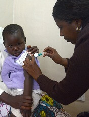
Photo by Caitlin Kleiboer
The European Medicines Agency’s Committee for Medicinal Products for Human Use (CHMP) has adopted a positive opinion of the malaria vaccine RTS,S (also known as RTS,S/AS01 or Mosquirix) for use in children ages 6 weeks to 17 months.
RTS,S is the first candidate vaccine for malaria to reach this milestone. The vaccine was designed to prevent malaria caused by the Plasmodium falciparum parasite.
The CHMP’s opinion on RTS,S is a final stage in the European Medicines Agency’s Article 58 procedure. This allows the CHMP, in cooperation with the World Health Organization (WHO), to give a scientific opinion on a medicinal product intended for markets outside the European Union. This assessment requires products to meet the same standards as those intended for use in the European Union.
Because the CHMP has issued a positive opinion of RTS,S, the WHO is now formulating a policy recommendation on use of the vaccine in national immunization programs.
The vaccine must also pass the WHO pre-qualification process and be approved by national regulatory authorities before it can be used in countries in sub-Saharan Africa, where P falciparum malaria is most prevalent.
About RTS,S
RTS,S aims to trigger the body’s immune system to defend against P falciparum when it first enters the human host’s bloodstream and/or when the parasite infects liver cells.
The vaccine is designed to prevent the parasite from infecting, maturing, and multiplying in the liver, after which time the parasite would re-enter the bloodstream and infect red blood cells, leading to disease symptoms.
The safety and efficacy of RTS,S has been evaluated in a large-scale, phase 3 trial. The CHMP’s recommendation was based mainly on the results of this study. Updated results were published in The Lancet last April.
According to that account, the trial included 15,459 young infants (aged 6 weeks to 12 weeks at first vaccination) and children (5 months to 17 months at first vaccination) from 11 sites across 7 sub-Saharan African countries (Burkina Faso, Gabon, Ghana, Kenya, Malawi, Mozambique, and United Republic of Tanzania) with varying levels of malaria transmission.
The subjects received RTS,S in 3 doses, 1 month apart. Some subjects received an additional booster dose 18 months later. Researchers compared subjects receiving RTS,S to those receiving a control vaccine.
Children who received 3 doses of RTS,S plus a booster had a 36% reduction in the number of clinical episodes of malaria at 4 years. Infants who received 3 doses of RTS,S plus a booster had a 26% reduction in the number of clinical malaria episodes over 3 years.
Children had a significantly lower incidence of severe malaria only if they received the booster dose of RTS,S. The vaccine (with or without a booster dose) did not confer the same benefit in infants.
Subjects who received RTS,S had more adverse events than subjects in the control group. This included meningitis and convulsions.
The road to approval
Two of the WHO’s independent advisory groups, the Strategic Advisory Group of Experts (SAGE) on Immunization and the Malaria Policy Advisory Committee (MPAC), are reviewing the evidence base for RTS,S and will make a joint policy recommendation for how it might be used with other tools to prevent malaria if RTS,S is approved by national regulatory authorities in sub-Saharan Africa.
The WHO has indicated that such a policy recommendation may be possible by end of this year.
Once the WHO policy recommendation is complete, GSK (the company developing RTS,S in partnership with the PATH Malaria Vaccine Initiative) will submit an application to the WHO for pre-qualification of RTS,S.
WHO pre-qualification involves a scientific assessment of the quality, safety, and efficacy of any new vaccine proposed for introduction in the WHO Expanded Programme on Immunization. A pre-qualification decision is used by the United Nations agencies and other large-scale public procurement agencies to help inform vaccine purchasing decisions.
Once a WHO pre-qualification is granted, GSK would then apply for marketing authorization in sub-Saharan Africa on a country-by-country basis. These regulatory and policy decisions would, if positive, enable countries to begin using RTS,S through their universal immunization program.
Both a WHO policy recommendation and WHO pre-qualification are requirements for Gavi, the Vaccine Alliance, to support eligible African countries introducing RTS,S into local immunization programs supported by UNICEF.
GSK has committed to a not-for-profit price for RTS,S. If the vaccine is approved, the price would cover the cost of
manufacturing RTS,S and a return of around 5% to be reinvested in
research and development for second-generation malaria vaccines or
vaccines against other neglected tropical diseases. ![]()
Photo by Caitlin Kleiboer
The European Medicines Agency’s Committee for Medicinal Products for Human Use (CHMP) has adopted a positive opinion of the malaria vaccine RTS,S (also known as RTS,S/AS01 or Mosquirix) for use in children ages 6 weeks to 17 months.
RTS,S is the first candidate vaccine for malaria to reach this milestone. The vaccine was designed to prevent malaria caused by the Plasmodium falciparum parasite.
The CHMP’s opinion on RTS,S is a final stage in the European Medicines Agency’s Article 58 procedure. This allows the CHMP, in cooperation with the World Health Organization (WHO), to give a scientific opinion on a medicinal product intended for markets outside the European Union. This assessment requires products to meet the same standards as those intended for use in the European Union.
Because the CHMP has issued a positive opinion of RTS,S, the WHO is now formulating a policy recommendation on use of the vaccine in national immunization programs.
The vaccine must also pass the WHO pre-qualification process and be approved by national regulatory authorities before it can be used in countries in sub-Saharan Africa, where P falciparum malaria is most prevalent.
About RTS,S
RTS,S aims to trigger the body’s immune system to defend against P falciparum when it first enters the human host’s bloodstream and/or when the parasite infects liver cells.
The vaccine is designed to prevent the parasite from infecting, maturing, and multiplying in the liver, after which time the parasite would re-enter the bloodstream and infect red blood cells, leading to disease symptoms.
The safety and efficacy of RTS,S has been evaluated in a large-scale, phase 3 trial. The CHMP’s recommendation was based mainly on the results of this study. Updated results were published in The Lancet last April.
According to that account, the trial included 15,459 young infants (aged 6 weeks to 12 weeks at first vaccination) and children (5 months to 17 months at first vaccination) from 11 sites across 7 sub-Saharan African countries (Burkina Faso, Gabon, Ghana, Kenya, Malawi, Mozambique, and United Republic of Tanzania) with varying levels of malaria transmission.
The subjects received RTS,S in 3 doses, 1 month apart. Some subjects received an additional booster dose 18 months later. Researchers compared subjects receiving RTS,S to those receiving a control vaccine.
Children who received 3 doses of RTS,S plus a booster had a 36% reduction in the number of clinical episodes of malaria at 4 years. Infants who received 3 doses of RTS,S plus a booster had a 26% reduction in the number of clinical malaria episodes over 3 years.
Children had a significantly lower incidence of severe malaria only if they received the booster dose of RTS,S. The vaccine (with or without a booster dose) did not confer the same benefit in infants.
Subjects who received RTS,S had more adverse events than subjects in the control group. This included meningitis and convulsions.
The road to approval
Two of the WHO’s independent advisory groups, the Strategic Advisory Group of Experts (SAGE) on Immunization and the Malaria Policy Advisory Committee (MPAC), are reviewing the evidence base for RTS,S and will make a joint policy recommendation for how it might be used with other tools to prevent malaria if RTS,S is approved by national regulatory authorities in sub-Saharan Africa.
The WHO has indicated that such a policy recommendation may be possible by end of this year.
Once the WHO policy recommendation is complete, GSK (the company developing RTS,S in partnership with the PATH Malaria Vaccine Initiative) will submit an application to the WHO for pre-qualification of RTS,S.
WHO pre-qualification involves a scientific assessment of the quality, safety, and efficacy of any new vaccine proposed for introduction in the WHO Expanded Programme on Immunization. A pre-qualification decision is used by the United Nations agencies and other large-scale public procurement agencies to help inform vaccine purchasing decisions.
Once a WHO pre-qualification is granted, GSK would then apply for marketing authorization in sub-Saharan Africa on a country-by-country basis. These regulatory and policy decisions would, if positive, enable countries to begin using RTS,S through their universal immunization program.
Both a WHO policy recommendation and WHO pre-qualification are requirements for Gavi, the Vaccine Alliance, to support eligible African countries introducing RTS,S into local immunization programs supported by UNICEF.
GSK has committed to a not-for-profit price for RTS,S. If the vaccine is approved, the price would cover the cost of
manufacturing RTS,S and a return of around 5% to be reinvested in
research and development for second-generation malaria vaccines or
vaccines against other neglected tropical diseases.
CHMP recommends eltrombopag for SAA

The European Medicines Agency’s Committee for Medicinal Products for Human Use (CHMP) has recommended approval for eltrombopag (Revolade) as a treatment for adults with severe aplastic anemia (SAA) who have had an insufficient response to immunosuppressive therapy (IST) and are not eligible to receive a hematopoietic stem cell transplant (HSCT).
If approved by the European Commission, eltrombopag would be the first treatment option in its class for these patients.
The European Commission will review the CHMP recommendation and is expected to deliver its final decision within 3 months. The decision will be applicable to all 28 European Union member states plus Iceland, Norway, and Liechtenstein.
Phase 2 trial of eltrombopag in SAA
The CHMP’s positive opinion of eltrombopag is mainly based on results of a phase 2 trial (NCT00922883). Results from this ongoing study were published in NEJM in 2012 and Blood in 2013.
The trial has enrolled 43 patients with SAA who had an insufficient response to at least one prior IST and who had a platelet count of 30 x 109/L or less.
At baseline, the median platelet count was 20 x 109/L, hemoglobin was 8.4 g/dL, the absolute neutrophil count was 0.58 x 10⁹/L, and absolute reticulocyte count was 24.3 x 10⁹/L.
Patients had a median age of 45 (range, 17 to 77), and 56% were male. The majority of patients (84%) received at least 2 prior ISTs.
Patients received eltrombopag at an initial dose of 50 mg once daily for 2 weeks. The dose increased over 2-week periods to a maximum of 150 mg once daily.
The study’s primary endpoint was hematologic response, which was initially assessed after 12 weeks of treatment. Treatment was discontinued after 16 weeks in patients who did not exhibit a hematologic response.
Forty percent of patients (n=17) experienced a hematologic response in at least one lineage—platelets, red blood cells (RBCs), or white blood cells—after week 12.
In the extension phase of the study, 8 patients achieved a multilineage response. Four of these patients subsequently tapered off treatment and maintained the response. The median follow up was 8.1 months (range, 7.2 to 10.6 months).
Ninety-one percent of patients were platelet-transfusion-dependent at baseline. Patients who responded to eltrombopag did not require platelet transfusions for a median of 200 days (range, 8 to 1096 days).
Eighty-six percent of patients were RBC-transfusion-dependent at baseline. Patients who responded to eltrombopag did not require RBC transfusions for a median of 208 days (range, 15 to 1082 days).
The most common adverse events (≥20%) associated with eltrombopag were nausea (33%), fatigue (28%), cough (23%), diarrhea (21%), and headache (21%).
Patients were also evaluated for cytogenetic abnormalities. Eight patients had a new cytogenetic abnormality after treatment, including 5 patients who had complex changes in chromosome 7.
Patients who develop new cytogenetic abnormalities while on eltrombopag may need to be taken off treatment.
About eltrombopag
Eltrombopag is already approved to treat SAA in the US and Canada. The drug recently gained approval in the US to treat children age 6 and older who have chronic immune thrombocytopenia and have had an insufficient response to corticosteroids, immunoglobulins, or splenectomy.
Eltrombopag is approved in more than 100 countries to treat adults with chronic immune thrombocytopenia who have had an inadequate response to or are intolerant of other treatments. The drug is approved in more than 45 countries for the treatment of thrombocytopenia in patients with chronic hepatitis C to allow them to initiate and maintain interferon-based therapy.
Eltrombopag is marketed under the brand name Promacta in the US and Revolade in most other countries. For more details on the drug, see the European Medicines Agency’s Summary of Product Characteristics.
FDA expands approval of carfilzomib
Photo courtesy of Amgen
The US Food and Drug Administration (FDA) has approved carfilzomib (Kyprolis) in combination with lenalidomide and dexamethasone to treat patients with relapsed multiple myeloma (MM) who have received 1 to 3 prior lines of therapy.
Carfilzomib already has accelerated approval from the FDA as monotherapy for MM patients who have received at least 2 prior therapies, including bortezomib and an immunomodulatory agent, and have demonstrated disease progression on or within 60 days of completing their last treatment.
The FDA’s expanded approval was based on results of the phase 3 ASPIRE trial.
The trial enrolled 792 patients with relapsed or refractory MM who had received 1 to 3 prior lines of therapy. The patients were randomized (1:1) to receive lenalidomide and dexamethasone with or without carfilzomib for 18 cycles.
Lenalidomide and dexamethasone were continued thereafter until disease progression. There was no planned cross-over from the control arm to treatment with carfilzomib.
There was a statistically significant prolongation of progression-free survival (PFS), as determined by an independent review committee, in the carfilzomib arm (hazard ratio=0.69, P=0.0001). The median PFS was 26.3 months in the 3-drug arm and 17.6 months in the 2-drug arm.
A treatment effect was observed across all subgroups tested, but the magnitude of the treatment effect was reduced in patients with higher tumor burden at baseline. The improvement in median PFS was 11 months for patients with International Staging System (ISS) Stage I disease, 8 months for ISS Stage II, and 2 months for ISS Stage III.
An interim analysis of overall survival, the key secondary endpoint, was conducted at the same time. The difference in overall survival did not reach the prespecified boundary for statistical significance.
The overall response rate (partial response or better) was 87% in the 3-drug arm and 67% in the 2-drug arm.
The safety profile of carfilzomib in the 3-drug combination was similar to that described on the current label.
Cardiovascular events, venous thromboembolic events (VTE), and thrombocytopenia occurred more frequently in the 3-drug arm than in the 2-drug arm. In cycles 1 to 12, the VTE rate was 13% and 6%, respectively, despite protocol-mandated use of thromboprophylaxis.
The revised labeling for carfilzomib includes new Warnings and Precautions for VTE, cardiac toxicities, acute renal failure, pulmonary toxicities, and hypertension. The increased safety risks, including mortality, for elderly patients is described. Detailed safety information in the prescribing information was also updated for the use of carfilzomib monotherapy.
The recommended dose schedule for carfilzomib has been revised for both monotherapy and combination treatment. For details, see the full prescribing information.
Carfilzomib is marketed as Kyprolis in the US by Onyx Pharmaceuticals, Inc., an Amgen subsidiary.
Analysis reveals potential therapeutic target for AML

The protein tetraspanin3 (Tspan3) plays a critical role in the development and progression of acute myeloid leukemia (AML), according to research published in Cell Stem Cell.
Investigators found that Tspan3, a cell surface molecule, is expressed in hematopoietic stem and progenitor cells as well as in leukemic cells.
Deleting Tspan3 did not affect normal hematopoiesis, but it prevented AML self-renewal and propagation in vitro and in vivo.
Inhibiting Tspan3 in patient samples led to decreased colony formation in vitro and hindered leukemic growth in primary patient-derived xenografts.
“We found that blocking this molecule leads to a very profound inhibition of leukemia growth,” said study author Tannishtha Reya, PhD, of the University of California San Diego in La Jolla.
These findings build on earlier work by Dr Reya and her colleagues, in which they identified the RNA binding protein Musashi 2 (Msi2) as a critical stem cell signal that is hijacked in several hematologic malignancies.
“We had this idea that analysis of the molecular programs controlled by Musashi 2 may identify new genes important for these leukemias,” Dr Reya said.
So the investigators conducted a genome-wide expression analysis of Msi2-deficient cancer stem cells from blast-crisis chronic myelogenous leukemia and AML. This revealed genes commonly regulated by Msi2 in both leukemias.
Tspan3 was one of the core genes controlled by Msi2. The Tspan3 protein is part of a large family of membrane proteins (the tetraspanin family) that are active in diverse cellular processes, including cell adhesion and proliferation, hematopoietic stem cell function, and blood formation.
“We are particularly excited about this work because, to our knowledge, this is the first demonstration of a requirement for Tspan3 in any primary cancer,” Dr Reya said.
To explore the connection further, the investigators generated the first Tspan3 knockout mouse. In testing, the team found that Tspan3 deletion impaired leukemia stem cell self-renewal and disease propagation and markedly improved survival in the mice.
In patient samples, Tspan3 inhibition blocked the growth of AML, which suggests Tspan3 is also important in human disease.
Dr Reya said these findings are particularly important because AML often doesn’t respond to current therapies. And because Tspan3 is a surface molecule, it is of great translational interest as a target for antibody-mediated therapy.
“There’s been great progress in pediatric leukemia research and treatment over the last few years,” Dr Reya said. “But unfortunately, children with acute myeloid leukemia are often poor responders to current treatments. So identifying new approaches to target this disease remains critically important.”