User login
Postmenopausal hot flashes cut by 93% with novel nonhormonal treatment
AT ENDO 2017
ORLANDO –
After 12 weeks of treatment with the oral small molecule, women had a 93% reduction in moderate to severe hot flashes, compared with a 23% reduction for those taking placebo (P less than .0001). The effects of fezolinetant were seen earlier as well, with an 88% reduction in hot flashes at 4 weeks into the trial, compared with a 12% reduction for the placebo group (P less than .001).
The eight-site study enrolled 87 patients in a double-blind, randomized, placebo-controlled trial that assessed hot flash frequency and severity at study weeks 4 and 12. Postmenopausal women had to have frequent, moderate to severe hot flashes to qualify for enrollment, and they had to be otherwise healthy.
To capture data for a secondary endpoint, participants also completed a quality of life questionnaire. Dr. Fraser and his coinvestigators tracked safety and pharmacokinetic data by measuring levels of luteinizing hormone, follicle-stimulating hormone, estradiol, and sex hormone–binding globulin at baseline and 12 weeks, as well.
Evenly divided between study arms, 80 patients completed the trial. Two patients withdrew because of adverse events, one patient violated inclusion criteria, and the others withdrew for a variety of reasons. Mean age was similar between the two groups, at 53.7 years for those on placebo and 53.3 years for the fezolinetant group. Other anthropometric characteristics were similar, as well.
At baseline, patients taking placebo experienced a mean 10.3 moderate to severe hot flashes daily, compared with the fezolinetant group at a mean of 11.5. By study week 4, 14 of the 40 patients in the active arm had zero hot flashes, compared with 2 of 40 in the in the placebo arm, when the intention-to-treat population was examined. Hot flash severity dropped by 70% (P less than .0001).
Quality of life was assessed with the Hot Flash Related Daily Interference Scale (HFRDIS). When the two groups were compared, a significant (P less than .001) reduction in HFRDIS score was seen by week 4 and continued through to week 12 in the group on active treatment. Lower HFRDIS scores mean improved hot flash–related quality of life.
Using the Leeds Sleep Evaluation Questionnaire allowed Dr. Fraser and his colleagues to ask about how long it took patients to get to sleep while they were participating in the study, compared with their normal sleep latency. Patients taking fezolinetant reported getting to sleep significantly more quickly (P less than .01) than the placebo group. They also reported better quality of sleep (P less than .001) and a better awakening experience (P less than .05). However, they did not report feeling better after awakening (P = .08).
Women taking fezolinetant also showed significant improvement, compared with the placebo group, on other quality of life questionnaires, the Greene Climacteric Scale and the Sheehan Disability Scale. By week 8, a significant (P less than .001) improvement was seen on both scales and specifically at week 4 on the Sheehan Disability Scale.
Among the hormone biomarkers that were followed in the study, only plasma luteinizing-hormone levels dropped, compared with patients’ baseline levels. These were reduced 20% 12 hours after one of the two daily 60-mg oral doses, and 50% at 3 hours post dose, a point at which maximum serum fezolinetant concentration would be seen. These were all statistically significant reductions and expected effects of the drug’s mechanism of action.
Further pharmacokinetic analysis, said Dr. Fraser, “supports testing of once-daily dosing for vasomotor symptoms,” given that, when the drug was tested in premenopausal women in phase I clinical trials, there was no difference in peak and trough drug concentration.
The safety profile was good overall, said Dr. Fraser. No patients in the fezolinetant group reported serious treatment-emergent adverse events. “More patients reported treatment-emergent adverse events in the placebo group than in the fezolinetant group,” he said.
The NK3R antagonist is also under investigation for use in the treatment of polycystic ovary syndrome and uterine fibroids.
The study was funded by Ogeda, which employs Dr. Fraser. Another author reported serving as a clinical adviser for Ogeda.
[email protected]
On Twitter @karioakes
Key clinical point: .
Major finding: Women on fezolinetant had a 93% drop in hot flash frequency, compared with a 23% reduction for those on placebo (P less than .0001).
Data source: A phase II randomized, double-blind, placebo-controlled clinical trial of 87 postmenopausal women with moderate to severe hot flashes.
Disclosures: The study was funded by Ogeda, which employs Dr. Fraser. Another author reported serving as a clinical adviser for Ogeda.
In PCOS, too much sitting means higher glucose levels
ORLANDO – Prolonged time spent sitting was associated with higher post–glucose tolerance test blood glucose levels among less-active women with polycystic ovary syndrome (PCOS).
The trend toward this effect persisted even after researchers controlled for age and body mass index, and was not seen in women who were more active.
The results showed a “compounded adverse metabolic effect of prolonged sitting time in women with PCOS who do not achieve exercise goals,” according to Eleni Greenwood, MD, and her colleagues at the University of California, San Francisco, department of obstetrics, gynecology, and reproductive sciences.
“In PCOS, insulin resistance is tissue specific and occurs in the skeletal muscle,” said Dr. Greenwood, a fellow in the reproductive endocrinology and infertility program at the University of California, San Francisco. Diet and exercise are primary interventions to help with these sequelae. In the general population, prolonged time spent being sedentary is associated with type 2 diabetes, cardiovascular disease, and even some cancers.
Because “the adverse effects of sitting are not reversed through exercise,” as Dr. Greenwood and her colleagues pointed out, the study sought to ascertain whether worse metabolic parameters would be seen in women with PCOS who had had more sedentary time, and whether the association would be independent of exercise status.
Accordingly, the investigators conducted a cross-sectional study of 324 women who met Rotterdam criteria for PCOS. The results were presented during a poster session at the annual meeting of the Endocrine Society.
Patients took the International Physical Activity Questionnaire, and responses were used to determine activity levels and amounts of sedentary times. Other measurements taken at the interdisciplinary clinic where the patients were seen included anthropometric measurements, as well as the results of serum lipid levels, fasting glucose and insulin levels, and the results of a 75-g, 2-hour oral glucose tolerance test (OGTT).
In their analysis, the investigators calculated homeostasis model assessment of insulin resistance (HOMA-IR), and used multivariable analysis to find correlations and eliminate potentially confounding variables. In a further attempt to eliminate confounders, Dr. Greenwood and her colleagues asked patients to stop hormonal contraceptive methods and insulin sensitizing medications 30 days before beginning the study.
The investigators looked at the women’s exercise status, meaning whether they had achieved the level of activity recommended by the U.S. Department of Health & Human Services. However, they also asked the women to report how sedentary they were, measured by the number of hours spent sitting in a day.
It would theoretically be possible for an individual to be very “active,” exercising vigorously for 2 hours each day, but also very “sedentary,” sitting for much of the rest of her waking hours.
Overall, two-thirds of the women (217, 67%) met the activity goals outlined by the HHS. That consisted of exercising enough to achieve 600 metabolic equivalents per week. If the women sat for more than 6 hours a day, they were judged to be sedentary. Of the inactive women, 35% (37) sat for 6 or fewer hours per day, compared with 44% (94) of the active women.
Though the results did not reach statistical significance, HOMA-IR and post-OGTT glucose levels tended to be lower among those who sat less (1.93 vs. 2.73, P = .10; 99 mg/dL vs. 107 mg/dL, P = .09).
Looking at just the inactive group, less sitting time was associated with significantly lower post-OGTT glucose levels (99.1 mg/dL vs. 117.6 mg/dL, P = .03). This difference was not seen among the group of women judged to be active.
“Our results indicate a compounded adverse metabolic effect of prolonged sitting time in women with PCOS who do not achieve exercise goals,” wrote Dr. Greenwood and her collaborators.
Because PCOS pathophysiology can be “disrupted” by an improvement in insulin sensitivity and overall metabolic health, “women with PCOS should be counseled regarding strategies for reducing sedentary time, in addition to improving exercise and diet, as a means of improving metabolic health,” they said.
Dr. Greenwood and her colleagues reported no relevant disclosures.
[email protected]
On Twitter @karioakes
AT ENDO 2017
Key clinical point:
Major finding: Less-active women who also had prolonged sitting time had significantly higher post–oral glucose tolerance test levels (99.1 mg/dL vs. 117.6 mg/dL, P = .03).
Data source: Cross-sectional study of 324 women who met Rotterdam criteria for PCOS.
Disclosures: None of the study authors reported relevant disclosures, and no external source of funding was reported.
One in four practitioners doing FNAs are endocrinologists
AT ENDO 2017
ORLANDO – Endocrinologists made up about one in four of the practitioners performing fine needle aspiration (FNA) biopsies between 2012 and 2014, according to a review of data from the Centers for Medicare & Medicaid Services.
Similarly, endocrinologists performed 25% of image-guided thyroid biopsies.
Endocrine surgeons represent only a small percentage of all practitioners performing head and neck ultrasound exams and image-guided FNA, lead author Mamoona Khokhar, MD, said during a poster presentation at the annual meeting of the Endocrine Society. This is true even though the more portable nature of ultrasound has made it easier for motivated surgeons to incorporate its use into their practice, she said.
Examining 3 years of data from a provider utilization and payment database, Dr. Khokhar and her colleagues identified the types of practitioners who performed head and neck ultrasound, as well as image-guided FNA.
In their analysis, the researchers broadly divided practitioners into surgeons and nonsurgeons. Overall, of the 14,750 median annual practitioners performing head and neck ultrasound between 2012 and 2014, 97.2% were nonsurgeon practitioners, reported Dr. Khokhar, an endocrine surgery fellow at Columbia University Medical Center in New York.
Of all practitioners performing head and neck ultrasound, most (81%) were radiologists. Endocrinologists made up 8% of the overall pool performing ultrasounds.
Breaking the surgeon group down further showed that endocrine surgeons represented 14.7% of surgeons performing head and neck ultrasound, meaning that they made up just 0.4% of the practitioner pool for this procedure. Just over half (52%) of the surgeons performing ultrasounds were otolaryngologists.
The number of practitioners performing image-guided FNA was smaller, at a median 3,695 per year during the study period. Surgeons made up 10.7% of this number. Of the surgeons who performed image-guided FNA, 10.5% were endocrine surgeons. Endocrine surgeons made up 1.1% of all practitioners who billed for FNA.
Again, radiologists made up the majority (58%) of the practitioners performing FNA, and one in four (25%) of practitioners performing FNAs were endocrinologists. Just 5% of the practitioners performing FNAs were otolaryngologists.
More endocrine surgeons performed ultrasound than advanced practice providers (nurse practitioners or physician assistants, 0.2%), pathologists (0.1%), and surgical oncologists (0.04%; P for all, less than .0001). However, advanced practice providers and pathologists both performed significantly more FNAs than did endocrine surgeons (2.1% and 1.8%, P less than .0001).
Although the raw proportion of endocrine surgeons billing for these procedures increased during the study period, the increases were not statistically significant. Dr. Khokhar and her colleagues found that the proportion of American Association of Endocrine Surgeons members who performed head and neck ultrasound grew from 59% in 2012 to 72% in 2014 (P = 0.37), while the proportion performing FNA also increased, from 36% in 2012 to 46% in 2014 (P = .40).
Surgeons, however, may face a number of obstacles in setting up office-based ultrasound, which can lead to underutilization by surgeons, Dr. Khokhar noted, adding that “the results of this study suggest that endocrine surgeons may not be fully utilizing this critical tool in their clinical practice.”
The authors reported no outside sources of funding, and had no relevant conflicts of interest.
[email protected]
On Twitter @karioakes
AT ENDO 2017
ORLANDO – Endocrinologists made up about one in four of the practitioners performing fine needle aspiration (FNA) biopsies between 2012 and 2014, according to a review of data from the Centers for Medicare & Medicaid Services.
Similarly, endocrinologists performed 25% of image-guided thyroid biopsies.
Endocrine surgeons represent only a small percentage of all practitioners performing head and neck ultrasound exams and image-guided FNA, lead author Mamoona Khokhar, MD, said during a poster presentation at the annual meeting of the Endocrine Society. This is true even though the more portable nature of ultrasound has made it easier for motivated surgeons to incorporate its use into their practice, she said.
Examining 3 years of data from a provider utilization and payment database, Dr. Khokhar and her colleagues identified the types of practitioners who performed head and neck ultrasound, as well as image-guided FNA.
In their analysis, the researchers broadly divided practitioners into surgeons and nonsurgeons. Overall, of the 14,750 median annual practitioners performing head and neck ultrasound between 2012 and 2014, 97.2% were nonsurgeon practitioners, reported Dr. Khokhar, an endocrine surgery fellow at Columbia University Medical Center in New York.
Of all practitioners performing head and neck ultrasound, most (81%) were radiologists. Endocrinologists made up 8% of the overall pool performing ultrasounds.
Breaking the surgeon group down further showed that endocrine surgeons represented 14.7% of surgeons performing head and neck ultrasound, meaning that they made up just 0.4% of the practitioner pool for this procedure. Just over half (52%) of the surgeons performing ultrasounds were otolaryngologists.
The number of practitioners performing image-guided FNA was smaller, at a median 3,695 per year during the study period. Surgeons made up 10.7% of this number. Of the surgeons who performed image-guided FNA, 10.5% were endocrine surgeons. Endocrine surgeons made up 1.1% of all practitioners who billed for FNA.
Again, radiologists made up the majority (58%) of the practitioners performing FNA, and one in four (25%) of practitioners performing FNAs were endocrinologists. Just 5% of the practitioners performing FNAs were otolaryngologists.
More endocrine surgeons performed ultrasound than advanced practice providers (nurse practitioners or physician assistants, 0.2%), pathologists (0.1%), and surgical oncologists (0.04%; P for all, less than .0001). However, advanced practice providers and pathologists both performed significantly more FNAs than did endocrine surgeons (2.1% and 1.8%, P less than .0001).
Although the raw proportion of endocrine surgeons billing for these procedures increased during the study period, the increases were not statistically significant. Dr. Khokhar and her colleagues found that the proportion of American Association of Endocrine Surgeons members who performed head and neck ultrasound grew from 59% in 2012 to 72% in 2014 (P = 0.37), while the proportion performing FNA also increased, from 36% in 2012 to 46% in 2014 (P = .40).
Surgeons, however, may face a number of obstacles in setting up office-based ultrasound, which can lead to underutilization by surgeons, Dr. Khokhar noted, adding that “the results of this study suggest that endocrine surgeons may not be fully utilizing this critical tool in their clinical practice.”
The authors reported no outside sources of funding, and had no relevant conflicts of interest.
[email protected]
On Twitter @karioakes
Key clinical point:
Major finding: Endocrinologists made up 25% of the practitioners performing FNAs.
Data source: A retrospective analysis of 3 years’ worth of data on head and neck ultrasound and fine needle aspiration from the Centers for Medicare & Medicaid Services.
Disclosures: None of the study authors reported relevant disclosures, and no external source of funding was reported.
Low-calorie sweeteners may allow more glucose to enter fat cells
ORLANDO – Consumption of low-calorie sweeteners was associated with upregulation of gene expression for glucose transporters in experiments using human mesenchymal stromal cells and adipose tissue, according to new research.
The effect was strongest in individuals with obesity.
Using human subcutaneous fat tissue taken from individuals who consumed low-calorie sweeteners, Sabyasachi Sen, MD, of George Washington University, Washington, and his colleagues showed that these cells had at least a twofold overexpression of glucose transporters. Also overexpressed were sweet taste receptors and adipogenic genes.
Dr. Sen and his colleagues first tested the effects of both physiologic and supraphysiologic doses of sucralose on human adipose-derived mesenchymal stromal cells (MSCs). In the presence of adipogenic media, sucralose was added to the MSCs for a period of 12 days.
When sucralose was added at a concentration of 0.2 millimoles (mM), the investigators detected “small but reproducible” increases in expression of adiponectin, as well as genes that encode for the production of fatty acid binding proteins (FABP4) and proteins implicated in body weight homeostasis (CEBPA). Increases ranged from 1.04- to 1.32-fold.
Dr. Sen explained that the 0.2 mM concentration approximates the amount of sucralose in the tissues of individuals with high low-calorie sweetener consumption.
These effects were more pronounced when the sucralose concentration was raised to a supraphysiologic 1 mM. Then, CEBPA expression increased by a factor of 3.45, adiponectin by 4.39, and FABP4 by 4.06. More intracellular fat droplets were seen with higher sucralose concentrations in a “dose dependent fashion,” said Dr. Sen and his colleagues.
The researchers also took subcutaneous fat biopsies from four individuals of healthy weight (body mass index [BMI] less than 24.9 kg/m2) and from four individuals with obesity (BMI greater than 24.9). Participants ranged in age from 36 years to 60 years.
Individuals in both groups had kept a 7-day food diary before the biopsies were taken, so Dr. Sen and his coinvestigators knew whether or not they had consumed low-calorie sweeteners and in what quantity. Reported sweeteners were sucralose, acesulfame-potassium, and aspartame.
The adipose tissue biopsies were examined for mRNA expression of adipogenic genes, as well as for glucose transporters and inflammatory markers.
In individuals with low-calorie sweetener exposure, the glucose transporter GLUT1 was overexpressed by 2- to 2.2-fold, while the glucose transporter GLUT4 was overexpressed by a factor of 2.7-4.3.
In addition, low-calorie sweetener consumption was associated with up to 2.5-fold overexpression of the sweet taste receptor TAS1R3. Sweet taste receptors are G protein–coupled receptors that are present in taste buds and in other tissues, serving as carbohydrate sensors.
Adipogenic genes were also overexpressed; a 1.5- to 2.4-fold increase was seen in PPARG, for example.
“This expression pattern was most apparent in obese individuals,” the investigators wrote in the abstract accompanying the study, which was presented at the annual meeting of the Endocrine Society.
Further and larger studies are called for, especially in individuals with diabetes and obesity, Dr. Sen said. “However, from our study, we believe that low-calorie sweeteners promote additional fat formation by allowing more glucose to enter the cells, and promote inflammation, which may be more detrimental to obese individuals.”
Dr. Sen and his collaborators reported no relevant disclosures.
[email protected]
On Twitter @karioakes
Key clinical point:
Major finding: Glucose transporters and sweet taste receptors were upregulated by 2.0- to 4.3-fold in participants who used low-calorie sweeteners.
Data source: In vitro examination of human adipose tissue-derived mesenchymal stromal cells, and examination of biopsied adipose tissue from eight individuals with varying low-calorie sweetener intake.
Disclosures: None of the study authors reported relevant disclosures, and no external source of funding was reported.
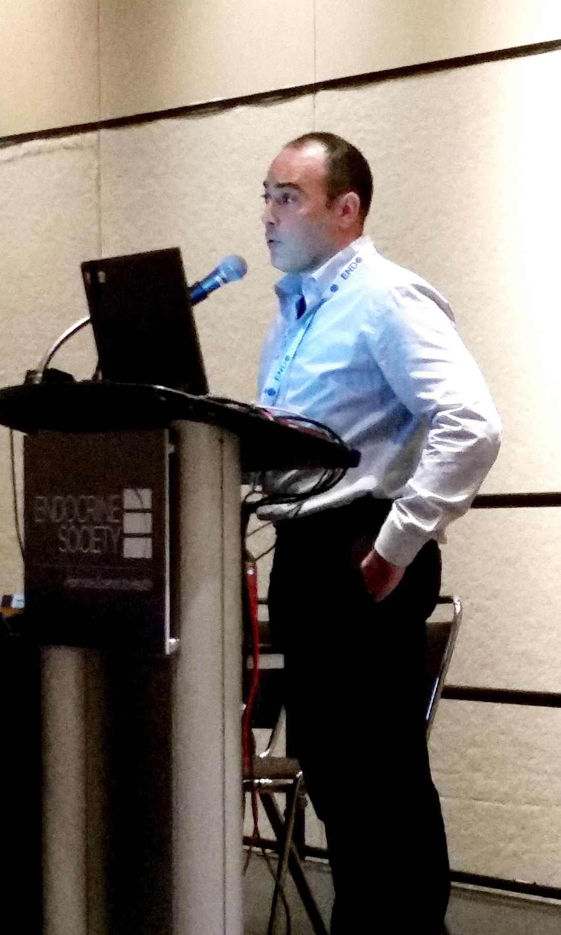
Neurokinin receptor antagonist nearly halves hot flashes
ORLANDO – A novel antagonist of a neurokinin pathway effectively reduced hot flashes in postmenopausal women, according to results of a phase II trial.
The neurokinin 3 receptor (NK3R) antagonist, termed MLE4901, cut the number of hot flashes nearly in half compared to the effect of a placebo, and also improved overall menopause-related quality of life symptoms.
The medication has the potential to fulfill an unmet clinical need, since 70% of women experience hot flashes and the median duration of symptoms is more than 7 years, Julia K. Prague, MD, said at the annual meeting of the Endocrine Society.
The study results were published concurrently with Dr. Prague’s presentation (Lancet. 2017 Apr 3. doi: 10.1016/S0140-6736[17]30823-1).
Evidence from postmortem studies of postmenopausal women, said Dr. Prague, pointed to NKB (neurokinin B)/NK3R signaling as playing a role in hot flashes. The brains of these women showed hypertrophy of NKB neurons and increased neuronal activity, as well as increased NKB gene expression. The same effects were seen in monkeys whose ovaries had been removed, and the effect was reversed when the monkeys were given estrogen. 
Also, previous work has shown that administering NKB to premenopausal women gave them hot flashes (Sci Rep. 2015 Feb 6;5:8466). The preclinical work laid the groundwork for the hypothesis that taking an oral NK3R antagonist could mitigate hot flashes in menopausal women.
The randomized, placebo-controlled, double-blind 2-way crossover study enrolled 37 healthy women aged 40-62 who were at least 12 months out from their last menses. To qualify for enrollment, they needed to be experiencing at least seven hot flashes per 24 hours; the actual mean total number in the study group was 13 per 24 hours and 85 per week. A total of 28 women completed the full protocol.
The mean age of the women was 55 years, and the mean body mass index was 25.8 kg/m2. Three quarters of the women were white.
The women had an initial 2-week baseline period, during which they kept a symptom diary, and then were randomized to receive either an oral NK3R antagonist or a placebo twice daily. Then, each patient went through a 2-week washout period, after which they were crossed over to the other arm of the study. Finally, patients were monitored for a further 2 weeks after taking the study drug.
The NK3R antagonist, said Dr. Prague, “significantly reduced the total weekly number of hot [flashes] compared to placebo in the fourth week of treatment.” Patients taking placebo experienced 49.01 hot flashes per week (range, 40.81-58.86), while those taking the NK3R antagonist had 19.35 (range, 15.99-23.42, P less than .0001). This amounted to a 45% reduction in number of hot flashes compared to placebo.
Dr. Prague, a clinical research fellow at Imperial College, London, said that the treatment effect size seen was similar regardless of whether patients received the active study drug or placebo first.
Using the Menopause-Related Quality of Life questionnaire, Dr. Prague and her colleagues determined that when women were taking the NK3R antagonist, in addition to a significant reduction in vasomotor symptoms, menopause-related psychosocial symptoms were reduced by 15% (P = .0083) and physical symptoms by 19% (P = .0002). As expected for the nonhormonal medication, she said, sexual symptoms were reduced by a nonsignificant 8% (P = .24).
A subset of patients received more intensive study, with external validation of hot flashes and a series of 3-day-long stays during which an intravenous catheter was placed for blood sampling every 10 minutes.
Data from this group of patients showed that, while the NK3R antagonist did not significantly affect the number of luteinizing hormone pulses detected compared with placebo (P = .41), it did increase the mean amplitude of each pulse (P = .0243). Also, the active drug made the luteinizing hormone pulses more orderly (P = .0006 compared to placebo).
The NK3R antagonist was well tolerated. Though no serious adverse events were seen, three patients taking the NK3R antagonist did have a transient and asymptomatic elevation of transaminases without hyperbilirubinemia at 28 days after starting treatment. The elevation resolved for all patients within 90 days of stopping the medication.
The novel drug could represent “a potentially practice-changing therapeutic,” said Dr. Prague, since it “significantly relieves hot [flash] symptoms without the need for estrogen exposure.” She added that planning is underway for longer studies involving more patients.
The study was supported by AstraZeneca and Millendo Therapeutics, which are involved with manufacturing MLE4901.
[email protected]
On Twitter @karioakes
ORLANDO – A novel antagonist of a neurokinin pathway effectively reduced hot flashes in postmenopausal women, according to results of a phase II trial.
The neurokinin 3 receptor (NK3R) antagonist, termed MLE4901, cut the number of hot flashes nearly in half compared to the effect of a placebo, and also improved overall menopause-related quality of life symptoms.
The medication has the potential to fulfill an unmet clinical need, since 70% of women experience hot flashes and the median duration of symptoms is more than 7 years, Julia K. Prague, MD, said at the annual meeting of the Endocrine Society.
The study results were published concurrently with Dr. Prague’s presentation (Lancet. 2017 Apr 3. doi: 10.1016/S0140-6736[17]30823-1).
Evidence from postmortem studies of postmenopausal women, said Dr. Prague, pointed to NKB (neurokinin B)/NK3R signaling as playing a role in hot flashes. The brains of these women showed hypertrophy of NKB neurons and increased neuronal activity, as well as increased NKB gene expression. The same effects were seen in monkeys whose ovaries had been removed, and the effect was reversed when the monkeys were given estrogen.
Also, previous work has shown that administering NKB to premenopausal women gave them hot flashes (Sci Rep. 2015 Feb 6;5:8466). The preclinical work laid the groundwork for the hypothesis that taking an oral NK3R antagonist could mitigate hot flashes in menopausal women.
The randomized, placebo-controlled, double-blind 2-way crossover study enrolled 37 healthy women aged 40-62 who were at least 12 months out from their last menses. To qualify for enrollment, they needed to be experiencing at least seven hot flashes per 24 hours; the actual mean total number in the study group was 13 per 24 hours and 85 per week. A total of 28 women completed the full protocol.
The mean age of the women was 55 years, and the mean body mass index was 25.8 kg/m2. Three quarters of the women were white.
The women had an initial 2-week baseline period, during which they kept a symptom diary, and then were randomized to receive either an oral NK3R antagonist or a placebo twice daily. Then, each patient went through a 2-week washout period, after which they were crossed over to the other arm of the study. Finally, patients were monitored for a further 2 weeks after taking the study drug.
The NK3R antagonist, said Dr. Prague, “significantly reduced the total weekly number of hot [flashes] compared to placebo in the fourth week of treatment.” Patients taking placebo experienced 49.01 hot flashes per week (range, 40.81-58.86), while those taking the NK3R antagonist had 19.35 (range, 15.99-23.42, P less than .0001). This amounted to a 45% reduction in number of hot flashes compared to placebo.
Dr. Prague, a clinical research fellow at Imperial College, London, said that the treatment effect size seen was similar regardless of whether patients received the active study drug or placebo first.
Using the Menopause-Related Quality of Life questionnaire, Dr. Prague and her colleagues determined that when women were taking the NK3R antagonist, in addition to a significant reduction in vasomotor symptoms, menopause-related psychosocial symptoms were reduced by 15% (P = .0083) and physical symptoms by 19% (P = .0002). As expected for the nonhormonal medication, she said, sexual symptoms were reduced by a nonsignificant 8% (P = .24).
A subset of patients received more intensive study, with external validation of hot flashes and a series of 3-day-long stays during which an intravenous catheter was placed for blood sampling every 10 minutes.
Data from this group of patients showed that, while the NK3R antagonist did not significantly affect the number of luteinizing hormone pulses detected compared with placebo (P = .41), it did increase the mean amplitude of each pulse (P = .0243). Also, the active drug made the luteinizing hormone pulses more orderly (P = .0006 compared to placebo).
The NK3R antagonist was well tolerated. Though no serious adverse events were seen, three patients taking the NK3R antagonist did have a transient and asymptomatic elevation of transaminases without hyperbilirubinemia at 28 days after starting treatment. The elevation resolved for all patients within 90 days of stopping the medication.
The novel drug could represent “a potentially practice-changing therapeutic,” said Dr. Prague, since it “significantly relieves hot [flash] symptoms without the need for estrogen exposure.” She added that planning is underway for longer studies involving more patients.
The study was supported by AstraZeneca and Millendo Therapeutics, which are involved with manufacturing MLE4901.
[email protected]
On Twitter @karioakes
AT ENDO 2017
Key clinical point:
Major finding: The neurokinin 3 receptor antagonist reduced hot flashes from a mean of 85 per week at baseline to a mean of 49 per week.
Data source: Phase II randomized, double-blind, placebo-controlled two-way crossover study of 37 postmenopausal women with moderate to severe hot flashes.
Disclosures: The study was funded by AstraZeneca and Millendo Therapeutics, which are involved with the manufacture of the medicine, termed MLE4901.
Obstacles plague prenatal cell-free DNA testing use
A recent California study examining cell-free DNA (cfDNA) testing suggests that the test generally performs well as a second-line screen for fetal aneuploidy. However, while the test itself is reliable, some physicians say gaps in patient education, limited availability of genetic counseling, and a lack of coordinated research and data collection are all standing in the way of its optimal utilization.
In some of the most recent data presented on cfDNA test performance, researchers in California found that the test had few false positives and was best at predicting Down syndrome, with a positive predictive value of 98.6%.

California offers prenatal screening as a public health program, with a structured system of screening tests and follow-up services, according to Dr. Currier, who is chief of the evaluation section of the state’s Genetic Disease Screening Program. First trimester screening includes screens for trisomies 18 and 21. During the second trimester, the screening adds on checks for neural tube defects and Smith-Lemli-Opitz syndrome, also known as 7-dehydrocholesterol reductase deficiency.

Overall, 20,852 patients were offered cfDNA screening during this period, and 63% (n = 12,960) went on to have the screen. Dr. Currier and his colleagues found that about a third of patients with a first- or second-trimester positive cfDNA test opted for diagnostic testing with either chorionic villus sampling or amniocentesis. If patients had a known karyotype, most were in agreement with the trisomy or sex-chromosome aneuploidies that had been picked up by cfDNA testing.
There were a small number of false-positive cfDNA results seen in the California data, Dr. Currier noted. Performance was best for Down syndrome, where cfDNA had a positive predictive value of 98.6%. Here, 214 of the 611 cfDNA positive results received diagnostic confirmation. Of those, 197 were true positives and 3 were false positives, while a karyotype other than trisomy 21 was revealed in 14 cases. Positive predictive values were lower in the less-common aneuploidies, he said.
A small number of test failures also occurred. Of the 148 failures reported, just 40 were followed by successful repeat tests.
“Cell-free DNA is a good second-tier screening test, but it does have false-positive and false-negative results, so pre- and post-test counseling addressing these limitations are essential,” Dr. Currier said.
Nancy Rose, MD, agrees with that assessment of the limitations of the cfDNA test. She helped write the American College of Obstetricians and Gynecologists 2015 opinion on cell-free DNA screening for aneuploidy, which states that conventional screening methods “remain the most appropriate choice for first-line screening for most women in the general obstetric population.”
While it is “perfectly reasonable” for general ob.gyns. to offer cfDNA screening, in her practice, “genetic counselors call out all the results for general providers because patients don’t really understand what a screen negative result is,” Dr. Rose said in an interview. “Certainly, screen positive results should go to a genetic counselor for sure.”
However, that level of follow-up may be hard to implement. “One issue that we’re all facing right now is that genetic counselors are a scarce commodity because of the competition with them working for labs or insurance companies,” said Dr. Rose, a maternal-fetal medicine specialist in Salt Lake City.
If the lack of appropriate counseling is an issue, are patients sometimes jumping the gun? The immediate past president of the Society for Maternal-Fetal Medicine, Mary Norton, MD, said maybe so.
When asked whether she thinks pregnancies are being terminated on the basis of cfDNA results alone, Dr. Norton said, “I absolutely think that is happening. How many is hard to know. I do know that we have many patients that we see who say, ‘My result here says that the chance of Down syndrome is greater than 99%,’ but that is not what that number means, and people don’t understand that.”
She added, “In some cases there are also ultrasound abnormalities, but there is no question that there is misunderstanding leading to the loss of normal fetuses.”
Patient misunderstandings about what’s entailed in first- and second-line screening tests persist, said Dr. Norton, professor of obstetrics and gynecology and reproductive sciences at the University of California, San Francisco. One example is ongoing hesitancy about amniocentesis. “Amnio has become very, very safe,” she said. “It’s much safer than it used to be. The miscarriage rate is nearly nonexistent.”
The care team needs to work to find time to educate patients about both older technologies and noninvasive prenatal testing (NIPT), Dr. Rose and Dr. Norton stressed.
“Even though ‘NIPT’ is easy to say – it rolls off the tongue – we really shouldn’t call it that. It’s misleading, because all of the other screening tests are noninvasive too,” Dr. Norton said. “I’ve had many patients who declined diagnostic testing, even though they want as much information as possible about what they should do.”
Dr. Rose added, “It’s a screening test; it’s not perfect. But screening tests are really what obstetricians do. They do pap smears; they do breast exams. So I think that isn’t the issue so much as that there is no time in an obstetrician’s office to really educate women about what they are accepting.”
Additional ethical issues revolving around the mother may arise in rare cases. Only about 10% of the DNA sampled is fetal, meaning that the rest is maternal DNA, Dr. Norton explained. “And they’re not separated,” she said. “So, the issue that is coming up is that they are finding things in the mother that are unanticipated, and women aren’t told that ahead of time.”
Some of these thorny – and still evolving – legal and ethical questions were addressed during a workshop at the 2017 Pregnancy Meeting. A working group that met there is developing a summary of their discussion for future publication.
For Dr. Rose, the larger issue is the lack of coordinated data collection and quality initiatives. She pointed out that the California outcomes study may not be generalizable to most of the rest of the country because most states don’t have such a coordinated public health approach to prenatal testing and data collection.
This reality is hampering knowledge advancement in the field, she said. When she was asked about terminations on the basis of cfDNA alone, Dr. Rose said, “Because there’s no outcome data on these patients, because there are no unbiased studies that cross through these companies, we actually don’t really know that on a national level. There are no unbiased outcome data. There may be small studies, but I think there are no national data to answer that question at the moment.”
Overall, Dr. Rose said that cfDNA screening is fairly reliable. “This is no different than what has been done before with serum analyte screening, and it’s probably a little bit better,” she said. “My feeling is the tragedy is really in the lack of companies either being able to work together or to have some unbiased funding so you could really actually know what test performance means.”
Dr. Norton reported receiving research funding from Natera and Ultragenyx. Dr. Rose reported no relevant disclosures. Dr. Currier’s study was funded by the California Department of Public Health, where he is employed.
[email protected]
On Twitter @karioakes
A recent California study examining cell-free DNA (cfDNA) testing suggests that the test generally performs well as a second-line screen for fetal aneuploidy. However, while the test itself is reliable, some physicians say gaps in patient education, limited availability of genetic counseling, and a lack of coordinated research and data collection are all standing in the way of its optimal utilization.
In some of the most recent data presented on cfDNA test performance, researchers in California found that the test had few false positives and was best at predicting Down syndrome, with a positive predictive value of 98.6%.
California offers prenatal screening as a public health program, with a structured system of screening tests and follow-up services, according to Dr. Currier, who is chief of the evaluation section of the state’s Genetic Disease Screening Program. First trimester screening includes screens for trisomies 18 and 21. During the second trimester, the screening adds on checks for neural tube defects and Smith-Lemli-Opitz syndrome, also known as 7-dehydrocholesterol reductase deficiency.
Overall, 20,852 patients were offered cfDNA screening during this period, and 63% (n = 12,960) went on to have the screen. Dr. Currier and his colleagues found that about a third of patients with a first- or second-trimester positive cfDNA test opted for diagnostic testing with either chorionic villus sampling or amniocentesis. If patients had a known karyotype, most were in agreement with the trisomy or sex-chromosome aneuploidies that had been picked up by cfDNA testing.
There were a small number of false-positive cfDNA results seen in the California data, Dr. Currier noted. Performance was best for Down syndrome, where cfDNA had a positive predictive value of 98.6%. Here, 214 of the 611 cfDNA positive results received diagnostic confirmation. Of those, 197 were true positives and 3 were false positives, while a karyotype other than trisomy 21 was revealed in 14 cases. Positive predictive values were lower in the less-common aneuploidies, he said.
A small number of test failures also occurred. Of the 148 failures reported, just 40 were followed by successful repeat tests.
“Cell-free DNA is a good second-tier screening test, but it does have false-positive and false-negative results, so pre- and post-test counseling addressing these limitations are essential,” Dr. Currier said.
Nancy Rose, MD, agrees with that assessment of the limitations of the cfDNA test. She helped write the American College of Obstetricians and Gynecologists 2015 opinion on cell-free DNA screening for aneuploidy, which states that conventional screening methods “remain the most appropriate choice for first-line screening for most women in the general obstetric population.”
While it is “perfectly reasonable” for general ob.gyns. to offer cfDNA screening, in her practice, “genetic counselors call out all the results for general providers because patients don’t really understand what a screen negative result is,” Dr. Rose said in an interview. “Certainly, screen positive results should go to a genetic counselor for sure.”
However, that level of follow-up may be hard to implement. “One issue that we’re all facing right now is that genetic counselors are a scarce commodity because of the competition with them working for labs or insurance companies,” said Dr. Rose, a maternal-fetal medicine specialist in Salt Lake City.
If the lack of appropriate counseling is an issue, are patients sometimes jumping the gun? The immediate past president of the Society for Maternal-Fetal Medicine, Mary Norton, MD, said maybe so.
When asked whether she thinks pregnancies are being terminated on the basis of cfDNA results alone, Dr. Norton said, “I absolutely think that is happening. How many is hard to know. I do know that we have many patients that we see who say, ‘My result here says that the chance of Down syndrome is greater than 99%,’ but that is not what that number means, and people don’t understand that.”
She added, “In some cases there are also ultrasound abnormalities, but there is no question that there is misunderstanding leading to the loss of normal fetuses.”
Patient misunderstandings about what’s entailed in first- and second-line screening tests persist, said Dr. Norton, professor of obstetrics and gynecology and reproductive sciences at the University of California, San Francisco. One example is ongoing hesitancy about amniocentesis. “Amnio has become very, very safe,” she said. “It’s much safer than it used to be. The miscarriage rate is nearly nonexistent.”
The care team needs to work to find time to educate patients about both older technologies and noninvasive prenatal testing (NIPT), Dr. Rose and Dr. Norton stressed.
“Even though ‘NIPT’ is easy to say – it rolls off the tongue – we really shouldn’t call it that. It’s misleading, because all of the other screening tests are noninvasive too,” Dr. Norton said. “I’ve had many patients who declined diagnostic testing, even though they want as much information as possible about what they should do.”
Dr. Rose added, “It’s a screening test; it’s not perfect. But screening tests are really what obstetricians do. They do pap smears; they do breast exams. So I think that isn’t the issue so much as that there is no time in an obstetrician’s office to really educate women about what they are accepting.”
Additional ethical issues revolving around the mother may arise in rare cases. Only about 10% of the DNA sampled is fetal, meaning that the rest is maternal DNA, Dr. Norton explained. “And they’re not separated,” she said. “So, the issue that is coming up is that they are finding things in the mother that are unanticipated, and women aren’t told that ahead of time.”
Some of these thorny – and still evolving – legal and ethical questions were addressed during a workshop at the 2017 Pregnancy Meeting. A working group that met there is developing a summary of their discussion for future publication.
For Dr. Rose, the larger issue is the lack of coordinated data collection and quality initiatives. She pointed out that the California outcomes study may not be generalizable to most of the rest of the country because most states don’t have such a coordinated public health approach to prenatal testing and data collection.
This reality is hampering knowledge advancement in the field, she said. When she was asked about terminations on the basis of cfDNA alone, Dr. Rose said, “Because there’s no outcome data on these patients, because there are no unbiased studies that cross through these companies, we actually don’t really know that on a national level. There are no unbiased outcome data. There may be small studies, but I think there are no national data to answer that question at the moment.”
Overall, Dr. Rose said that cfDNA screening is fairly reliable. “This is no different than what has been done before with serum analyte screening, and it’s probably a little bit better,” she said. “My feeling is the tragedy is really in the lack of companies either being able to work together or to have some unbiased funding so you could really actually know what test performance means.”
Dr. Norton reported receiving research funding from Natera and Ultragenyx. Dr. Rose reported no relevant disclosures. Dr. Currier’s study was funded by the California Department of Public Health, where he is employed.
[email protected]
On Twitter @karioakes
FROM THE PREGNANCY MEETING
Infants’ head circumference larger with PCOS moms on metformin
Women with polycystic ovarian syndrome (PCOS) who took metformin had newborns with larger heads than the offspring of women with PCOS who took a placebo, though PCOS offspring were, on average, shorter than newborns in a reference population, according to a recent study.
Metformin is commonly prescribed to women with PCOS, and though metformin passes the placental barrier and can reach therapeutic concentrations in the umbilical cord blood, it hasn’t been proven teratogenic, said Anna Hjorth-Hansen, MD, of the internal medicine department at Levanger Hospital, Norway.

The study was a post hoc analysis of data from the PregMet study, which was run from 2005 to 2009, and compared metformin to placebo, in combination with diet and lifestyle changes, for women with PCOS, said Dr. Hjorth-Hansen. The PregMet study tested the hypothesis that women with PCOS who received metformin from the first trimester until delivery had fewer pregnancy complications overall than women who did not.
Though women receiving placebo in the PregMet study had no more eclampsia, preterm delivery, or gestational diabetes than those who received metformin, the newborns who had in utero metformin exposure had significantly larger head circumference.
Dr. Hjorth-Hansen said that the study looked at in utero growth and anthropometric measurements at birth of infants born to women taking metformin, to determine whether metformin could affect fetal growth and newborn anthropometrics.
Ultrasound examination was used to measure crown-rump length, biparietal diameter (BPD), and mean abdominal diameter (MAD). At birth, head circumference (HC), length, and weight were measured.
Maternal characteristics were comparable between the metformin (131 patients) and placebo (127 patients) groups, said Dr. Hjorth-Hansen. Specifically, there were no significant differences between groups in terms of PCOS phenotype, blood glucose levels, and parity.
Infants born to women who took metformin had, on average, a larger BPD at 32 weeks gestation, compared with the infants whose mothers took placebo (86.1 mm versus 85.2 mm, P = .027). This larger head size was also seen at birth (mean HC, metformin, 35.6 cm; placebo, 35.0 cm; P = .007).
There were no significant differences between the groups in MAD or weight, either as assessed by ultrasound at 32 weeks gestation or as measured at birth.
Although the two groups did not differ in length at birth, the aggregate study population of infants born to women with PCOS was shorter than a large Swedish reference population.
When Dr. Hjorth-Hansen and her colleagues stratified the results by maternal body mass index (BMI), looking at babies born to women with BMIs below 25 kg/m2, compared with those with BMI of 25 kg/m2 and greater, they saw no differences in infant anthropometric measurements for women who had taken placebo.
However, when the investigators dichotomized maternal BMI for the metformin group, they found that infants born to the higher BMI group had a larger head size (P = .022), and were heavier (P = .002) and longer (P = .003) than infants born to women with BMIs less than 25.
“Metformin resulted in a larger head size, traceable already in utero,” said Dr. Hjorth-Hansen. However, she said, there’s a “PCOS effect” that results in the offspring of women with the condition to have a shorter body, compared with offspring of women without PCOS.
Dr. Hjorth-Hansen reported no relevant disclosures.
[email protected]
On Twitter @karioakes
Women with polycystic ovarian syndrome (PCOS) who took metformin had newborns with larger heads than the offspring of women with PCOS who took a placebo, though PCOS offspring were, on average, shorter than newborns in a reference population, according to a recent study.
Metformin is commonly prescribed to women with PCOS, and though metformin passes the placental barrier and can reach therapeutic concentrations in the umbilical cord blood, it hasn’t been proven teratogenic, said Anna Hjorth-Hansen, MD, of the internal medicine department at Levanger Hospital, Norway.
The study was a post hoc analysis of data from the PregMet study, which was run from 2005 to 2009, and compared metformin to placebo, in combination with diet and lifestyle changes, for women with PCOS, said Dr. Hjorth-Hansen. The PregMet study tested the hypothesis that women with PCOS who received metformin from the first trimester until delivery had fewer pregnancy complications overall than women who did not.
Though women receiving placebo in the PregMet study had no more eclampsia, preterm delivery, or gestational diabetes than those who received metformin, the newborns who had in utero metformin exposure had significantly larger head circumference.
Dr. Hjorth-Hansen said that the study looked at in utero growth and anthropometric measurements at birth of infants born to women taking metformin, to determine whether metformin could affect fetal growth and newborn anthropometrics.
Ultrasound examination was used to measure crown-rump length, biparietal diameter (BPD), and mean abdominal diameter (MAD). At birth, head circumference (HC), length, and weight were measured.
Maternal characteristics were comparable between the metformin (131 patients) and placebo (127 patients) groups, said Dr. Hjorth-Hansen. Specifically, there were no significant differences between groups in terms of PCOS phenotype, blood glucose levels, and parity.
Infants born to women who took metformin had, on average, a larger BPD at 32 weeks gestation, compared with the infants whose mothers took placebo (86.1 mm versus 85.2 mm, P = .027). This larger head size was also seen at birth (mean HC, metformin, 35.6 cm; placebo, 35.0 cm; P = .007).
There were no significant differences between the groups in MAD or weight, either as assessed by ultrasound at 32 weeks gestation or as measured at birth.
Although the two groups did not differ in length at birth, the aggregate study population of infants born to women with PCOS was shorter than a large Swedish reference population.
When Dr. Hjorth-Hansen and her colleagues stratified the results by maternal body mass index (BMI), looking at babies born to women with BMIs below 25 kg/m2, compared with those with BMI of 25 kg/m2 and greater, they saw no differences in infant anthropometric measurements for women who had taken placebo.
However, when the investigators dichotomized maternal BMI for the metformin group, they found that infants born to the higher BMI group had a larger head size (P = .022), and were heavier (P = .002) and longer (P = .003) than infants born to women with BMIs less than 25.
“Metformin resulted in a larger head size, traceable already in utero,” said Dr. Hjorth-Hansen. However, she said, there’s a “PCOS effect” that results in the offspring of women with the condition to have a shorter body, compared with offspring of women without PCOS.
Dr. Hjorth-Hansen reported no relevant disclosures.
[email protected]
On Twitter @karioakes
FROM ENDO 2017
Key clinical point:
Major finding: Infants born to women with PCOS who took metformin during pregnancy had larger head circumferences at birth than those born to women with PCOS who took placebo (35.6 cm versus 35.0 cm, P = .007).
Data source: Retrospective analysis of data from 258 women in the PregMet study.
Disclosures: Dr. Hjorth-Hansen reported no relevant disclosures.
Three factors linked to rhinovirus pneumonia in HCT patients
ORLANDO – For patients who have received hematopoietic cell transplants, a rhinovirus infection can become much more than a cold.
“It holds true that rhinovirus is just as likely to be associated with mortality as are other respiratory viruses” among HCT recipients, Alpana Waghmare, MD, said at the combined annual meetings of the Center for International Blood & Marrow Transplant Research and the American Society for Blood and Marrow Transplantation.
In a new retrospective study, Dr. Waghmare and her coinvestigators found that the median time for a rhinovirus infection to progress from an upper to a lower respiratory tract infection was about 2 weeks among post-HCT patients.
Clinical and demographic risk factors for progression to lower respiratory tract infection included higher levels of steroid use (2 mg/kg per day or more) before developing the upper respiratory infection, a low white blood cell count, and a low monocyte count, said Dr. Waghmare, an infectious disease specialist and professor of pediatrics at the University of Washington, Seattle.
Of 3,445 HCT patients treated at the university center during the 6-year study, 732 patients (21%) were positive for human rhinovirus. Patients were classified as having upper respiratory infections if they had a PCR-positive nasal swab.
Patients were classed in one of three categories for potential lower respiratory infections: Proven lower respiratory infections were those detected by bronchoalveolar lavage or biopsy in patients who had a new radiographic abnormality. Probable lower respiratory infections were those with positive findings on bronchoalveolar lavage or biopsy but without radiographic changes. In possible lower respiratory infections, patients had upper tract virus detected on nasal swabs but did have a new radiographic abnormality.
Among the patients positive for human rhinovirus, 85% (665 patients) presented with upper respiratory infections and 15% (117 patients) with lower respiratory tract infections. By day 90, 16% of patients progressed from upper to lower respiratory tract infections. The median time to progression was 13.5 days. Progression to proven lower respiratory tract infection affected 5% of the HCT recipients.
In multivariable analytic models, a minimum white blood cell count of 1,000 or less was associated with a hazard ratio (HR) of 2.21 for progression to lower respiratory tract infection. A minimum monocyte count of 1,000 or less was associated with a HR of 3.66 for progression to lower respiratory tract infection.
The model also found a HR of 3.37 for lower respiratory tract infection with steroid use of 2 mg/kg per day or more. The patient’s conditioning regimen and donor type were not significantly associated with risk of progression to lower respiratory infection.
Viral copathogens, prior respiratory virus episodes, and the duration of time since HCT were not associated with risk of progress to lower respiratory infections. Neither were patient age, baseline lung function, and the year the transplant occurred.
“These data provide an initial framework for patient risk stratification and the development of rational prevention and treatment strategies in HCT recipients,” she said.
Dr. Waghmare reported receiving research funding from Aviragen, the maker of vapendavir, an investigational drug for human rhinovirus infection, and Gilead Sciences.
[email protected]
On Twitter @karioakes
AT THE BMT TANDEM MEETINGS
Key clinical point:
Major finding: Of 3,445 HCT patients, 732 patients (21%) were positive for human rhinovirus.
Data source: Single-center, 6-year retrospective study of 732 HCT patients with human rhinovirus infection.
Disclosures: Dr. Waghmare reported receiving research funding from Aviragen, the maker of vapendavir, an investigational drug for human rhinovirus infection, and Gilead Sciences.
Off-the-shelf T cells an option for post-HCT viral infections
ORLANDO – Infusions of banked multivirus-specific T lymphocytes were associated with complete or partial responses in 93% of 42 patients who had undergone hematopoietic cell transplants and had drug-refractory viral illnesses. Further, these patients experienced minimal new or reactivated graft-versus-host disease (GVHD).
Viral infections cause nearly 40% of deaths after alternative donor hematopoietic cell transfer (HCT), Ifigeneia Tzannou, MD, said at the combined annual meetings of the Center for International Blood & Marrow Transplant Research and the American Society of Blood and Marrow Transplantation. Banked, “off-the-shelf” donor virus-resistant T cells can be an alternative to antiviral drugs, which are far from universally effective and may have serious side effects.
“Traditionally, we have generated T cells for infusion from the stem cell donor” by isolating and then stimulating and expanding the peripheral blood mononuclear cells for about 10 days ex vivo, said Dr. Tzannou. At that point, the clonal multivirus-resistant T cells can then be transferred to the recipient.
Donor-derived T cells have been used to prevent and treat Epstein-Barr virus (EBV), cytomegalovirus (CMV), adenovirus (AdV), BK virus (BKV), and human herpes virus 6 (HHV6) infections. The approach has been safe, reconstituting antiviral immunity and clearing disease effectively, with a 94% response rate reported in one recent study. However, said Dr. Tzannou, donor-derived virus-specific T cells (VSTs) have their limitations. Donors are increasingly younger and cord blood is being used more commonly, so there are growing numbers of donors who are seronegative for pathogenic viruses. In addition, the 10 days of production time and the additional week or 10 days required for release means that donor-derived VSTs can’t be urgently used.
The concept of banked third party VST therapy came about to address those limitations, said Dr. Tzannou of Baylor College of Medicine, Houston.
In a banked VST scenario, donor T cells with specific multiviral immunity are human leukocyte antigen (HLA) typed, expanded, and cryopreserved. A post-HCT patient with drug-refractory viral illness can receive T cells that are partially matched at HLA –A, HLA-B, or HLA-DR. Dr. Tzannou said that her group has now generated a bank of 59 VST lines to use in clinical testing of the third party approach.
In the study, Dr. Tzannou and her colleagues included both pediatric and adult post-allo-HCT patients with refractory EBV, CMV, AdV, BKV, and/or HHV6 infections. All had either failed a 14-day trial of antiviral therapy or could not tolerate antivirals. Patients could not be on more than 0.5 mg/kg per day of prednisone; they had to have an absolute neutrophil count above 500 per microliter and hemoglobin greater than 8 g/dL. Patients were excluded if they had acute GVHD of grade 2 or higher. There had to be a compatible VST line available that matched both the patient’s illness and HLA typing.
Patients initially received 20,000,000 VST cells per square meter of body surface area. If the investigators saw a partial response, patients could receive additional VST doses every 2 weeks.
Of the 42 patients infused, 23 received one infusion and 19 required two or more infusions. Seven study participants had two viral infections; 18 had CMV, 2 had EBV, 9 had AdV, 17 had BKV, and 3 had HHV6.
Dr. Tzannou and her colleagues tracked the virus-specific T cells and viral load for particular viruses. Virus-specific peripheral T cell counts also rose measurably and viral load plummeted within 2 weeks of VST infusions for most patients.
Overall, 93% of patients met the primary outcome measure of achieving complete or partial response; a partial response was defined as a 50% or better decrease in the viral load and/or clinical improvement.
All of the 17 BKV patients treated to date had tissue disease; 15 had hemorrhagic cystitis and 2 had nephritis. All responded to VSTs, and all of those with hemorrhagic cystitis had symptomatic improvement or resolution.
Overall, the safety profile for VST was good, said Dr. Tzannou. Four patients developed grade 1 acute cutaneous GVHD within 45 days of infusion; one of these developed de novo, but resolved with topical steroids. Another patient had a flare of gastrointestinal GVHD when immunosuppresion was being tapered. One more patient had a transient fever post infusion that resolved spontaneously, said Dr. Tzannou.
Next steps include a multicenter registration study, said Dr. Tzannou, who reports being a consultant for ViraCyte, which helped fund the study.
[email protected]
On Twitter @karioakes
AT THE 2017 BMT TANDEM MEETINGS
Key clinical point:
Major finding: With banked multivirus-specific T cells, viral illnesses either improved or resolved in 93% of 42 patients.
Data source: Clinical trial of 42 postallogeneic hematopoietic cell transfer patients who had any of five viral illnesses and had either failed a 14-day trial of antiviral therapy or could not tolerate antivirals.
Disclosures: Dr. Tzannou is a consultant for Incyte, which partially funded the trial and is developing third-party VSTs.
Bioidentical hormone replacement fares well in phase III trial
ORLANDO – An oral combination of naturally-occurring estrogen and progesterone was found safe and effective for treatment of hot flashes in postmenopausal women with an intact uterus.
The phase III trial results represent another step toward approval of a formulation of bioidentical hormone therapy (HT) by the Food and Drug Administration.
“No similar combined HT has been approved in the U.S.; however, compounded bioidentical HT is estimated to have become the most prevalent HT by U.S. prescription volume,” Rogerio Lobo, MD, professor of obstetrics and gynecology at Columbia University, New York, wrote in an abstract accompanying the study. He presented his findings at the annual meeting of the Endocrine Society.
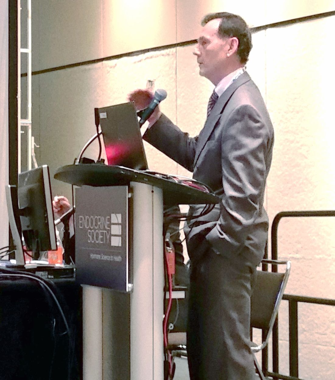
The study enrolled 1,835 patients, of whom 89% completed the efficacy portion of the study. The estrogen-progesterone combination significantly reduced hot flashes, compared with placebo (P less than .05 for all doses at 12 weeks), with the higher two of four different combination doses resulting in significant differences by study week 4. Menopause-related quality of life was also significantly improved by study week 12 for all doses (P less than .05, compared with placebo).
Up to 39 million prescriptions annually may be written for up to 2.5 million women in the United States, Dr. Lobo said. None of the currently available formulations of 17 beta-estradiol and progesterone are FDA approved. The medication studied – dubbed TX-001HR and produced by TherapeuticsMD – combines the two hormones in an oral capsule.
The REPLENISH trial was designed to evaluate the efficacy and safety of four different dose combinations of estradiol (E2) and progesterone (P4), compared with placebo, to treat moderate to severe vasomotor symptoms in postmenopausal women.
The phase III randomized, double-blind, placebo-controlled trial of the E2/P4 combination in postmenopausal women with an intact uterus had an efficacy portion of the study that lasted 12 weeks; endometrial safety was followed for 1 year in a smaller subset of patients.
The dose-ranging study design randomized women 1:1:1:1:1 to one of four combinations of E2 and P4, or to placebo. The four active treatment groups received either 1.0 mg E2/100 mg P4, 0.5 mg E2/100 mg P4, 0.5 mg E2/50 mg P4, or 0.25 mg E2/50 mg P4. There was no active comparator.
The safety portion of the study could include women whose vasomotor severity did not qualify them for the efficacy substudy; there was no placebo in this arm of the study.
Women participating in the vasomotor menopausal symptom (VMS) portion of the study kept a daily symptom diary and completed the Menopause-Specific Quality of Life (MENQOL) questionnaire as an objective measure of menopause-related symptomatology.
The study’s primary efficacy endpoints were VMS frequency and severity, tracked by measuring the mean change from baseline at study weeks 4 and 12. The secondary endpoint was the mean change in VMS frequency and severity week to week, compared with baseline. Patients were included in the modified intention-to-treat population if they took at least one dose of study drug and had at least 5 days of baseline diary data as well as at least 4 days of diary data in one on-treatment week.
The safety cohort included all women who took at least one capsule of the study drug, and tracked the incidence of endometrial hyperplasia out to 12 months for those who participated in the extended safety portion of the trial. The secondary endpoint was the incidence of other adverse events and serious adverse events.
All four dose combinations “provided statistically and clinically significant reduction in the weekly frequency of moderate to severe VMS from baseline at weeks 4 and 12, compared with placebo,” Dr. Lobo said. The lone exception, he said, was the lowest dose combination, which didn’t produce significant VMS reduction until study week 6.
Looking at the week-by-week improvement measure, the 1.0 mg E2/100 mg P4 and the 0.5 mg E2/100 mg P4 formulations improved VMS severity at weeks 4 and 12, compared with placebo.
Quality of life as measured by the MENQOL was significantly improved by all doses by study week 12, compared with placebo. Participants also reported significant improvement on the vasomotor domain of the MENQOL.
There was no endometrial hyperplasia in any study subject, nor were any malignancies detected in any study participant, Dr. Lobo said. The most frequently reported treatment-emergent adverse events were headaches, nasopharyngitis, breast tenderness, upper respiratory tract infection, nausea, back pain, and abdominal pain. Though seven serious treatment-emergent adverse events were considered treatment-related, “no unexpected safety signals were observed,” Dr. Lobo said.
To be included, postmenopausal women aged 40-65 years needed to have an intact uterus and be generally healthy, with a body mass index of less than 35 kg/m2. They also underwent an endometrial biopsy before participating. Their VMS had to occur at least seven times daily, or 50 times in a week, and be moderate to severe in intensity.
Patients with endometrial hyperplasia or melanoma, as well as women with uterine, endometrial, ovarian, or breast cancer, were excluded from the study, as were women with cardiovascular, hepatic, or renal disorders. Women with diabetes and those with thyroid disorders also were excluded.
Though women could have used sex hormone–containing or –modifying medications, they had to cease those medications for a variable washout period before beginning the study. The mean age of study participants was 55, and their mean BMI was 27. Two-thirds of the women were white.
“TX-001HR, if approved, would be a new oral hormone therapy option for postmenopausal women with moderate to severe vasomotor symptoms with an intact uterus,” Dr. Lobo said.
The drug, he said, could present an option in bioidentical hormones – one that has been evaluated for safety and efficacy – for women who are currently using “less regulated and unapproved compounded bioidentical hormone therapy.”
Dr. Lobo reported receiving research support from TherapeuticsMD, which funded the study.
[email protected]
On Twitter @karioakes
ORLANDO – An oral combination of naturally-occurring estrogen and progesterone was found safe and effective for treatment of hot flashes in postmenopausal women with an intact uterus.
The phase III trial results represent another step toward approval of a formulation of bioidentical hormone therapy (HT) by the Food and Drug Administration.
“No similar combined HT has been approved in the U.S.; however, compounded bioidentical HT is estimated to have become the most prevalent HT by U.S. prescription volume,” Rogerio Lobo, MD, professor of obstetrics and gynecology at Columbia University, New York, wrote in an abstract accompanying the study. He presented his findings at the annual meeting of the Endocrine Society.
The study enrolled 1,835 patients, of whom 89% completed the efficacy portion of the study. The estrogen-progesterone combination significantly reduced hot flashes, compared with placebo (P less than .05 for all doses at 12 weeks), with the higher two of four different combination doses resulting in significant differences by study week 4. Menopause-related quality of life was also significantly improved by study week 12 for all doses (P less than .05, compared with placebo).
Up to 39 million prescriptions annually may be written for up to 2.5 million women in the United States, Dr. Lobo said. None of the currently available formulations of 17 beta-estradiol and progesterone are FDA approved. The medication studied – dubbed TX-001HR and produced by TherapeuticsMD – combines the two hormones in an oral capsule.
The REPLENISH trial was designed to evaluate the efficacy and safety of four different dose combinations of estradiol (E2) and progesterone (P4), compared with placebo, to treat moderate to severe vasomotor symptoms in postmenopausal women.
The phase III randomized, double-blind, placebo-controlled trial of the E2/P4 combination in postmenopausal women with an intact uterus had an efficacy portion of the study that lasted 12 weeks; endometrial safety was followed for 1 year in a smaller subset of patients.
The dose-ranging study design randomized women 1:1:1:1:1 to one of four combinations of E2 and P4, or to placebo. The four active treatment groups received either 1.0 mg E2/100 mg P4, 0.5 mg E2/100 mg P4, 0.5 mg E2/50 mg P4, or 0.25 mg E2/50 mg P4. There was no active comparator.
The safety portion of the study could include women whose vasomotor severity did not qualify them for the efficacy substudy; there was no placebo in this arm of the study.
Women participating in the vasomotor menopausal symptom (VMS) portion of the study kept a daily symptom diary and completed the Menopause-Specific Quality of Life (MENQOL) questionnaire as an objective measure of menopause-related symptomatology.
The study’s primary efficacy endpoints were VMS frequency and severity, tracked by measuring the mean change from baseline at study weeks 4 and 12. The secondary endpoint was the mean change in VMS frequency and severity week to week, compared with baseline. Patients were included in the modified intention-to-treat population if they took at least one dose of study drug and had at least 5 days of baseline diary data as well as at least 4 days of diary data in one on-treatment week.
The safety cohort included all women who took at least one capsule of the study drug, and tracked the incidence of endometrial hyperplasia out to 12 months for those who participated in the extended safety portion of the trial. The secondary endpoint was the incidence of other adverse events and serious adverse events.
All four dose combinations “provided statistically and clinically significant reduction in the weekly frequency of moderate to severe VMS from baseline at weeks 4 and 12, compared with placebo,” Dr. Lobo said. The lone exception, he said, was the lowest dose combination, which didn’t produce significant VMS reduction until study week 6.
Looking at the week-by-week improvement measure, the 1.0 mg E2/100 mg P4 and the 0.5 mg E2/100 mg P4 formulations improved VMS severity at weeks 4 and 12, compared with placebo.
Quality of life as measured by the MENQOL was significantly improved by all doses by study week 12, compared with placebo. Participants also reported significant improvement on the vasomotor domain of the MENQOL.
There was no endometrial hyperplasia in any study subject, nor were any malignancies detected in any study participant, Dr. Lobo said. The most frequently reported treatment-emergent adverse events were headaches, nasopharyngitis, breast tenderness, upper respiratory tract infection, nausea, back pain, and abdominal pain. Though seven serious treatment-emergent adverse events were considered treatment-related, “no unexpected safety signals were observed,” Dr. Lobo said.
To be included, postmenopausal women aged 40-65 years needed to have an intact uterus and be generally healthy, with a body mass index of less than 35 kg/m2. They also underwent an endometrial biopsy before participating. Their VMS had to occur at least seven times daily, or 50 times in a week, and be moderate to severe in intensity.
Patients with endometrial hyperplasia or melanoma, as well as women with uterine, endometrial, ovarian, or breast cancer, were excluded from the study, as were women with cardiovascular, hepatic, or renal disorders. Women with diabetes and those with thyroid disorders also were excluded.
Though women could have used sex hormone–containing or –modifying medications, they had to cease those medications for a variable washout period before beginning the study. The mean age of study participants was 55, and their mean BMI was 27. Two-thirds of the women were white.
“TX-001HR, if approved, would be a new oral hormone therapy option for postmenopausal women with moderate to severe vasomotor symptoms with an intact uterus,” Dr. Lobo said.
The drug, he said, could present an option in bioidentical hormones – one that has been evaluated for safety and efficacy – for women who are currently using “less regulated and unapproved compounded bioidentical hormone therapy.”
Dr. Lobo reported receiving research support from TherapeuticsMD, which funded the study.
[email protected]
On Twitter @karioakes
Key clinical point:
Major finding: Four different dose combinations of 17 beta estradiol and progesterone improved hot flashes and menopause-related quality of life, compared with placebo (P less than .05 for all).
Data source: Multicenter randomized, double-blind, placebo-controlled study of 1,835 postmenopausal women with an intact uterus.
Disclosures: Dr. Lobo reported receiving research funding from TherapeuticsMD, which sponsored the study.