User login
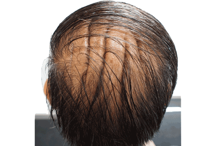
A 54-year-old man presented with a 2-year history of unusual skin folds on the scalp with deep furrows in an anteroposterior direction, located in parieto-occipital regions (Figure 1). A clinical diagnosis of cutis verticis gyrata was made.
CUTIS VERTICIS GYRATA: THE DIFFERENTIAL DIAGNOSIS
Cutis verticis gyrata (“bulldog scalp”) is a rare condition, with a prevalence of 0.026 to 0.1 per 100,000,1 primary and secondary forms, and a male preponderance.2 It is characterized by excessive soft-tissue proliferation with the formation of ridges on the scalp similar in appearance to cerebral cortex gyri.
Primary essential cutis verticis gyrata is extremely rare with no associated abnormalities. Primary nonessential cutis verticis gyrata is associated with neurologic manifestations (microcephaly, seizure, cerebral palsy, mental retardation) and ophthalmologic changes (cataract, strabismus, retinitis pigmentosa, blindness).
Cutis verticis gyrata can also be secondary to conditions such as pachydermoperiostosis, Rosenthal-Kloepfer syndrome, tuberous sclerosis, and insulin resistance syndrome.3 It may occur in fragile X syndrome, Noonan syndrome, Turner syndrome, Beare-Stevenson syndrome, and Ehlers-Danlos syndrome.
When cutis verticis gyrata presents at age 50 or later, acromegaly, amyloidosis, myxedema, paraneoplastic syndromes, and drug-related lipodystrophy from antiretroviral drugs or tyrosine kinase inhibitors should be excluded. Other conditions included in the differential diagnosis are inflammatory diseases of the scalp (psoriasis, pemphigus) and nevoid abnormalities (nevus sebaceous, nevus of Ota, cerebriform nevus).4 The male preponderance suggests a genetic determination and an endocrine cause, but the pathophysiology remains unknown.
MANAGEMENT IN OUR PATIENT
Further evaluation in our patient showed bossing of the frontal bone, coarse facial features, and acral enlargement suggestive of acromegaly. The diagnosis was confirmed by elevated levels of growth hormone and insulin-like growth factor 1.
Magnetic resonance imaging of the pituitary gland revealed a pituitary adenoma 11 × 6 × 8 mm. After treatment of the adenoma with stereotactic radiosurgery, the scalp soft-tissue thickness decreased but persisted.
Overgrowth of the scalp manifesting as cutis verticis gyrata in acromegaly is not uncommon.2,4 The severity or duration of acromegaly is not correlated with the presence and severity of cutis verticis gyrata.4
Besides treatment of acromegaly, good scalp hygiene is necessary to avoid the accumulation of secretions in the furrows. Surgery for scalp reduction is only required for cosmetic reasons.5
- Akesson HO. Cutis verticis gyrata and mental deficiency in Sweden. I. Epidemiologic and clinical aspects. Acta Med Scand 1964; 175:115–127.
- Polan S, Butterworth T. Cutis verticis gyrata: a review with report of seven new cases. Am J Ment Defic 1953; 57:613–631.
- Larsen F, Birchall N. Cutis verticis gyrata: three cases with different aetiologies that demonstrate the classification system. Australas J Dermatol 2007; 48:91–94.
- Kolawole TM, AI Orainy IA, Patel PJ, Fathuddin S. Cutis verticis gyrata: its computed tomographic demonstration in acromegaly. Eur J Radiol 1998; 27:145–148.
- Garden JM, Robinson JK. Essential primary cutis verticis gyrata. Treatment with the scalp reduction procedure. Arch Dermatol 1984; 120:1480–1483.
A 54-year-old man presented with a 2-year history of unusual skin folds on the scalp with deep furrows in an anteroposterior direction, located in parieto-occipital regions (Figure 1). A clinical diagnosis of cutis verticis gyrata was made.
CUTIS VERTICIS GYRATA: THE DIFFERENTIAL DIAGNOSIS
Cutis verticis gyrata (“bulldog scalp”) is a rare condition, with a prevalence of 0.026 to 0.1 per 100,000,1 primary and secondary forms, and a male preponderance.2 It is characterized by excessive soft-tissue proliferation with the formation of ridges on the scalp similar in appearance to cerebral cortex gyri.
Primary essential cutis verticis gyrata is extremely rare with no associated abnormalities. Primary nonessential cutis verticis gyrata is associated with neurologic manifestations (microcephaly, seizure, cerebral palsy, mental retardation) and ophthalmologic changes (cataract, strabismus, retinitis pigmentosa, blindness).
Cutis verticis gyrata can also be secondary to conditions such as pachydermoperiostosis, Rosenthal-Kloepfer syndrome, tuberous sclerosis, and insulin resistance syndrome.3 It may occur in fragile X syndrome, Noonan syndrome, Turner syndrome, Beare-Stevenson syndrome, and Ehlers-Danlos syndrome.
When cutis verticis gyrata presents at age 50 or later, acromegaly, amyloidosis, myxedema, paraneoplastic syndromes, and drug-related lipodystrophy from antiretroviral drugs or tyrosine kinase inhibitors should be excluded. Other conditions included in the differential diagnosis are inflammatory diseases of the scalp (psoriasis, pemphigus) and nevoid abnormalities (nevus sebaceous, nevus of Ota, cerebriform nevus).4 The male preponderance suggests a genetic determination and an endocrine cause, but the pathophysiology remains unknown.
MANAGEMENT IN OUR PATIENT
Further evaluation in our patient showed bossing of the frontal bone, coarse facial features, and acral enlargement suggestive of acromegaly. The diagnosis was confirmed by elevated levels of growth hormone and insulin-like growth factor 1.
Magnetic resonance imaging of the pituitary gland revealed a pituitary adenoma 11 × 6 × 8 mm. After treatment of the adenoma with stereotactic radiosurgery, the scalp soft-tissue thickness decreased but persisted.
Overgrowth of the scalp manifesting as cutis verticis gyrata in acromegaly is not uncommon.2,4 The severity or duration of acromegaly is not correlated with the presence and severity of cutis verticis gyrata.4
Besides treatment of acromegaly, good scalp hygiene is necessary to avoid the accumulation of secretions in the furrows. Surgery for scalp reduction is only required for cosmetic reasons.5
- Akesson HO. Cutis verticis gyrata and mental deficiency in Sweden. I. Epidemiologic and clinical aspects. Acta Med Scand 1964; 175:115–127.
- Polan S, Butterworth T. Cutis verticis gyrata: a review with report of seven new cases. Am J Ment Defic 1953; 57:613–631.
- Larsen F, Birchall N. Cutis verticis gyrata: three cases with different aetiologies that demonstrate the classification system. Australas J Dermatol 2007; 48:91–94.
- Kolawole TM, AI Orainy IA, Patel PJ, Fathuddin S. Cutis verticis gyrata: its computed tomographic demonstration in acromegaly. Eur J Radiol 1998; 27:145–148.
- Garden JM, Robinson JK. Essential primary cutis verticis gyrata. Treatment with the scalp reduction procedure. Arch Dermatol 1984; 120:1480–1483.

