User login
SAN DIEGO – Age-accelerated changes in certain aspects of cognition, including psychomotor speed and verbal memory, appear to occur in aging persons with childhood-onset epilepsy, compared with healthy controls, according to preliminary results from an ongoing study.
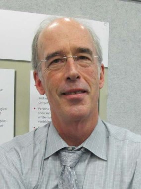
"One thing that remains unclear is how people with childhood-onset epilepsies age over the decades in terms of brain structure and function," Bruce Hermann, Ph.D., said in an interview during a poster session at the annual meeting of the American Epilepsy Society.
"There has been much interest in the issues of cognitive and brain aging in the general population, and, of course, in disorders such as dementia and Alzheimer’s disease, but unknown is how individuals with childhood-onset epilepsy may age over the decades. There are very few population-based cohorts available to address that question."
Dr. Hermann, who directs of the Charles Matthews Neuropsychology Lab in the department of neurology at the University of Wisconsin, Madison, is examining this question in collaboration with Dr. Matti Sillanpää, professor emeritus of child neurology at the University of Turku, Finland.
Together they are studying Dr. Sillanpää’s well known population-based cohort of children with epilepsy and controls. Followed since their youth over the decades, the research participants now range in age from 45 to 62 years. These subjects are returning for a comprehensive battery of cognitive tests, neuroimaging, EEG, and clinical interview to evaluate their aging processes. Cognitive tests administered including the Alzheimer’s Disease Assessment Scale–Cognitive Subscale (ADAS-Cog), the Wechsler Adult Intelligence Scale–III (WAIS-III), the Boston Naming Test, the Controlled Oral Word Association Test, the Clock Drawing Test, the Wechsler Memory Scale–Revised (WMS-R), the Rey Auditory Verbal Learning Test, the Trail Making Test, and the Beck Depression Inventory. The follow-up visits also include structural MRI and PET amyloid and glucose imaging.
The mean age of patients examined to date is 57 years, and they are compared with a group of similarly aged matched controls. Dr. Hermann reported that in the first group of participants to be examined, 26 epilepsy patients performed significantly worse than did 31 controls on measures of psychomotor speed based on the WAIS-III (P less than .05) and in delayed verbal memory based on the WMS-R (P = .031).
"Secondary analyses revealed that patients with persisting abnormal EEG and either active epilepsy/seizures into adulthood had more adversely affected cognition compared to those whose epilepsy completely remitted," the researchers wrote in their poster.
Results from the PET imaging studies are pending, Dr. Hermann said, but preliminary results from the MRI studies demonstrated that there were no differences in cortical thickness between the epilepsy and control groups after correction for multiple comparisons and adjustment for age and gender. However, compared with the control group, the epilepsy group demonstrated a significant increase in the white matter hypointensity index (P less than .05) and a trend toward larger left and right lateral ventricles (P = .10). "What’s surprising is that their MRI results look as good as they do, compared with controls," Dr. Hermann said. "We thought we’d see more pathology and age-accelerated changes."
Dr. Sillanpää’s entire cohort is in the process of returning for study and the results should shed new light on the issues of cognitive and brain aging in childhood epilepsy.
The study was funded by the Finnish Government Research Fund and Citizens United for Research in Epilepsy (CURE). Dr. Hermann said that he had no relevant financial conflicts to disclose.
AT THE ANNUAL MEETING OF THE AMERICAN EPILEPSY SOCIETY
Major Finding: A cohort of 26 patients with childhood-onset epilepsy who were now aged 45-62 years performed significantly worse than did 31 controls on measures of psychomotor speed based on the Wechsler Adult Intelligence Scale-III (P less than .05) and in delayed verbal memory based on the Wechsler Memory Scale-Revised (P = .031).
Data Source: A study of 26 epilepsy patients with a mean age of 57 years who have been followed since childhood and who were compared with 31 similarly aged matched controls.
Disclosures: The study was funded by the Finnish Research Fund and Citizens United for Research in Epilepsy (CURE). Dr. Hermann said that he had no relevant financial conflicts to disclose.

