User login
HCV Hub
AbbVie
acid
addicted
addiction
adolescent
adult sites
Advocacy
advocacy
agitated states
AJO, postsurgical analgesic, knee, replacement, surgery
alcohol
amphetamine
androgen
antibody
apple cider vinegar
assistance
Assistance
association
at home
attorney
audit
ayurvedic
baby
ban
baricitinib
bed bugs
best
bible
bisexual
black
bleach
blog
bulimia nervosa
buy
cannabis
certificate
certification
certified
cervical cancer, concurrent chemoradiotherapy, intravoxel incoherent motion magnetic resonance imaging, MRI, IVIM, diffusion-weighted MRI, DWI
charlie sheen
cheap
cheapest
child
childhood
childlike
children
chronic fatigue syndrome
Cladribine Tablets
cocaine
cock
combination therapies, synergistic antitumor efficacy, pertuzumab, trastuzumab, ipilimumab, nivolumab, palbociclib, letrozole, lapatinib, docetaxel, trametinib, dabrafenib, carflzomib, lenalidomide
contagious
Cortical Lesions
cream
creams
crime
criminal
cure
dangerous
dangers
dasabuvir
Dasabuvir
dead
deadly
death
dementia
dependence
dependent
depression
dermatillomania
die
diet
direct-acting antivirals
Disability
Discount
discount
dog
drink
drug abuse
drug-induced
dying
eastern medicine
eat
ect
eczema
electroconvulsive therapy
electromagnetic therapy
electrotherapy
epa
epilepsy
erectile dysfunction
explosive disorder
fake
Fake-ovir
fatal
fatalities
fatality
fibromyalgia
financial
Financial
fish oil
food
foods
foundation
free
Gabriel Pardo
gaston
general hospital
genetic
geriatric
Giancarlo Comi
gilead
Gilead
glaucoma
Glenn S. Williams
Glenn Williams
Gloria Dalla Costa
gonorrhea
Greedy
greedy
guns
hallucinations
harvoni
Harvoni
herbal
herbs
heroin
herpes
Hidradenitis Suppurativa,
holistic
home
home remedies
home remedy
homeopathic
homeopathy
hydrocortisone
ice
image
images
job
kid
kids
kill
killer
laser
lawsuit
lawyer
ledipasvir
Ledipasvir
lesbian
lesions
lights
liver
lupus
marijuana
melancholic
memory loss
menopausal
mental retardation
military
milk
moisturizers
monoamine oxidase inhibitor drugs
MRI
MS
murder
national
natural
natural cure
natural cures
natural medications
natural medicine
natural medicines
natural remedies
natural remedy
natural treatment
natural treatments
naturally
Needy
needy
Neurology Reviews
neuropathic
nightclub massacre
nightclub shooting
nude
nudity
nutraceuticals
OASIS
oasis
off label
ombitasvir
Ombitasvir
ombitasvir/paritaprevir/ritonavir with dasabuvir
orlando shooting
overactive thyroid gland
overdose
overdosed
Paolo Preziosa
paritaprevir
Paritaprevir
pediatric
pedophile
photo
photos
picture
post partum
postnatal
pregnancy
pregnant
prenatal
prepartum
prison
program
Program
Protest
protest
psychedelics
pulse nightclub
puppy
purchase
purchasing
rape
recall
recreational drug
Rehabilitation
Retinal Measurements
retrograde ejaculation
risperdal
ritonavir
Ritonavir
ritonavir with dasabuvir
robin williams
sales
sasquatch
schizophrenia
seizure
seizures
sex
sexual
sexy
shock treatment
silver
sleep disorders
smoking
sociopath
sofosbuvir
Sofosbuvir
sovaldi
ssri
store
sue
suicidal
suicide
supplements
support
Support
Support Path
teen
teenage
teenagers
Telerehabilitation
testosterone
Th17
Th17:FoxP3+Treg cell ratio
Th22
toxic
toxin
tragedy
treatment resistant
V Pak
vagina
velpatasvir
Viekira Pa
Viekira Pak
viekira pak
violence
virgin
vitamin
VPak
weight loss
withdrawal
wrinkles
xxx
young adult
young adults
zoloft
financial
sofosbuvir
ritonavir with dasabuvir
discount
support path
program
ritonavir
greedy
ledipasvir
assistance
viekira pak
vpak
advocacy
needy
protest
abbvie
paritaprevir
ombitasvir
direct-acting antivirals
dasabuvir
gilead
fake-ovir
support
v pak
oasis
harvoni
VIDEO: Endoscopic pyloromyotomy works for gastroparesis when meds don’t
SAN DIEGO – Gastric peroral endoscopic myotomy, a novel procedure for gastroparesis, restored gastric emptying in 30 refractory patients at Johns Hopkins University, Baltimore, and elsewhere in the largest series to date for the technique.
Drug therapy had failed, and Botox injections and transpyloric stenting weren’t helping much. On gastric emptying scans (GES), patients had around 40% of solid meals in their stomachs at 4 hours. Their gastroparesis was related mostly to diabetes and postoperative complications, but about a quarter of the cases were idiopathic.
Twenty-six patients (87%) responded to gastric peroral endoscopic myotomy (G-POEM) during a median follow-up of 5.5 months. Nausea, vomiting, and abdominal pain resolved or improved in most. On repeat GES in 17 patients, emptying time normalized in about half and improved in a third. Overall, patients had 17% of solid meals in their stomachs at 4 hours. G-POEM took an average of 72 minutes, and patients were in the hospital for about 3 days. One patient in the series developed pneumoperitoneum, and another had a prepyloric ulcer.
“The problem with transpyloric stents is that they migrate,” said investigator Dr. Mouen A. Khashab, director of therapeutic endoscopy at Johns Hopkins University. “G-POEM offers a permanent solution with few side effects. You have to be good at doing POEM in the esophagus first, as a prerequisite.”
In an interview at the annual Digestive Disease Week, Dr. Khashab explained the procedure in detail, as well as how he incorporates it into his practice and the patient population most likely to benefit.
AT DDW® 2016
HIV/HCV coinfection further raises inflammatory markers
Individuals with HIV and hepatitis C virus coinfection show significantly higher levels of particular inflammatory cytokines linked to liver damage, according to a study published online in HIV Medicine.
Researchers examined a range of markers of systemic inflammation in 79 HIV-infected patients – 42 of whom were coinfected with hepatitis C – and all of whom had been on antiretroviral therapy and had plasma viral levels suppressed below 50 HIV-1 RNA copies per ml, and compared them to 20 healthy controls.
Their analysis initially revealed that the individuals with HIV/HCV coinfection had higher plasma levels of interleukin-6, interferon gamma-induced protein (IP)-10, monocyte/macrophage markers neopterin and sCD163, and soluble tumour necrosis factor receptor-II than individuals with HIV infection alone.
However after adjusting for duration of HIV infection and gender, researchers only found a significant difference between HIV/HCV coinfected individuals and those with HIV alone in plasma levels of IP-10, sCD163 and neopterin (HIV Medicine. 2016 May 17. doi:10.1111/hiv.12357).
The study also undertook to correlate these markers with hepatic damage, and found a highly significant and consistent relationship between aspartate aminotransferase, alanine aminotransferase and AST-to platelet-ratio index, and plasma IP-10, sCD163 and neopterin.
“Although a contribution of ART-induced hepatotoxicity in the setting of HCV/HIV coinfection cannot be excluded, a simpler and more plausible explanation is that the observed effects are related to HCV/HIV-mediated liver damage,” wrote Dr. Konstantin V. Shmagel, from the Institute of Ecology and Genetics of Microorganisms at the Russian Academy of Sciences in Perm, Russia.
“The relationship among these indices remains incompletely understood but it is possible that processes taking place in the liver may play a role in alteration of CD4 T-cell recovery during ART in the setting of HCV/HIV coinfection.”
The analysis also showed the inflammatory markers IL-6, neopterin and sCD14 were also elevated – although not to the same degree – in individuals with HIV monoinfection compared to the healthy controls.
“The drivers of persistent inflammation in treated HIV infection and HIV/HCV coinfection are not entirely clear but, in these settings, damage to the gut epithelial barrier has been implicated in promoting translocation of microbial products from the gut lumen into the systemic circulation,” the authors wrote.
This study was supported by the National Institute of Allergy and Infectious Diseases, the Center for AIDS Research at Case Western Reserve University, and the Russian Science Foundation. No conflicts of interest were declared.
FROM HIV MEDICINE
Key clinical point: Coinfection with HIV and hepatitis C virus is associated with elevated levels of particular cytokines, that also correspond to indicators of hepatic damage.
Major finding: Individuals with HIV/HCV coinfection have higher plasma levels of IP-10, sCD163 and neopterin than individuals with HIV alone.
Data source: A study in 79 HIV-infected patients – 42 of whom were coinfected with hepatitis C – and 20 healthy controls.
Disclosures: This study was supported by the National Institute of Allergy and Infectious Diseases, the Center for AIDS Research at Case Western Reserve University, and the Russian Science Foundation. No conflicts of interest were declared.
VIDEO: Accelerated infliximab dosing halved colectomy rate in severe ulcerative colitis
SAN DIEGO – Inpatient infliximab rescue for severe, steroid-refractory ulcerative colitis is more likely to work if patients receive a second infusion 3 days after the first, according to a review of 55 University of Michigan, Ann Arbor, patients.
The traditional approach is one inpatient 5-mg/kg infusion, followed by either colectomy or subsequent outpatient infusions, depending on response. In 2013, physicians at the university began offering a second infusion at 72 hours to patients whose C-reactive protein (CRP) levels did not drop below 0.7 mg/dL after their first infusion, and they also began opting more often for 10-mg/kg dosing.
The review found that 90-day colectomy-free survival was 50% in the 16 accelerated-dosing patients, up from 10.2% in the 36 patients treated with the traditional approach (P less than .001). The finding has led to a new, more aggressive infliximab protocol for inpatient ulcerative colitis.
Among patients who did undergo colectomies, postoperative complications were similar between the two groups. But for reasons that are not clear, 30-day postoperative readmission rates were higher in accelerated patients (58% vs. 25%).
In an interview at the annual Digestive Disease Week, lead investigator Dr. Shail Govani of the University of Michigan explained the thinking behind the new approach, how CRP/albumin ratios come into play, and how to counsel patients in light of the findings.
The video associated with this article is no longer available on this site. Please view all of our videos on the MDedge YouTube channel
AT DDW 2016
VIDEO: Daily fecal transplants put refractory ulcerative colitis into remission
SAN DIEGO – When fecal transplants don’t work for ulcerative colitis, it’s probably because they aren’t used often enough, according to investigators from the University of New South Wales, Sydney.
The researchers made sure that wasn’t the case in their own double-blind trial, the largest to date of fecal microbiota transplantation (FMT) for ulcerative colitis. Forty-one patients with active, mild to moderate disease (Mayo score 4-10) that was resistant to standard medications were randomized to 150-mL, self-administered fecal enemas 5 days a week for 8 weeks, and 40 others to placebo enemas. Patients were tapered off steroids at study entrance, and each FMT patient received stool from 3-7 unrelated donors.
Steroid-free clinical remission and endoscopic remission or response were achieved in 11 FMT patients (27%), compared with 3 (8%) placebo patients (P = .02). A total of 18 treated patients (44%) and 8 placebo patients (20%) had steroid-free clinical remissions, while 22 treated patients (54%) and 9 patients in the placebo group (23%) had some type of positive clinical response (P less than .01).
Patients were in their mid-30s, on average, with disease durations of about 6 years. More than half were men. About one-quarter were on oral steroids, and more than half were on oral 5-aminosalicylic acid medications, which were allowed during the study. Almost half were on methotrexate or other oral immunomodulators, which were also allowed. Patients were excluded if they had been on a biologic in the previous 12 weeks.
Afterward, 37 patients in the placebo arm opted for open-label FMT. Results were similar, with steroid-free clinical remission and endoscopic remission or response in 10 patients (27%), clinical remission in 17 (46%), and endoscopic remission in 9 (24%).
The anatomical extent of disease did not affect outcome, but patients with more severe endoscopic disease and those on steroids at study entrance didn’t do as well. Three patients flared during the trial, one in the placebo arm and two in the FMT arm, one of whom required colectomy.
The investigators were surprised by the magnitude of the benefit, given the mixed results in previous investigations with less frequent dosing. But they were not surprised that FMT worked.
“In ulcerative colitis, the microbiota appear to be the antigenic driver, so it makes sense that correcting the disturbance” helps, said lead investigator Dr. Sudarshan Paramsothy, a gastroenterologist at the University of New South Wales.
Dr. Paramsothy and his colleagues have a hunch they can do even better. They are looking into the microbiologic factors of donors and patients that influence response, with the ultimate goal of matching the best donor to the best patient. They’re examining maintenance therapy, too; “it’s one thing to induce remission, it’s another thing to maintain remission,” Dr. Paramsothy said.
In an interview at the annual Digestive Disease Week, he explained the technique, the thinking behind it, future directions, and how to counsel patients in light of the findings.
The video associated with this article is no longer available on this site. Please view all of our videos on the MDedge YouTube channel
AT DDW 2016
Disappointing results for GI bleed prevention in high-risk aspirin users
SAN DIEGO – Neither a proton pump inhibitor nor an H2 antagonist is an optimal choice for users of low-dose aspirin with previously confirmed ulcer bleeding, according to data from a 12-month randomized trial.
“This is one of the largest clinical trials focusing on aspirin users with a history of ulcer bleeding. Previous trials of aspirin users were mostly endoscopic trials. The important message here is that – while PPI may be useful as gastroprotection for patients with a history of ulcer bleed – it seems that neither treatment is sufficiently protective,” Dr. Francis Chan said at the annual Digestive Disease Week.
Findings from earlier research by the same investigators showed that cotreatment with aspirin and a PPI seemed to be effective as secondary prevention for aspirin-induced ulcer bleeding. He noted that PPIs have a warning for high-risk aspirin users, so alternatives are being sought.
In the present randomized trial, even with PPI prophylaxis, 7.9% of these high-risk aspirin users developed recurrent bleed or endoscopic ulcers versus 12.4% of the group treated with an H2 antagonist, a nonsignificant difference.
The prospective, randomized double-blind trial randomized 270 patients in a 1:1 ratio to 1 year of treatment with either 20 mg rabeprazole (a PPI) once daily or 40 mg of the H2 antagonist famotidine once daily. All patients’ ulcers had healed, and all tested negative for Helicobacter pylori prior to randomization. Study participants were taking 80 mg of aspirin daily.
Patients were followed for 12 months. Endoscopy was repeated if there was suspicion of recurrent bleed or they reached 12 months of treatment.
The primary endpoint was a composite of upper gastrointestinal bleed or recurrent ulcer. Secondary endpoints included a composite of recurrent bleed, ulcers visible on endoscopy, and early withdrawal due to severe dyspepsia; lower GI bleeding; and cardiothrombotic events.
Study participants had a mean age of 73 years. At baseline, all patients were negative for H. pylori and hepatitis B virus infection. Both treatment arms were comparable for indication for aspirin. A history of coronary disease was noted in 37% of the PPI group and 40.2% of the H2 antagonist group. A history of cerebrovascular disease was present in 35.5% and 37.1%, respectively.
The source of previous bleeding was comparable in the two groups.
In an intent-to-treat analysis that included all patients who took at least one dose of study medication as well as those who underwent endoscopy at 12 months, 24 cases of suspected bleeding were found: 14 in the PPI group (1 confirmed) and 10 in the H2 antagonist group (4 confirmed). The rate of recurrent bleeding was 5.1% for the PPI and 8.1% for the H2 antagonist.
Lower GI bleeding was reported in 11 patients (8.9%) on the PPI and 6 patients (5%) on the H2 antagonist.
Cardiothrombotic events were reported in five patients: two on the PPI (1.6%) and three on the H2 antagonist (2.5%) .
“We didn’t see any trends for cardiovascular bleeding in either group,” Dr. Chan noted.
An audience member asked what the best way is to treat these patients, given that neither drug provided adequate protection against upper GI bleeding.
“The answer is, I don’t know. We need more study of high-risk patients, and we should study a combination of PPI plus misoprostol. In the absence of larger studies, I currently treat my patients with PPI plus low-dose misoprostol,” he said.
AT DDW® 2016
Key clinical point: Neither a PPI nor an H2 antagonist provided sufficient gastroprotection in high-risk aspirin users.
Major finding: The rate of recurrent bleeding or endoscopic ulcers was 7.9% with a PPI versus 12.4% with an H2 antagonist.
Data source: A randomized, controlled trial of 270 patients with a previous history of ulcers.
Disclosures: Dr. Chan has received financial support from Pfizer and Eisai.
Visceral hypersensitivity an independent contributor to GI symptoms
SAN DIEGO – Visceral hypersensitivity may be an independent contributor to gastrointestinal symptoms in patients with functional GI disorders, a study showed.
The association was independent of the subjects’ tendency to report non-GI symptoms and independent of psychological distress.
“We found a gradual increase in GI symptom severity related to hypersensitivity tertiles in irritable bowel syndrome and dyspepsia, and small but significant correlations between pain/discomfort thresholds and GI symptom severity across several large patient groups from different countries,” said presenting author Dr. Magnus Simren. “This association was independent of the tendency to report GI symptoms or comorbid anxiety and depression.”
The relationship between visceral hypersensitivity and GI symptoms has been questionable, with some studies suggesting an association between visceral hypersensitivity and symptom severity and pain in irritable bowel syndrome (IBS), dyspepsia, and other GI disorders.
However, some negative studies have failed to confirm this association, explained Dr. Simren. “The thinking is that reporting GI symptoms and severity of symptoms may reflect a general tendency to report symptoms that [are] explained by comorbid psychological stress,” he noted.
The study Dr. Simren presented at the annual Digestive Disease Week sought to determine the association between visceral hypersensitivity and GI symptoms and symptom severity, adjusted for psychological distress (anxiety and depression) as well as a tendency to report symptoms.
The study enrolled five patient cohorts with functional GI disorders at three different centers. Subjects underwent balloon distension (a validated method to determine GI hypersensitivity) and completed questionnaires to assess GI symptom severity, somatization, anxiety, and depression.
The five cohorts were the Belgian functional dyspepsia cohort (n = 242), IBS cohort 1 in the United States (n = 243), U.S. IBS cohort 2 (n = 159), Swedish IBS cohort 1 (n = 353), and Swedish IBS cohort 2 (n = 147). Subjects were divided into sensitivity tertiles based on pain/discomfort thresholds.
Dr. Simren pointed out that the three different countries used questionnaires with subtle differences to measure GI symptoms, anxiety, and depression. Therefore, the total scores of all GI questionnaires were recalculated to z scores for comparisons.
The barostat protocols also differed among the three countries, and different pain thresholds were used, he noted.
For statistical analysis, sensitivity tertiles (low, medium, and high) were constructed based on pain/discomfort thresholds. GI symptom severity was compared between tertiles and adjusted for anxiety and depression as well as for non-GI symptom reporting.
Then GI hypersensitivity (high and low thresholds) was correlated with symptom severity total scores and pain/discomfort thresholds. Significant differences in GI symptom severity were found between high and low threshold hypersensitivity tertiles across all five cohorts. These differences remained after investigators controlled for the presence of anxiety or depression. The same association was seen when investigators controlled for reporting of non-GI symptoms.
Dr. Simren acknowledged that these barostat tests are used in a research setting. “But you can use this information in managing patients. We would need a simpler test for clinical utility,” he said.
“These findings confirm that visceral hypersensitivity is a contributor to symptom generation in functional GI disease,” Dr. Simren said.
AT DDW® 2016
Ocaliva approved for primary biliary cholangitis
Obeticholic acid (Ocaliva) has been granted accelerated approval for use in combination with ursodeoxycholic acid (UDCA) for the treatment of primary biliary cholangitis in adults with an inadequate response to UDCA, and for use as a single therapy in adults unable to tolerate UDCA, the U.S. Food and Drug Administration announced.
Obeticholic acid should not be used in patients with complete biliary obstruction.
Given orally, obeticholic acid binds to the farnesoid X receptor (FXR), a receptor found in cells of the liver and intestine. FXR is a key regulator of bile acid metabolic pathways. Obeticholic acid increases bile flow from the liver and suppresses bile acid production in the liver, thus reducing exposure to toxic levels of bile acids.
Obeticholic acid was shown to reduce levels of alkaline phosphatase, which was used as a surrogate endpoint to predict clinical benefit, including an improvement in transplant-free survival. The FDA’s accelerated approval program allows approval based on a surrogate endpoint that is reasonably likely to predict clinical benefit. The manufacturer will conduct confirmatory clinical trials to examine any improvements in survival, progression to cirrhosis, or other disease-related symptoms.
After 12 months, in a controlled clinical trial with 216 participants, the proportion of participants achieving reductions in levels of alkaline phosphatase was higher among treated participants than in participants given placebo. Pruritus, fatigue, abdominal pain and discomfort, arthralgia, oropharyngeal pain, dizziness, and constipation were the drug’s most common side effects in the study.
Obeticholic acid is manufactured by Intercept Pharmaceuticals.
Monitored anesthesia care for endoscopy on the rise even without financial incentives
SAN DIEGO – Monitored anesthesia care (MAC) for outpatient endoscopy is on the rise in the United States, presumably because of financial incentives for fee-for-service gastroenterology (GI) practices. However, a new study found that use of MAC is increasing in Veteran’s Health Administration (VHA) facilities, an environment free of financial incentives.
The increase in VHA hospitals is much smaller than in the country as a whole, but the study suggests that there are additional drivers for use of MAC that need to be more fully explored.
Over the past two decades, the rate of MAC use for outpatient endoscopy in fee-for-service practices has increased by at least 30%. Over the study period, the rate of MAC use for endoscopy procedures doubled in VHA facilities from 5.7% in 2000 to 11.1% in 2013, with a larger proportion of increase between 2011 and 2013.
“The increase in MAC use in fee-for-service practices is thought to be driven by financial gain. With an anesthesiologist on hand for an endoscopy procedure, the practice can bill double [duplicative billing with separate billing codes], essentially getting double reimbursement. The VHA has little incentive to increase use of MAC for financial gain. We wanted to study use of MAC in the VHA environment to determine if there are other factors involved,” explained Dr. Megan A. Adams of the University of Michigan, Ann Arbor.
In an interview, Dr. Adams explained that American Society for Gastrointestinal Endoscopy guidelines for MAC use are relatively broad and diffuse. They state that MAC should be considered for patients with anticipated intolerance to standard sedatives, certain cardiopulmonary morbidities, and the potential for airway compromise.
“These will need to be more specific in the future,” she said.
The retrospective cohort study she reported on at the annual Digestive Disease Week was based on national VHA data from more than 1,700 sites of care, with about 300,000 endoscopies performed each year.
“A large variation of MAC use was observed across study facilities, particularly in the later years of the study period,” Dr. Adams explained.
The investigators developed a model based on 122 VHA facilities, 2.1 million patient encounters, and the time of event to analyze patient-level and provider-level predictors of MAC using multilevel random effects logistical regression analysis.
Patient-level factors associated with the increased use of MAC included female gender (35% increase), body mass index greater than 35 kg/m2 (20% increase), obstructive sleep apnea (50% increase), opioid use (17% increase), and benzodiazepine use (13%). Charlson comorbidity scores were associated with a significant increase, compared with healthy patients, with a score of 3 having a 30% increased likelihood of MAC use.
Provider-level predictors of MAC use were related to facilities. Outside of the GI endoscopy suite, endoscopy procedures were about four times more likely to be performed with MAC, and surgeons were about 50% more likely to use MAC.
“The variation in MAC use is largely explained by facility factors. Potential facility factors could be academic versus nonacademic setting, differences in how sites triage care, and differences in local policy. Patient-level factors were relatively weak influences. We will need to explore provider-level [facility] factors more fully to understand the specifics,” she said.
“We will need to align incentives to promote more appropriate use of MAC tailored to patient factors,” Dr. Adams said.
“Payment reforms are looming. CMS will probably remove duplicative reimbursement for MAC. The country will follow CMS. This will affect the financial drivers of MAC, but not necessarily the nonfinancial drivers,” she predicted.
Dr. Adams had no financial disclosures.
Reflecting on the economic landscape of sedation for endoscopy, Dr. John Vargo of the Cleveland Clinic noted that the winds of change are brewing. “Fee for service is dead. We will probably get into a provider-led integrated network,” he noted.
“The proportion of GI procedures with anesthesia has doubled, while the payment for anesthesia providers has tripled. There are regional disparities. Essentially, all growth is among commercially insured patients, and most patients receiving anesthesia are low risk,” he said.
“The business model has changed. The anesthesiologist is an employee. GIs bill for those codes and bank the difference,” he told listeners.
Currently, the scale of the cost is $1.5 million per life-year gained for propofol anesthesia and $9-$21 million per colon cancer case prevented, Dr. Vargo told listeners.
“Anesthesia codes are misvalued. They are being reviewed, and I suspect they will go down. I predict we will see decreasing price pressure. The devil is in the details. In 3+ million outpatient colonoscopies, anesthesia complications are about 15% higher,” he stated.
“There is not a positive argument for anesthesia assistance in healthy patients undergoing colonoscopy. Moderate sedation is not dead. Not everyone likes propofol-mediated sedation. Patient satisfaction is equivalent, while propofol gets patients in and out more quickly,”
“Let’s not throw benzodiazepines out of our armamentarium. Conscious sedation is still the only universally accepted combination available to GIs who practice sedation,” he stated.
“As we get more competition, we will see a resurgence of GI-administered propofol as well as sedation,” he predicted.
SAN DIEGO – Monitored anesthesia care (MAC) for outpatient endoscopy is on the rise in the United States, presumably because of financial incentives for fee-for-service gastroenterology (GI) practices. However, a new study found that use of MAC is increasing in Veteran’s Health Administration (VHA) facilities, an environment free of financial incentives.
The increase in VHA hospitals is much smaller than in the country as a whole, but the study suggests that there are additional drivers for use of MAC that need to be more fully explored.
Over the past two decades, the rate of MAC use for outpatient endoscopy in fee-for-service practices has increased by at least 30%. Over the study period, the rate of MAC use for endoscopy procedures doubled in VHA facilities from 5.7% in 2000 to 11.1% in 2013, with a larger proportion of increase between 2011 and 2013.
“The increase in MAC use in fee-for-service practices is thought to be driven by financial gain. With an anesthesiologist on hand for an endoscopy procedure, the practice can bill double [duplicative billing with separate billing codes], essentially getting double reimbursement. The VHA has little incentive to increase use of MAC for financial gain. We wanted to study use of MAC in the VHA environment to determine if there are other factors involved,” explained Dr. Megan A. Adams of the University of Michigan, Ann Arbor.
In an interview, Dr. Adams explained that American Society for Gastrointestinal Endoscopy guidelines for MAC use are relatively broad and diffuse. They state that MAC should be considered for patients with anticipated intolerance to standard sedatives, certain cardiopulmonary morbidities, and the potential for airway compromise.
“These will need to be more specific in the future,” she said.
The retrospective cohort study she reported on at the annual Digestive Disease Week was based on national VHA data from more than 1,700 sites of care, with about 300,000 endoscopies performed each year.
“A large variation of MAC use was observed across study facilities, particularly in the later years of the study period,” Dr. Adams explained.
The investigators developed a model based on 122 VHA facilities, 2.1 million patient encounters, and the time of event to analyze patient-level and provider-level predictors of MAC using multilevel random effects logistical regression analysis.
Patient-level factors associated with the increased use of MAC included female gender (35% increase), body mass index greater than 35 kg/m2 (20% increase), obstructive sleep apnea (50% increase), opioid use (17% increase), and benzodiazepine use (13%). Charlson comorbidity scores were associated with a significant increase, compared with healthy patients, with a score of 3 having a 30% increased likelihood of MAC use.
Provider-level predictors of MAC use were related to facilities. Outside of the GI endoscopy suite, endoscopy procedures were about four times more likely to be performed with MAC, and surgeons were about 50% more likely to use MAC.
“The variation in MAC use is largely explained by facility factors. Potential facility factors could be academic versus nonacademic setting, differences in how sites triage care, and differences in local policy. Patient-level factors were relatively weak influences. We will need to explore provider-level [facility] factors more fully to understand the specifics,” she said.
“We will need to align incentives to promote more appropriate use of MAC tailored to patient factors,” Dr. Adams said.
“Payment reforms are looming. CMS will probably remove duplicative reimbursement for MAC. The country will follow CMS. This will affect the financial drivers of MAC, but not necessarily the nonfinancial drivers,” she predicted.
Dr. Adams had no financial disclosures.
AT DDW® 2016
Key clinical point: Monitored anesthesia care during endoscopy appears to be driven by factors other than financial gain.
Major finding: In the VHA, with little financial incentive, use of MAC doubled over a 13-year period.
Data source: Large retrospective cohort study using national VHA administrative data.
Disclosures: Dr. Adams had no financial disclosures.
Duodenal resurfacing achieves metabolic benefits in type 2 diabetes
SAN DIEGO – A first-in-human study suggests that a novel technique holds promise for the treatment of diabetes, reducing the complications, associated morbidity, and economic burden of this disease. Duodenal mucosal resurfacing (DMR) achieved a significant reduction in hemoglobin A1c (HbA1c) levels as well as a robust reduction in the liver enzymes aspartate aminotransferase and alanine aminotransferase in patients with poorly controlled type 2 diabetes.
DMR entails thermal ablation of the duodenal mucosa via minimally invasive endoscopy. According to the pioneer behind this technique, Dr. Harith Rajagopalan, who is the founder and CEO of Fractyl Laboratories, in Waltham, Mass., DMR can be compared to laser resurfacing of the skin to remove actinic keratosis. The procedure removes the surface cells of the duodenum, which are insulin resistant, and they heal and become insulin sensitive.
The procedure does not involve surgery or implants, and takes about 60 minutes.
“We hope that once the procedure is done, patients will adopt lifestyle interventions. In the future, we plan to study whether DMR will have even greater potential if patients adapt to a healthy lifestyle,” he said.
During the annual Digestive Disease Week, Dr. Rajagopalan detailed experience with the first 39 humans treated with DMR. All patients had poorly controlled type 2 diabetes and were taking at least one antidiabetic medication. The average age was 53 years, mean body mass index was 31 kg/m2, and 50% were taking metformin. DMR was performed after 2 weeks of a low-calorie diet.
In this single-arm study, DMR reduced HbA1c by 1.2%, reduced ALT from 40 to 27 IU/L, and reduced AST from 32 to 22 IU/L over a period of 6 months. Patients with the highest entry levels of AST and ALT had the greatest reductions.
Of the 39 patients, 28 had a 9-cm ablation of the duodenum, and in these patients, the metabolic benefits were even more robust. Thus, 9 cm was identified as the optimal surface area for ablation in future studies.
The procedure was well tolerated, with minimal gastrointestinal symptoms. Adverse events were mostly mild and mainly abdominal pain due to air for the first 2 days following DMR. Three episodes of duodenal stenosis were reported over the first 6 weeks post procedure.
“These resolved with endoscopic balloon dilatation. We have since improved the procedure, and have not seen subsequent events,” Dr Rajagopalan said.
“We know that bariatric surgeries, particularly those that prevent contact of nutrients with mucosa, improve metabolic measures of metabolism in type 2 diabetes, including indicators of fatty liver disease. Revita resurfaces the duodenal mucosa. We have shown in this first-in-human study that the procedure appears to be safe, that the optimal length of the segment to be resurfaced is 9 cm, and that the procedure is associated with metabolic benefits that persist through 6 months after the mucosa heals,” Dr Rajagopalan said.
Longer-term data are needed beyond 6 months, he noted. The investigators will have 12-month data from this study in a few months, and a multicenter trial is now being conducted in Europe in patients with type 2 diabetes treated with DMR and 9-cm resurfacing of the duodenal mucosa.
“These early results raise the intriguing possibility that our intervention might be used not only in type 2 diabetes, but also in patients with metabolic liver disease and other insulin resistance–mediated diseases,” he said.
Insulin resistance plays a central role across a range of disorders, affecting many different organs, including the liver and gastrointestinal tract. Insulin resistance leads to inherent complications and associated syndromes. Just as polycystic ovarian syndrome is now called metabolic reproductive syndrome, nonalcoholic fatty liver disease and nonalcoholic steatohepatitis could be called metabolic liver disease, he suggested.
“Although some patients treated with DMR lost a modest amount of weight, the liver enzyme levels were still reduced. The weight really didn’t budge, yet the liver enzymes were reduced, which was surprising. This forces the question whether obesity really causes type 2 diabetes, or is the cause of insulin resistance independent of weight and are there other contributing factors than obesity,” he said.
“This study shows the potential for a single-point upper GI intervention that can exert broad metabolic effects in glycemia and fatty liver disease,” he commented.
“Even though the duodenal mucosa regenerates over months 1-3, there appears to be a sustained effect of DMR. However, we need controlled studies that prevent medication adjustments that affect glycemic signals,” Dr Rajagopalan noted.
AT DDW® 2016
Key clinical point: Duodenal mucosal resurfacing appears to convert insulin-resistant cells to insulin-sensitive cells.
Major finding: DMR reduced HbA1c by 1.2%, reduced ALT from 40 to 27 IU/L, and reduced AST from 32 to 22 IU/L over a period of 6 months.
Data source: A single-arm study in 39 patients with type 2 diabetes.
Disclosures: The study was sponsored by Fractyl.
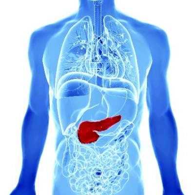
VIDEO: Asymptomatic pancreatic cysts rarely became malignant
Only 1% of adults with asymptomatic neoplastic pancreatic cysts developed invasive pancreatic adenocarcinoma after more than 5 years of follow-up, according to a multicenter retrospective study reported in the June issue of Clinical Gastroenterology and Hepatology.
Furthermore, there were no malignant conversions among patients lacking American Gastroenterological Association high-risk features – that is, mural nodules, dilated pancreatic ducts, or cysts measuring more than 3 cm, said Dr. Wilson Kwong at the University of California San Diego Health Sciences in La Jolla. “There is a very low risk of malignant transformation of asymptomatic neoplastic pancreatic cysts after 5 years,” he and his associates wrote.
Up to 20% of cross-sectional imaging studies reveal incidental pancreatic cysts, the researchers noted. Cysts with neoplastic features are recommended for indefinite surveillance, even though there is little or no data on their natural history and malignant potential beyond 5- 10 years, they added. Therefore, they studied 310 patients who underwent endoscopic ultrasound of pancreatic cysts at an academic medical center, a Veterans’ Affairs hospital, and two community health care systems in California between 2002 and 2010. The most common age at enrollment was 66 years, 60% of patients were women, and the median follow-up period was 87 months (range, 60 to 189 months). A total of 90% of patients were followed for 5-10 years, while 10% were followed for more than 10 years (Clin Gastroenterol Hepatol. 2016 Feb 10. doi: 10.1016/j.cgh.2015.11.013).
Source: American Gastroenterological Association
In all, three patients developed invasive pancreatic malignancies after 6, 8, and 11 years of follow-up, for an overall conversion rate of 1%. Conversion rates by subgroup were 0% for patients with no high-risk AGA features, 1% (one case) for patients with one high-risk feature, and 15% (two cases) for patients with two high-risk features. “Because the risk of malignant transformation beyond 5 years is lower than the 1.4% mortality risk of pancreatic resection at high-volume centers, the argument can be made that discontinuing surveillance after 5 years is justified,” the researchers said. Specifically, surveillance could be discontinued after 5 years for neoplastic pancreatic cysts with up to one high-risk feature, particularly if patients have significant comorbidities that increase their risk of imminent death from other causes, they added. In contrast, healthy patients in their 60s and 70s might benefit from long-term surveillance given their longer life expectancy, they said. “Among patients with two high-risk features who remain surgically fit, discussion of surgery or surveillance beyond 5 years should be considered,” they emphasized.
A total of two patients developed high-grade dysplasia – a risk factor for invasive pancreatic cancer – but even so, the aggregate rate of cancer and high-grade dysplasia was 1.6%, only slightly higher than the fatality rate associated with pancreatic resection, the researchers noted. By excluding patients with recent acute pancreatitis (because of the likelihood of pseudocysts), they might have inadvertently excluded “a small number” of patients with pancreatic intraductal papillary mucinous neoplasms, they added.
The University of California San Diego Health Care System supported the study. The investigators had no disclosures.
Kwong et al. present important data demonstrating a low risk of malignant transformation for pancreas cysts followed for more than 5 years, which is similar to the risk of surgical resection. Mortality from nonpancreatic causes was found to be eightfold higher than mortality from pancreatic cancer. The goal of pancreas cyst surveillance is to prevent death from pancreatic cancer, currently accomplished by identifying high-risk cysts for surgical resection. When evaluating the utility of surveillance, patient and cyst characteristics can be considered.
Elderly patients with multiple comorbidities are unlikely to benefit from long-term surveillance as they may be poor surgical candidates and are unlikely to die from the malignant progression of a pancreas cyst. Healthy patients with a family history of pancreatic cancer and/or identifiable genetic risk factors, however, may benefit from long-term surveillance. Although demonstrated to be infrequent, cysts that have been stable for 5-10 years rarely may progress to cancer. The presence of more than one high-risk cyst feature increased the risk of progression from approximately 1% to 15%. The study of larger groups of cysts with morphologic high-risk features is required. The addition of molecular and genetic cyst and patient features has the potential to assist in risk stratification.
Clarifying which cysts and patients are likely to benefit from surveillance and resection is of increasing importance as high-resolution, cross-sectional imaging identifies greater numbers of pancreas cysts.
Dr. Harry R. Aslanian, AGAF, is director, Advanced Endoscopy Fellowship, and associate professor, Yale University, New Haven, Conn. He is a consultant for Boston Scientific and Olympus.
Only 1% of adults with asymptomatic neoplastic pancreatic cysts developed invasive pancreatic adenocarcinoma after more than 5 years of follow-up, according to a multicenter retrospective study reported in the June issue of Clinical Gastroenterology and Hepatology.
Furthermore, there were no malignant conversions among patients lacking American Gastroenterological Association high-risk features – that is, mural nodules, dilated pancreatic ducts, or cysts measuring more than 3 cm, said Dr. Wilson Kwong at the University of California San Diego Health Sciences in La Jolla. “There is a very low risk of malignant transformation of asymptomatic neoplastic pancreatic cysts after 5 years,” he and his associates wrote.
Up to 20% of cross-sectional imaging studies reveal incidental pancreatic cysts, the researchers noted. Cysts with neoplastic features are recommended for indefinite surveillance, even though there is little or no data on their natural history and malignant potential beyond 5- 10 years, they added. Therefore, they studied 310 patients who underwent endoscopic ultrasound of pancreatic cysts at an academic medical center, a Veterans’ Affairs hospital, and two community health care systems in California between 2002 and 2010. The most common age at enrollment was 66 years, 60% of patients were women, and the median follow-up period was 87 months (range, 60 to 189 months). A total of 90% of patients were followed for 5-10 years, while 10% were followed for more than 10 years (Clin Gastroenterol Hepatol. 2016 Feb 10. doi: 10.1016/j.cgh.2015.11.013).
Source: American Gastroenterological Association
In all, three patients developed invasive pancreatic malignancies after 6, 8, and 11 years of follow-up, for an overall conversion rate of 1%. Conversion rates by subgroup were 0% for patients with no high-risk AGA features, 1% (one case) for patients with one high-risk feature, and 15% (two cases) for patients with two high-risk features. “Because the risk of malignant transformation beyond 5 years is lower than the 1.4% mortality risk of pancreatic resection at high-volume centers, the argument can be made that discontinuing surveillance after 5 years is justified,” the researchers said. Specifically, surveillance could be discontinued after 5 years for neoplastic pancreatic cysts with up to one high-risk feature, particularly if patients have significant comorbidities that increase their risk of imminent death from other causes, they added. In contrast, healthy patients in their 60s and 70s might benefit from long-term surveillance given their longer life expectancy, they said. “Among patients with two high-risk features who remain surgically fit, discussion of surgery or surveillance beyond 5 years should be considered,” they emphasized.
A total of two patients developed high-grade dysplasia – a risk factor for invasive pancreatic cancer – but even so, the aggregate rate of cancer and high-grade dysplasia was 1.6%, only slightly higher than the fatality rate associated with pancreatic resection, the researchers noted. By excluding patients with recent acute pancreatitis (because of the likelihood of pseudocysts), they might have inadvertently excluded “a small number” of patients with pancreatic intraductal papillary mucinous neoplasms, they added.
The University of California San Diego Health Care System supported the study. The investigators had no disclosures.
FROM CLINICAL GASTROENTEROLOGY AND HEPATOLOGY
Key clinical point: Asymptomatic neoplastic pancreatic cysts rarely become malignant, especially in the absence of multiple American Gastroenterological Association high-risk features.
Major finding: Only 1% of patients developed invasive pancreatic adenocarcinoma after more than 5 years of surveillance.
Data source: A multicenter retrospective study of 310 patients who underwent endoscopic ultrasound evaluations of pancreatic cysts.
Disclosures: The University of California San Diego Health Care System supported the study. The investigators had no disclosures.
