User login
Intra-Arterial Embolization With Fillers Is Rare, But Severe
WASHINGTON — Intra-arterial embolization during filler injection is rare, but it can happen to even the most experienced physician, said Dr. Claudio DeLorenzi.
Intra-arterial embolization occurs when a filler needle enters an artery. During injection, the filler flows retrograde in the vessel. Once the pressure from injection is stopped, the product is carried through the vasculature and the results can be disastrous, Dr. DeLorenzi said at the annual meeting of the American Society for Aesthetic Plastic Surgery.
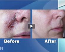
Local and distal necrosis can occur following intra-arterial embolization. Typically the whole angiosome--the vascular territory of skin and underlying muscles, tendons, nerves, and bones--is affected, resulting in full-thickness necrosis.
The first sign of trouble is severe pain, followed typically by a bluish livedo pattern.
This can progress to hemorrhagic blisters, necrotic eschar, and late scarring. "The severity of some of these complications is extreme by any measure," said Dr. DeLorenzi, who is a plastic surgeon in Kitchener, Ont.
"So if you see this reticular bluish pattern, it means that you need to take action," he said. A crash cart for this complication allows for an immediate response. The cart should contain hyaluronidase--which can reverse the effects of hyaluronic acid fillers--aspirin, nitroglycerine paste, and heat compresses.
Dr. DeLorenzi recommended having a low threshold for using hyaluronidase. Recommendations in the literature on the amount to use vary from 25 to 150 units.
Hyaluronidase should be injected diffusely in the affected area, which should then be massaged to distribute the filler. "Even if you've selected a non-hyaluronic acid filler, there may be some role for using hyaluronidase. You may get some improvement in circulation," he said.
Increased risk for intra-arterial embolization appears to be associated with large volumes of filler, small sharp needles, high pressure, deeper injections, and compound type.
"Prevention, of course, is best," said Dr. DeLorenzi. To avoid intra-arterial embolization, he recommends using a small bolus (less than 0.1 cc), a blunt cannula, low pressure, slow injection; knowing the relevant anatomy; being prepared; and using epinephrine prior to the filler procedure (this may reduce the vessel size and the risk of hitting a vessel).
Dr. Delorenzi reported that he is a speaker for Allergan Inc.
Expertise Is Essential for Effective Laser Tattoo Removal
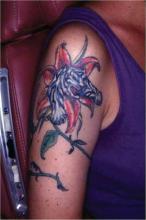
NAPLES, Fla. — Patients present with different reasons for tattoo removal—such as regret following a crazy night or a change in relationship status. The number of options for laser tattoo removal, however, is more limited, Dr. Albert J. Nemeth said.
Specific Q-switched Nd:YAG laser wavelengths are good at shattering tattoo ink but must be applied specific to the ink color. Also, do not use non-Q-switched Nd:YAG lasers or other lasers designed for different dermatologic indications, Dr. Nemeth said at the annual meeting of the Florida Society of Dermatology and Dermatologic Surgery.
In general, 95% of amateur tattoos take one to three laser treatment sessions for removal. In contrast, six or more sessions are often required to remove most professional tattoos. Variables include the depth at which pigment was placed and type of ink. "Tattoo depth can vary. That is important to conceptualize when treating these tattoos," Dr. Nemeth said.
Tattoos applied by a friend or family member and those inked professionally share a common challenge for dermatologists—a lack of regulation on what can be used for pigment. "It's really quite interesting in terms of the secret concoctions that I've heard … cigarette ash; for some reason Crest toothpaste seems to be a magic ingredient; and—I don't understand why—but urine seems to be important in there also," Dr. Nemeth said in an interview.
|
An important caveat: If the patient reports or displays any symptoms of allergic dermatitis to the ink (constant oozing or crusting), lasers can worsen those symptoms. Physical removal of the tattoo is the only option for these patients, said Dr. Nemeth, a private-practice dermatologist in Clearwater, Fla., and FSDDS president. Dermabrasion, for example, is an option "if you are extraordinarily proficient at that."
An estimated 14% of all adults in the United States have at least one tattoo, according to a Harris Poll by Harris Interactive in 2008. A predominance of tattooing among a younger segment of the population means dermatologists are likely to see more and more patients with tattoos.
Removal of tattoos via laser can be life changing and/or life saving for some patients. For example, former gang members might feel safer after removal of an identifying tattoo, said Dr. Nemeth, also of the department of dermatology and cutaneous surgery at the University of South Florida, Tampa.
For removal of green tattoo inks, Dr. Nemeth uses a 694-nm Q-switched ruby or 755-nm Q-switched alexandrite laser. To shatter black, blue, sky blue, or darker inks, he employs a 1064-nm Q switched Nd:YAG laser. If a patient's tattoo features red, orange, or purple inks, these can be effectively treated with a 532-nm Q-switched Nd:YAG laser, he said. Also be aware that laser therapy can darken tattoo inks if they contain iron oxide or titanium oxide.
Avoid the temptation to use lasers for other indications. "An EpiTouch ruby laser made for hair removal should not be used for tattoo removal," Dr. Nemeth said. "This is an important take-home message: Just because the wavelength is close, it does not mean the indications are the same."
Also, intense pulsed light systems are not appropriate for this indication, Dr. Nemeth said. "If your salesperson told you IPL can be used to remove tattoos, [he] lied to you. IPL cannot be used to remove a tattoo, in my humble opinion."
The Q-switched lasers have afforded patients a much better aesthetic outcome following tattoo removal. "It used to be every patient knew they were switching their tattoo for a scar, "Dr. Nemeth said. "Now you can get some very, very nice results."
Dr. Nemeth had no relevant financial disclosures.
Repeat Suction Enhances Subcision Efficacy for Depressed Facial Scars
A novel technique that combines subcision and suction proved safe and effective for promoting significant and lasting improvement of mild to severe depressed facial scars in a study of 58 patients.
In 46 patients who strictly followed the study protocol of subcision followed by frequent suctioning at the time of postsubcision depression recurrence, the depth and size of scars decreased significantly by a mean of nearly 72% at 6-month follow-up, according to assessment by two investigators, and by 75% according to patient assessment.
An improvement of at least 80% occurred in about 28% of patients, according to investigator assessment, and in 42%, according to patient assessment, Dr. S. Aalami Harandi of the Parsian Laser Clinic, Bandar Abbas, Iran, and colleagues reported online in the June 9 issue of the Journal of the European Academy of Dermatology and Venereology.
Mean improvement at 6 months in 12 patients who started suction late or had long intervals between suction sessions was 44% by investigator assessment and 49% by patient assessment, the investigators reported (J. Eur. Acad. Dermatol. Venereol. 2010 June 9 [doi:10.1111/ j.1468-3083.2010.03711.x]).
The study participants - 34 women and 24 men aged 16-44 years - had depressed acne scars of various types, including rolling, superficial boxcar, deep boxcar, and pitted, as well as scars from chicken pox, trauma, and surgery. Superficial dermal undermining was performed on 1-70 scars per patient using mainly 23-gauge needles. Suctioning was initiated on the third day following subcision, and was performed at least every other day for 2 weeks, per protocol.
The best results (80% or better improvement) were seen in 24 of the 46 patients on protocol who had the most frequent suctioning (almost daily versus every other day interval in the first week of suction period), the investigators noted.
Although subcision is a safe, valuable, and practical method in itself, it has only mild to moderate efficacy because of the frequency of depression recurrence. In the investigators' experience, recurrence with subcision alone generally starts 2-5 days after subcision, with rapid progression of re-depression for up to 10 days, and gradual progression of re-depression for about 1 week more; therefore, the protocol for this study involved repeated suctioning at the recurrence period.
The addition of suctioning, which prevents redepression by "induction of repeated haemorrhage in dermal pocket, delay in healing, and more new connective tissue formation at the scar area," appears to improve the efficacy of subcision alone.
The combined subcision and suction treatment in this study was associated with "significant" (greater than 60% improvement) and "excellent" (80% improvement or greater) efficacy, the investigators said.
Bruising occurred in all cases, but resolved within 12 days, and any discoloration that occurred resolved within 2 months; hypertrophic scarring occurred in 1.7% of treated scars (22 scars in six patients), which was mostly due to sub-epidermal like undermining( technical error), and was managed successfully in all cases; and hemorrhagic papules and pustules occurred in 5.6% of subcised scars, and were treated successfully with drainage and topical antibiotics or steroids.
Advantages of the subcision-suction method include ease of application, low cost, short down-time, applicability for various skin types (most had type III in this study), applicability for various scar types, lack of significant complications, and "remarkable and persistent improvement in short time without injury to the skin surface," the investigators reported.
"It seems that this method has the potential to be used as the first step for acne and other depressed scars management," they wrote, adding that since multistep treatment is necessary for optimal correction of acne scars, treatment may involve the use of other techniques or repeat subcision-suction after several months.
Further study of this technique is warranted, particularly given the prevalence of the problem of depressed scars of the face, they noted.
The investigators had no conflicts of interest to declare.
Lidocaine-Delivery System, Emla 5% Comparable for Pain Control
PHOENIX — Patients reported less or a comparable amount of pain during needling roller treatment for upper lip rhytids when lidocaine was delivered with a jet-phoresis system, compared with an application of Emla 5% cream, results from a small study of 20 patients showed.
During a poster session at the annual meeting of the American Society for Laser Medicine and Surgery, researchers presented results from a study designed to compare administration of lidocaine with the jet-phoresis system, compared with the topical cream for pain control in patients scheduled for needling roller procedures for upper lip rhytids.
For the study, Dr. Michael Gold, a dermatologist who practices in Nashville, Tenn., and Dr. Ram Burvin, a plastic surgeon who practices in Tel-Aviv, had patients serve as their own control. The mean age of the patients was 56 years, and all were female.
The researchers treated half (left or right) of each patient's upper lip with Emla 5% cream for 45 minutes and the contralateral portion of the lip with lidocaine 3% jet phoresis for 5 minutes. They used a visual analog scale to measure pain elicited by application of a needling roller across the upper lip.
Each patient again served as her own control 12-16 weeks later when the treatments (lidocaine 3% with jet phoresis vs. Emla 5%) were repeated on the opposite lip sides for the same durations, so that in all, there were 40 full-lip applications of the two treatments. Different readings for the left and right sides were registered in some of the patients.
Of the total 40 treatments, pain control with lidocaine 3% with jet phoresis and Emla 5% was comparable in 19 applications, it was better with the lidocaine 3% with jet phoresis in 14 of the applications, and better with Emla 5% in 7 applications
The delivery device, the JetPeel 3, uses pressurized gas at supersonic velocities to deliver saline or other liquid nutrients through special handpieces into the superficial layers of the skin. It was cleared by the Food and Drug Administration in 2006 for delivery of saline into the skin.
The researchers received honoraria from TavTech Ltd., maker of the jet-phoresis system, to conduct the study.
Ultrasound-Assisted Lipoplasty Helps Recontour Jowls
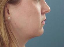
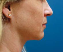
WASHINGTON — The removal of fat using ultrasound-assisted lipoplasty may be all it takes to recontour the jowls of younger patients and can complement techniques to recontour the jowls of older and more difficult-to-treat patients, said Dr. James C. Grotting.
Dr. Grotting has been using ultrasound-assisted liposuction (UAL) to recontour the jowls, "especially in the younger round-faced patient - who doesn't require a face lift - and the difficult, older, heavy-jowled patient."
"I think it's important to say that UAL is simply a tool that helps evenly and precisely remove the excess fat that contributes to the formation of the jowls," Dr. Grotting said at the annual meeting of the American Society for Aesthetic Plastic Surgery. "It's my opinion that UAL allows a little more control and may stimulate a modicum of skin retraction." This control helps to minimize the creation of visible lines, furrows, and other irregularities.
Dr. Grotting, a practicing plastic surgeon in Birmingham, Ala., grasps the fat to reposition and observe it to decide if he needs to reduce the amount of fat.
When performing UAL, he doubles the epinephrine injected, and hand-tunnels using the spatulated facial cannula to create the facial plane. Low power ultrasound helps provide additional control. An incision is then made right in front of the earlobe because "if you get behind the ear lobe, sometimes you get behind the platysma and you could potentially damage the mandibular branch," he said.
The area to be contoured is just the jowl area, extending down into the neck. "It's important to make sure that you're in the subcutaneous plane," he said.
"I've found this to be a very effective way to treat men - particularly men who are not interested in face lifts." He recommends being more aggressive when contouring the jowls in men because of the thickness of the skin. "I generally will come to the jowl from below."
Dr. Grotting did not have any relevant conflicts to disclose.
Tips for Treating Acne Scarring in Darker Skinned Patients
PHOENIX — Educate darker skinned patients who seek treatment for acne scars that there is no remedy to make the scars completely disappear.
"Depending on the patient's skin type, the sensitivity of their skin, and how aggressively you treat them, the risk of hyperpigmentation can be relatively modest, or well over 50%. The expected degree of improvement, on the other hand, even with multiple modalities and multiple treatments, is 40%-50%. I think it's very important to explain that," said Dr. Murad Alam at the annual meeting of the American Society for Laser Medicine and Surgery.
Dr. Alam, chief of cutaneous and aesthetic surgery at Northwestern University, Chicago, said that clinicians face certain challenges in treating acne scars in patients of color, including the risk of exacerbation of active acne, risk of focal or diffuse hyperpigmentation or hypopigmentation, risk of nodularity or surface texture change, and risk of minimal effect.
To mitigate risks, Dr. Alam considers oral antibiotics in patients who have any degree of active acne, "even if they get one or two acne pimples once in a blue moon," he said. "If the acne is more than very mild, you may wish to target that as the primary goal and defer treatment of the acne scarring until the acne is under good control."
If the acne is mild, "you can start oral antibiotics at least 1 month before the acne scarring intervention, so they do have something on board to reduce the risk of an acne flare," he said. "You may also consider pretreatment with bleaching agents. I'm personally not that convinced that pre-treatment is that helpful, but post-treatment with bleaching agents is of definite efficacy in mitigating postinflammatory hyperpigmentation."
As for treatment, nonablative resurfacing with mid-infrared lasers, including 1320-nm, 1450-nm, and 1540-nm devices, has been shown to be effective in patients with lighter skin. "This heating process causes collagen remodeling, and can have a modest effect on so-called rolling scars, which can be quite disfiguring," he said.
Another option is ablative resurfacing with non-CO2 fractional lasers such as the 1550-nm laser. "This is one of the most gentle devices in this category, but even so you have risks of postinflammatory hyperpigmentation," Dr. Alam said. "I like to err on the side of being very modest with regard to fluences. It's much better to do more treatments than to push each individual treatment at the risk of having pigmentary abnormalities."
A more aggressive approach is ablative resurfacing with CO2 fractional lasers, which "should be restricted to patients who are of lighter skin type. If they do choose this [modality], they need to understand the significant risk of postinflammatory hyperpigmentation. I would say that virtually every patient of skin of color who undergoes this treatment will have some degree of postinflammatory hyperpigmentation. In some cases they might consider that worth it if it makes their scarring better and if it can be managed after treatment so it eventually goes away."
Perhaps the most beneficial treatment for acne scars in patients of color, Dr. Alam said, is subdermal manipulation.
In one procedure, known as subcision, clinicians insert a needle with a sphere-like tip, often an 18-guage Nokor needle, underneath the skin. "By debriding the underside of the skin, you can cause some of the acne scars to float upward," he explained. "You want to ensure very good hemostasis before doing this—lidocaine with epinephrine—because you want to avoid bruising during the procedure. If done properly, this can result in modest improvement of rolling scars, and it can be done repeatedly."
Dermal fillers can be used as an adjunct. About a month after subcision procedures Dr. Alam considers collagen for fine defects, hyaluronic acid for medium defects, and calcium hydroxylapatite for deeper defects.
The best way to develop a treatment plan for acne scarring, he said, is to assess the patient's commitment to improvement and their tolerance for adverse events.
"How much annoyance and disfigurement are they willing to tolerate?" Dr. Alam asked. "If both of these are low, you might wish to restrict yourself to subcision with or without fillers, because if done properly, that almost eliminates the risk of adverse events like hyperpigmentation, and it does provide some modest improvement with relatively little cost."
If the patient is highly committed to achieving improvement but is wary of adverse events, "then you might consider subcision and fillers, followed by nonablative laser or repeated low energy non-CO2 fractional laser treatments."
In those rare patients with a high tolerance for adverse events, he said, consider CO2 fractional laser treatments "at very modest settings."
Dr. Alam said that he had no relevant financial conflicts.
Cosmeceuticals Enhance Cosmetic Procedures for Melasma
SANTA MONICA, Calif. - Melasma is notoriously difficult to eliminate, but adding cosmeceuticals to the treatment can improve chemical peel results, according to Dr. Cherie M. Ditre.
"I use a combination of retinols, combinations of antioxidants such as green tea, along with hydroquinones," Dr. Ditre said in an interview at a cosmetic dermatology seminar sponsored by Skin Disease Education Foundation (SDEF).
"Those three together are a powerhouse. And, occasionally, I'll add in alpha-hydroxy acids to actually help increase permeation through the skin."
The exact combinations depend on the procedure planned, said Dr. Ditre director of the University of Pennsylvania Health System's Skin Enhancement Center in Radnor.
If she is planning a chemical peel, for example, she has the patients "prepped first in the morning with an alpha-hydroxy acid cleanser starting at 10% and moving up. And then I also have them use hydroquinone at 4%. I can also titrate that in office to 6% or 8% depending on what they need. And then I go with an antioxidant such as green tea and a sunscreen."
She prefers sunscreens containing titanium dioxide and zinc oxide, and recommends Anthelios, which contains Mexoryl, and is a Food and Drug Administration-approved sun filter.
For the evening, Dr. Ditre instructs her chemical peel patients to use an alpha-hydroxy acid cleanser with a retinol that she titrates based on skin type and condition. She mixes that with an antioxidant such as green tea.
"I do that for a period of about 2 weeks prior to the chemical peel," she said. "The chemical peel that I'm presently using is a combination of a 1% retinol with 14% hydroquinone, and we leave it on as a masque for about 5-8 hours to wash off at home. And then, for the following 2 weeks, they use a regimen of a retinol and hydroquinone along with their sunscreen and a calming lotion," after which patients will return for follow-up.
Dr. Ditre said she has never seen an adverse reaction to green tea, which she described as "very gentle." Retinols are another story, however.
"Some of the retinols can approximate retinoic acid, so you have to be careful in very sensitive skin patients," she said. "I think that starting with the 2X [concentration] is better for sensitive skin patients. And that's why you do it as a prep prior to actually doing procedures, to make sure it's agreeable with them prior to doing a more invasive procedure."
Dr. Ditre reported having no disclosures. SDEF and this news organization are owned by Elsevier.
Burn Scar Treatment Called ‘Work in Progress’
PHOENIX — Burn scars rank as one of the most difficult dermatologic conditions to treat, Dr. Jill S. Waibel said at the annual meeting of the American Society for Laser Medicine and Surgery.
"Burn scars are the worst we see in clinical medicine," said Dr. Waibel, a dermatologist with a laser practice in Miami. "I believe that if we can treat a burn scar, we can treat any scar."
Under normal circumstances, wounded skin re-epithelizes from hair follicles and dermal glands, but because burn scars are often partially or completely deprived of their epidermal appendages, "healing is severely affected," she noted.
Thanks to surgical advances in the past decade, survival of burn patients has risen from about 30% to 95%. The types of scars they present with include hypertrophic, keloid, contracture, and atrophic.
Current efforts to treat burn scars fall into one of two camps: prevention of scar formation and late reconstruction of mature scars.
"We do have a model for a scarless wound," Dr. Waibel said. "A fetus in utero does not scar. We don't understand that process. There are also a number of topical applications to prevent scars at the time of the wound. Over 200 cytokines are involved in wound healing."
Research efforts are also under way on laser-assisted skin healing with a diode laser, she said, which alters the wound-healing process by thermal stress.
Current treatment for late reconstruction of mature scars includes surgery, followed by laser combination therapy. "I think fractional therapy is the … standard, but I really don't think we understand the mechanism of action in laser and scar reduction," Dr. Waibel commented. "I think we break it down into two areas: either fractional versus thermal, or probably it's fractional and thermal. The thermal effects are the most interesting. How much heat is required for the most constructive healing versus too much thermal injury? We need to look more at what that [ideal] temperature is."
She tells her burn scar patients to consider their treatment as a "work in progress" and asks them to give her a year before they start to assess efficacy. In 2005, her first burn patient underwent five treatments with a 1550-nm, nonablative, erbium-fiber fractional laser; intralesional Kenalog (triamcinolone); and a shave biopsy.
"We see functional improvement as well as cosmetic, especially with contracture scars, and we're working on some range-of-motion studies right now," Dr. Waibel said.
In a study presented at the society's 2009 meeting, Dr. Waibel and her associates presented results from a proof-of-concept study of 10 patients who had burn scars that were treated with a 1550-nm, nonablative, erbium-fiber fractional laser.
Objective scoring by blinded investigators of photos taken pretreatment and at 3 months posttreatment indicated that 78% of patients had excellent to moderate results.
Dr. Waibel acknowledged certain limitations in current efforts to improve treatment for patients with burn scars, including the need for surgery for anatomical fixes and the lack of understanding of the processes of scar formation and the laser effects on scars. "We need better technology, and we need to maximize treatment modalities," she said. "We really need to develop a scar laser. All of the lasers that we use for scars right now were invented for wrinkles."
Dr. Waibel has conducted research for Solta Medical and Sciton, and she has received honoraria from Lumenis for lectures.
"Peach Pit" Décolleté Defect Can Be Repaired With Fillers
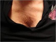
SANTA MONICA, CALIF. — The volume loss and wrinkled skin that appears in the décolleté area of some older women can be significantly softened with filler followed by fractionated laser treatments, Dr. Joel L. Cohen said at a cosmetic dermatology seminar sponsored by Skin Disease Education Foundation.
This cosmetic defect, which Dr. Cohen has dubbed "the peach pit," responds well to injections of hyaluronic acid fillers along with fractionated laser sessions.
"Over the course of many years, I've noticed that many patients become concerned about their décolleté area," said Dr. Cohen, director of AboutSkin Dermatology and DermSurgery, Englewood, Colo., in an interview. "I've tried botulinum toxin [type A]. And despite some reports of efficacy, I haven't been able to actually see this type of efficacy myself. People have topically treated it with retinoids and although that can be helpful, there are patients who have more severe crumpling of the skin."
Instead, he uses Juvéderm (Allergan) or Restylane (Medicis). With such hyaluronic acid fillers, "If you do get lumps or bumps, or the patients aren't happy with it, you could inject the enzyme hyaluronidase and make it go away," he said. "It's easier to mold out little contour irregularities with these types of agents as well."
Using only topical anesthesia, Dr. Cohen injects two or sometimes three syringes of the hyaluronic acid product, after which he asks a female assistant to massage it in. He follows up a few weeks later with two or more treatments with a fractionated laser, often combined with a light erbium laser peel for more texture improvement.
Pharmaceuticals Inc. Skin Disease Education Foundation (SDEF) and this news organization are owned by Elsevier.
Novel Device Uses Ultrasound to Treat Acne
PHOENIX – An investigational intense-therapy ultrasound device safely and effectively treated mild to moderate acne in a preliminary study.
The device, manufactured by Xthetix Inc., was used to treat 5-15 active acne lesions on one side of the face for 5 consecutive days in 18 women and 7 men with a mean age of 26 years. Of the patients, about 84% showed significant improvement after five treatment sessions.
According to a poster presented during the annual meeting of the American Society for Laser Medicine and Surgery, energy generated by the device is delivered to the level of the sebaceous gland, "raising the temperature 5-15 degrees Celsius above ambient skin temperature. It is designed not to cause thermal coagulation, but to increase the tissue temperature for a period of time sufficient to produce a therapeutic effect."
In an interview, lead investigator Dr. Bill Halmi said that although there are a few devices currently on the market that use heat to reduce the duration of an inflammatory acne papule, the machine uses ultrasound to heat the targeted area.
"Putting the energy intradermally into the pilosebaceous unit is the unique aspect of the device," he said. "It changes the inner workings of the gland, kills bacteria, [and] peaks the inflammatory process, among other things."
Each treatment session lasted about 15 minutes. The contralateral side of the face did not receive any ultrasound treatment and served as the control.
The researchers took photographs on all visit days and a masked reviewer conducted posttreatment and facial acne assessments at every visit. Each patient completed daily pain and acne assessments.
Dr. Halmi, who practices dermatology in Phoenix, reported that the greatest percentage of lesion clearance (30%) occurred on day 4 of treatment. "This differs from 'blue light' devices that expect to see improvements after at least 1 week of treatment," he said. "Going into the study we really weren't certain that we would see an immediate effect from the device. We were certainly pleased to discover that this ultrasound device measurably reduced the duration of an acne lesion."
A majority of patients (84%) reported moderate to significant improvement of their treated sides during the course of treatment, while 40% experienced mild transient erythema that lasted less than 30 minutes. The average pain score was 3 on a scale of 1-10, with 10 being the most severe.
The findings suggest that "there is a role for heat in reducing the duration of an acne lesion," Dr. Halmi said. "The advantage ultrasound has is that it can target that heat to the depth desired."
He cautioned that the study is preliminary and that, while encouraging, "repeated studies using more patients will help confirm our findings. Additionally, the role of ultrasound in the prophylactic treatment of acne remains to be revealed."
Dr. Halmi disclosed that he is a paid consultant for Xthetix.
