User login
Inhibiting integrin-mediated activation might help treat, reverse chronic pancreatitis
Integrins that bind arginine-glycine-aspartic acid (RGD) activated stellate cells in the pancreas, inducing pancreatic fibrogenesis in mice, researchers reported in the July issue of Cellular and Molecular Gastroenterology and Hepatology.
“Small-molecule antagonists of this interaction might be developed for the treatment of pancreatic fibrotic diseases,” wrote Dr. Barbara Ulmasov and her associates at Saint Louis University.
Cytokine transforming growth factor beta, or TGFB, plays a “central” role in the activation of pancreatic stellate cells and the promotion of fibrogenesis, both in the pancreas and in other organs, the investigators noted. Because latent TGFB is “abundantly present” in the pancreas and most other tissues, it might be more important to control the activation of TGFB than its expression, they added. Studies of other organs have shown that TGFB is activated when integrins of the av family bind the RGD sequence, but no one had determined whether this was true for pancreatic fibrogenesis, Dr. Ulmasov and her associates asserted (Cell Mol Gastroenterol Hepatol. 2016 Mar 16. doi: 10.1016/j.jcmgh.2016.03.004).
Therefore, they repeatedly injected female C57BL/6 mice with cerulein to induce pancreatic fibrogenesis, and gave a group of control mice sterile saline instead. The mice then received continuous infusions of a small molecule called CWHM-12, which is an antagonist of RGD-integrin that is known to prevent both pulmonary and hepatic fibrosis in mice. After euthanizing the mice, the researchers measured pancreatic parenchymal atrophy, fibrosis, and activation of pancreatic stellate cells. They also studied TGFB activation in an established line of pancreatic stellate cells from rats.
Pancreatic stellate cells expressed messenger RNAs encoding RGD-binding integrins, the investigators found. The mice that received cerulein had higher levels of these integrins than the mice that received saline, and the cerulein group also had more disrupted acinar cell architecture, tubular complexes, and infiltrations of inflammatory cells. Mice that received prophylactic CWHM-12 had only somewhat less acinar cell loss and atrophy than the control mice, and had similar levels of inflammatory cell infiltration, but had “dramatically” lower levels of pancreatic fibrosis, the researchers said. Even if mice received CWHM-12 several days after starting cerulein, they still had less fibrosis and activation of TGFB than if they received saline, they noted.
The established line of pancreatic stellate cells “could robustly activate endogenously produced TGFB,” the investigators also reported. Furthermore, CWHM-12 “potently blocked TGFB activation,” unlike the control compound. Taken together, the findings illustrate the “critical role of RGD-binding integrins in chronic pancreatitis, and the promising potential to arrest or possibly even reverse pancreatic fibrosis using a pharmacologic approach to inhibiting integrin-mediated TGFB activation,” the researchers concluded.
The National Pancreas Foundation and the Frank R. Burton Memorial Fund supported the study. Dr. Ulmasov had no disclosures. Three coinvestigators reported being consultants and/or holding equity in Integrin Therapeutics, Nimbus Therapeutics, Bristol-Myers Squibb, Janssen, Mitsubishi Tanabe, Conatus, and Scholar Rock.
Integrins are transmembrane proteins that organize epithelial cells and transmit signals from the tissue matrix. These proteins consist of two subunits that partner to form more than 20 specific combinations, are induced upon tissue injury, and act as signaling molecules that mediate inflammatory and wound-healing responses. The utility of targeting integrins has been established by drugs such as vedolizumab, which targets specific integrins to dampen injury in inflammatory bowel disease.

|
Dr. Chuhan Chung |
Specific integrins also mediate profibrotic responses by activating TGF-beta, the major fibrogenic cytokine. An arginine-glycine-aspartic acid (RGD) integrin-binding motif found on the TGF-beta molecule triggers this activation upon interaction with specific integrins. Blocking the RGD-integrin interaction reduces fibrosis in multiple organs including the lung, liver, and kidney.
In the current issue of Cellular and Molecular Gastroenterology and Hepatology, Ulmasov et al. report on the use of a synthetic peptide (CWHM-12) in a model of chronic pancreatitis. This peptide mimetic antagonizes the RGD interaction with integrins, thereby limiting TGF-beta activation. Using a cerulein-induced pancreatic fibrosis model, the authors demonstrated that CWHM-12 inhibits pancreatic stellate cell (PSC) activation and generation of active TGF-beta. CWHM-12 suppressed fibrosis when administered prior to cerulein injection, and to a lesser extent, during the course of generating fibrosis. The alpha-v-beta1 integrin was identified as a critical integrin and was expressed at high levels in the murine pancreas, primary PSCs, and further in cerulein-induced pancreatitis.

|
Dr. Fred Gorelick |
Limitations of the current study warrant comment. The most obvious is that this model does not generate true chronic pancreatitis, because unlike chronic pancreatitis, fibrosis resolves spontaneously. Proof of effect in more robust chronic pancreatitis models is needed. Off-target effects are also suggested by the finding that serum white blood counts were significantly higher with CWHM-12. Finally, the chronicity and unpredictability of human chronic pancreatitis make this preclinical study an early starting point for determining whether CWHM-12 has true “clinical legs.”
Dr. Chuhan Chung and Dr. Fred Gorelick are in the department of medicine, Yale University, New Haven, Conn., and the VA Connecticut Healthcare System, West Haven.
Integrins are transmembrane proteins that organize epithelial cells and transmit signals from the tissue matrix. These proteins consist of two subunits that partner to form more than 20 specific combinations, are induced upon tissue injury, and act as signaling molecules that mediate inflammatory and wound-healing responses. The utility of targeting integrins has been established by drugs such as vedolizumab, which targets specific integrins to dampen injury in inflammatory bowel disease.
|
|
Dr. Chuhan Chung |
Specific integrins also mediate profibrotic responses by activating TGF-beta, the major fibrogenic cytokine. An arginine-glycine-aspartic acid (RGD) integrin-binding motif found on the TGF-beta molecule triggers this activation upon interaction with specific integrins. Blocking the RGD-integrin interaction reduces fibrosis in multiple organs including the lung, liver, and kidney.
In the current issue of Cellular and Molecular Gastroenterology and Hepatology, Ulmasov et al. report on the use of a synthetic peptide (CWHM-12) in a model of chronic pancreatitis. This peptide mimetic antagonizes the RGD interaction with integrins, thereby limiting TGF-beta activation. Using a cerulein-induced pancreatic fibrosis model, the authors demonstrated that CWHM-12 inhibits pancreatic stellate cell (PSC) activation and generation of active TGF-beta. CWHM-12 suppressed fibrosis when administered prior to cerulein injection, and to a lesser extent, during the course of generating fibrosis. The alpha-v-beta1 integrin was identified as a critical integrin and was expressed at high levels in the murine pancreas, primary PSCs, and further in cerulein-induced pancreatitis.
|
|
Dr. Fred Gorelick |
Limitations of the current study warrant comment. The most obvious is that this model does not generate true chronic pancreatitis, because unlike chronic pancreatitis, fibrosis resolves spontaneously. Proof of effect in more robust chronic pancreatitis models is needed. Off-target effects are also suggested by the finding that serum white blood counts were significantly higher with CWHM-12. Finally, the chronicity and unpredictability of human chronic pancreatitis make this preclinical study an early starting point for determining whether CWHM-12 has true “clinical legs.”
Dr. Chuhan Chung and Dr. Fred Gorelick are in the department of medicine, Yale University, New Haven, Conn., and the VA Connecticut Healthcare System, West Haven.
Integrins that bind arginine-glycine-aspartic acid (RGD) activated stellate cells in the pancreas, inducing pancreatic fibrogenesis in mice, researchers reported in the July issue of Cellular and Molecular Gastroenterology and Hepatology.
“Small-molecule antagonists of this interaction might be developed for the treatment of pancreatic fibrotic diseases,” wrote Dr. Barbara Ulmasov and her associates at Saint Louis University.
Cytokine transforming growth factor beta, or TGFB, plays a “central” role in the activation of pancreatic stellate cells and the promotion of fibrogenesis, both in the pancreas and in other organs, the investigators noted. Because latent TGFB is “abundantly present” in the pancreas and most other tissues, it might be more important to control the activation of TGFB than its expression, they added. Studies of other organs have shown that TGFB is activated when integrins of the av family bind the RGD sequence, but no one had determined whether this was true for pancreatic fibrogenesis, Dr. Ulmasov and her associates asserted (Cell Mol Gastroenterol Hepatol. 2016 Mar 16. doi: 10.1016/j.jcmgh.2016.03.004).
Therefore, they repeatedly injected female C57BL/6 mice with cerulein to induce pancreatic fibrogenesis, and gave a group of control mice sterile saline instead. The mice then received continuous infusions of a small molecule called CWHM-12, which is an antagonist of RGD-integrin that is known to prevent both pulmonary and hepatic fibrosis in mice. After euthanizing the mice, the researchers measured pancreatic parenchymal atrophy, fibrosis, and activation of pancreatic stellate cells. They also studied TGFB activation in an established line of pancreatic stellate cells from rats.
Pancreatic stellate cells expressed messenger RNAs encoding RGD-binding integrins, the investigators found. The mice that received cerulein had higher levels of these integrins than the mice that received saline, and the cerulein group also had more disrupted acinar cell architecture, tubular complexes, and infiltrations of inflammatory cells. Mice that received prophylactic CWHM-12 had only somewhat less acinar cell loss and atrophy than the control mice, and had similar levels of inflammatory cell infiltration, but had “dramatically” lower levels of pancreatic fibrosis, the researchers said. Even if mice received CWHM-12 several days after starting cerulein, they still had less fibrosis and activation of TGFB than if they received saline, they noted.
The established line of pancreatic stellate cells “could robustly activate endogenously produced TGFB,” the investigators also reported. Furthermore, CWHM-12 “potently blocked TGFB activation,” unlike the control compound. Taken together, the findings illustrate the “critical role of RGD-binding integrins in chronic pancreatitis, and the promising potential to arrest or possibly even reverse pancreatic fibrosis using a pharmacologic approach to inhibiting integrin-mediated TGFB activation,” the researchers concluded.
The National Pancreas Foundation and the Frank R. Burton Memorial Fund supported the study. Dr. Ulmasov had no disclosures. Three coinvestigators reported being consultants and/or holding equity in Integrin Therapeutics, Nimbus Therapeutics, Bristol-Myers Squibb, Janssen, Mitsubishi Tanabe, Conatus, and Scholar Rock.
FROM CELLULAR AND MOLECULAR GASTROENTEROLOGY AND HEPATOLOGY
Key clinical point: Small molecules that inhibit the interaction between integrins and RGD (arginine-glycine-aspartic acid) might effectively treat chronic fibrosing pancreatic diseases.
Major finding: Continuous infusion with an active RGD peptidomimetic reduced atrophy and loss of pancreatic acinar cells and helped prevent pancreatic fibrosis, activation of pancreatic stellate cells, and expression of genes regulated by cytokine transforming growth factor beta.
Data source: A study of C57BL/6 female mice with chronic pancreatitis induced by repeated administration of cerulein.
Disclosures: The National Pancreas Foundation and the Frank R. Burton Memorial Fund supported the study. Dr. Ulmasov had no disclosures. Three coinvestigators reported being consultants and/or holding equity in Integrin Therapeutics, Nimbus Therapeutics, Bristol Myers Squibb, Janssen, Mitsubishi Tanabe, Conatus, and Scholar Rock.

Early sustained viral response linked to better outcomes among HCV patients
Among patients with hepatitis C virus infection who were in compensated cirrhosis, sustained viral response was associated with significantly lower rates of liver decompensation, hepatocellular carcinoma, and liver-related death, even in the presence of clinically meaningful portal hypertension.
But patients in stage 2 cirrhosis were more likely than were stage 1 patients to develop liver decompensation and to die of hepatocellular carcinoma, regardless of SVR, said Dr. Vito Di Marco and his associates at the University of Palermo, Italy. “The available evidence, including our own, suggests that it is opportune to treat HCV as early as possible, in order to reduce progression to stages of cirrhosis in which a viral cure may be less likely lead to ultimate achievement of a major benefit,” they wrote. The report is in the July issue of Gastroenterology.
Clinically significant portal hypertension causes esophageal varices, a “landmark” indicator of stage 2 cirrhosis, the researchers noted. To parse the effects of SVR by cirrhosis stage, they followed all 444 HCV patients with compensated cirrhosis who were treated with pegylated interferon a2b and ribavirin at their tertiary liver centers between 2001 and 2009 (Gastroenterology 2016 March 31. doi: 10.1053/j.gastro.2016.03.036).
The cohort included 218 patients without esophageal varices (that is, patients in stage 1 cirrhosis) and 226 patients with esophageal varices (stage 2 cirrhosis), the researchers said. The groups were demographically similar at baseline, and had similar body mass indices and rates of comorbidities. Rates of SVR were 31% for patients in stage 1 cirrhosis and 18% for patients in stage 2 cirrhosis (P = .003).
Among stage 1 patients, achieving SVR was associated with a lower risk of liver decompensation (0% versus 7%; P = .009), a lower annual rate of hepatocellular carcinoma (0.7% versus 2.9%; P = .002), and a lower risk of liver-related death (3% versus 12%; P = .03). Stage 1 cirrhosis patients also were about 75% less likely to develop esophageal varices if they achieved SVR than if they did not (hazard ratio [HR], 0.23; 95% confidence interval [CI], 0.11 to 0.48; P less than .001).
Eradicating HCV infection also was associated with better clinical outcomes among patients in stage 2 cirrhosis, according to the investigators. For example, those who achieved SVR had a 0.9% annual rate of hepatocellular carcinoma, compared with 3.6% for those who did not (P = .002). And only 2% of stage 2 patients who achieved SVR died from hepatocellular carcinoma, compared to 18% of those who did not (P = .003).
But regardless of SVR, patients with stage 2 cirrhosis were nearly three times more likely to develop liver compensation (HR, 2.8; 95% CI, 1.73 to 4.59) and about 1.75 times more likely to die of liver-related causes (HR, 1.77; 95% CI, 1.12 to 2.80) compared with patients in stage 1 cirrhosis, the investigators emphasized. Also, achieving SVR did not prevent stage 2 patients from developing more esophageal varices (HR, 1.58; 95% CI, 0.33 to 1.03).
The researchers acknowledged several study limitations. “We have performed many comparisons with respect to multiple endpoints, which could have increased the likelihood of inference errors that can result from multiple comparisons,” they wrote. “Furthermore, we cannot exclude the lack of power as an explanation for some of our results.” In addition, pegylated interferon–based therapies have low rates of SVR, and the study was limited to patients who could tolerate the regimen, they added. Nonetheless, the results indicate that eradicating HCV is clinically beneficial even in advanced compensated cirrhosis, but that early treatment helps maximize these outcomes, they concluded. “Pegylated interferon–free direct-acting antiviral combo regimens will ultimately increase the rate of virological cure to almost universal effectiveness,” they added. “It remains to be assessed whether the effectiveness of these new regimens will be automatically translated into a universal clinical benefit.”
The Italian Ministry of Health funded the study. The investigators had no disclosures.
FROM GASTROENTEROLOGY
Key clinical point: Patients with hepatitis C virus infections may have the best long-term outcomes if they achieve sustained viral response before they develop clinically significant portal hypertension.
Major finding: Patients with stage 2 cirrhosis, regardless of SVR, were at significantly greater risk of liver decompensation (hazard ratio, 2.8; P less than .001) and liver-related death (HR, 1.77; P = .015) compared with stage 1 cirrhosis patients.
Data source: A single-center prospective observational study of 444 patients with HCV and compensated cirrhosis, including 218 with stage 1 disease and 226 with stage 2 disease.
Disclosures: The Italian Ministry of Health funded the study. The investigators had no disclosures.
Immunosuppressive regimens did not affect risk of cancer recurrence in meta-analysis
Among patients with immune-mediated diseases and a history of malignancy, cancer recurrence rates were similar regardless of whether they received tumor necrosis factor inhibitors, traditional immunosuppressants, or no immunosuppression, according to a meta-analysis of 16 cohort and case-control studies.
“However, there is a need for larger studies to prospectively monitor for cancer recurrence and new malignancies in this population to better inform our practice,” wrote Dr. Edward Shelton of Massachusetts General Hospital in Boston, and his associates. The report is in the July issue of Gastroenterology.
Patients with immune-mediated diseases often have a history of malignancy, raising questions about the effects of therapies that target the immune system, the researchers noted. However, relevant studies have been “small with few events, preventing robust estimates of risk,” they said. They searched Medline, Embase, and conference proceedings through April 2015 for studies of the risk of primary or recurrent cancer among patients with a history of malignancy who were exposed to thiopurines, methotrexate, or anti-TNF agents, or no immunosuppression. Among the 16 studies meeting these criteria, seven included patients with rheumatoid arthritis, seven included patients with inflammatory bowel disease, one included both diseases, and one included patients with psoriasis. Twelve were cohort studies, one was a case-control study, and three were case series. The resulting meta-analysis comprised 11,702 patients who contributed a total of 31,258 person-years of follow-up (Gastroenterology 2016 May 13. doi: 10.1053/j.gastro.2016.03.037).
Rates of cancer recurrence were statistically similar among patients who received no immunosuppression, anti-TNF therapy, traditional immune modulators, or combination regimens (P greater than .1 for differences among groups). Numerically, however, the risk of recurrent cancer was highest with combination immunotherapy (54.5 cases per 1,000 person-years of follow-up), compared with 37.5 cases per 1,000 person-years for patients who did not receive immunosuppressive treatment, 36.2 cases per 1,000 person-years for patients who received traditional immunomodulator monotherapy, and 33.8 cases per 1,000 person-years for patients who received anti-TNF agents. Analyses of subgroups of patients with new or recurrent cancers, or various types of therapies, and of specific immune-mediated diseases yielded “similar results, with no increase in risk,” said the researchers. Furthermore, rates of cancer recurrence did not statistically differ depending on whether patients started immunosuppression less than or more than 6 years after their index cancer (P = .43).
“Treatment decisions after a cancer diagnosis are complex, and must take into account the natural history of cancer, histologic type and stage, time from diagnosis, and course of underlying chronic inflammatory disease,” the researchers noted. “Our findings suggest that anti-TNF therapy, conventional immunosuppressant therapy, or combination immunosuppression are not associated with an increased risk of cancer recurrence in this population.”
The researchers cited several limitations. Notably, three of the studies included only 20 patients, and the studies included variable index cancers and methods for detecting recurrent cancers. “It also is possible, and likely, that patients who were at high risk for recurrence were not recommenced on immunosuppression, and consequently our findings may be more applicable to a population in which the patient and physicians were comfortable with re-initiation of therapy,” they said.
The National Institutes of Health supported several of the study investigators. Dr. Shelton had no disclosures. Five coinvestigators disclosed relationships with a number of pharmaceutical companies.
As the proportion of inflammatory bowel disease (IBD) patients who are elderly rises, so does the occurrence of coexisting cancers accompanying advanced age. Experience from the posttransplant population has prompted concerns that use of immunosuppression may heighten the risk of new or recurrent cancers in individuals who have a prior history of cancer.

|
| Dr. Geoffrey Nguyen |
This apprehension may partly explain why elderly IBD patients are much less likely to be treated with immunosuppressive therapies than their younger counterparts are. Prior studies in IBD populations have not shown any association between use of immunosuppressants and recurrent cancer in individuals with prior cancer. However, these studies may have been underpowered to detect such associations.
Dr. Shelton and his colleagues performed a meta-analysis that draws upon more than 16 studies of patients with immune-mediated diseases with prior history of cancer. The investigators confirmed no association between immunosuppression and recurrent or new cancers. Despite some methodologic limitations, the study’s findings support a body of literature that suggests no increased risk of recurrent malignancy with immunosuppression in immune-mediated diseases, including IBD.
The findings also provide additional support for recent ECCO clinical guidelines, which recommend that immunosuppressive therapy can be initiated in patients with cancer after a 2- to 5-year cancer-free interval, depending on the risk of recurrence associated with specific types of cancers. A key point is that the decision to use immunosuppressants in IBD patients with prior cancer should be made with input from an oncologist and be individualized by taking into account disease course, surgical alternatives, and risk of cancer recurrence.
Dr. Geoffrey Nguyen, Mount Sinai Centre for Inflammatory Bowel Disease, University of Toronto. He has no conflicts of interest.
As the proportion of inflammatory bowel disease (IBD) patients who are elderly rises, so does the occurrence of coexisting cancers accompanying advanced age. Experience from the posttransplant population has prompted concerns that use of immunosuppression may heighten the risk of new or recurrent cancers in individuals who have a prior history of cancer.
|
|
| Dr. Geoffrey Nguyen |
This apprehension may partly explain why elderly IBD patients are much less likely to be treated with immunosuppressive therapies than their younger counterparts are. Prior studies in IBD populations have not shown any association between use of immunosuppressants and recurrent cancer in individuals with prior cancer. However, these studies may have been underpowered to detect such associations.
Dr. Shelton and his colleagues performed a meta-analysis that draws upon more than 16 studies of patients with immune-mediated diseases with prior history of cancer. The investigators confirmed no association between immunosuppression and recurrent or new cancers. Despite some methodologic limitations, the study’s findings support a body of literature that suggests no increased risk of recurrent malignancy with immunosuppression in immune-mediated diseases, including IBD.
The findings also provide additional support for recent ECCO clinical guidelines, which recommend that immunosuppressive therapy can be initiated in patients with cancer after a 2- to 5-year cancer-free interval, depending on the risk of recurrence associated with specific types of cancers. A key point is that the decision to use immunosuppressants in IBD patients with prior cancer should be made with input from an oncologist and be individualized by taking into account disease course, surgical alternatives, and risk of cancer recurrence.
Dr. Geoffrey Nguyen, Mount Sinai Centre for Inflammatory Bowel Disease, University of Toronto. He has no conflicts of interest.
Among patients with immune-mediated diseases and a history of malignancy, cancer recurrence rates were similar regardless of whether they received tumor necrosis factor inhibitors, traditional immunosuppressants, or no immunosuppression, according to a meta-analysis of 16 cohort and case-control studies.
“However, there is a need for larger studies to prospectively monitor for cancer recurrence and new malignancies in this population to better inform our practice,” wrote Dr. Edward Shelton of Massachusetts General Hospital in Boston, and his associates. The report is in the July issue of Gastroenterology.
Patients with immune-mediated diseases often have a history of malignancy, raising questions about the effects of therapies that target the immune system, the researchers noted. However, relevant studies have been “small with few events, preventing robust estimates of risk,” they said. They searched Medline, Embase, and conference proceedings through April 2015 for studies of the risk of primary or recurrent cancer among patients with a history of malignancy who were exposed to thiopurines, methotrexate, or anti-TNF agents, or no immunosuppression. Among the 16 studies meeting these criteria, seven included patients with rheumatoid arthritis, seven included patients with inflammatory bowel disease, one included both diseases, and one included patients with psoriasis. Twelve were cohort studies, one was a case-control study, and three were case series. The resulting meta-analysis comprised 11,702 patients who contributed a total of 31,258 person-years of follow-up (Gastroenterology 2016 May 13. doi: 10.1053/j.gastro.2016.03.037).
Rates of cancer recurrence were statistically similar among patients who received no immunosuppression, anti-TNF therapy, traditional immune modulators, or combination regimens (P greater than .1 for differences among groups). Numerically, however, the risk of recurrent cancer was highest with combination immunotherapy (54.5 cases per 1,000 person-years of follow-up), compared with 37.5 cases per 1,000 person-years for patients who did not receive immunosuppressive treatment, 36.2 cases per 1,000 person-years for patients who received traditional immunomodulator monotherapy, and 33.8 cases per 1,000 person-years for patients who received anti-TNF agents. Analyses of subgroups of patients with new or recurrent cancers, or various types of therapies, and of specific immune-mediated diseases yielded “similar results, with no increase in risk,” said the researchers. Furthermore, rates of cancer recurrence did not statistically differ depending on whether patients started immunosuppression less than or more than 6 years after their index cancer (P = .43).
“Treatment decisions after a cancer diagnosis are complex, and must take into account the natural history of cancer, histologic type and stage, time from diagnosis, and course of underlying chronic inflammatory disease,” the researchers noted. “Our findings suggest that anti-TNF therapy, conventional immunosuppressant therapy, or combination immunosuppression are not associated with an increased risk of cancer recurrence in this population.”
The researchers cited several limitations. Notably, three of the studies included only 20 patients, and the studies included variable index cancers and methods for detecting recurrent cancers. “It also is possible, and likely, that patients who were at high risk for recurrence were not recommenced on immunosuppression, and consequently our findings may be more applicable to a population in which the patient and physicians were comfortable with re-initiation of therapy,” they said.
The National Institutes of Health supported several of the study investigators. Dr. Shelton had no disclosures. Five coinvestigators disclosed relationships with a number of pharmaceutical companies.
FROM GASTROENTEROLOGY
Key clinical point: Among patients with immune-mediated diseases and a history of cancer, rates of cancer recurrence did not statistically differ according to whether or not they received immunosuppression regimens, or regimen type.
Major finding: Numerically, the rate was highest with combination immunotherapy (54.5 cases per 1,000 person-years of follow-up), vs. 37.5 cases per 1,000 person-years for no immunosuppression, 36.2 cases per 1,000 person-years for traditional immunomodulator monotherapy, and 33.8 cases per 1,000 person-years for anti-TNF agents.
Data source: A systematic review and meta-analysis of 16 studies and 11,702 patients with rheumatoid arthritis, inflammatory bowel disease, and psoriasis.
Disclosures: The National Institutes of Health supported several of the study investigators. Dr. Shelton had no disclosures. Five coinvestigators disclosed relationships with a number of pharmaceutical companies.

Psychological therapies eased IBS for at least 6-12 months
Adults with irritable bowel syndrome (IBS) who underwent psychotherapy improved more than about 75% of controls, and the effect “remained significant and medium in magnitude” for at least 6-12 months, according to a meta-analysis of 41 randomized, controlled trials reported in the July issue of Clinical Gastroenterology and Hepatology.
The finding “is particularly noteworthy because of the typically recurrent, persistent nature of IBS symptoms,” said Kelsey Laird, a doctoral student of clinical psychology at Vanderbilt University in Nashville, Tenn., together with her associates there. “Future research is needed to compare the longevity of treatment effects for psychotherapy with pharmacologic therapies, such as antidepressants,” they added. “Although it is beyond the scope of this review, it is also important to consider the mechanisms by which psychotherapies improve GI symptoms and to determine the ‘active ingredients’ responsible for this effect.”

Up to 16% of individuals in the United States have IBS, and treating it costs anywhere from $950 million to $1.35 billion per year, the researchers noted. Other meta-analyses found psychotherapy about as effective as antidepressants for treating IBS-related GI symptoms over the short term, but its long-term efficacy was unknown, they added. Therefore, they searched PubMed, PsycINFO, Science Direct, and ProQuest through Aug. 15, 2015, identifying randomized controlled trials of psychological therapy and active or nonactive comparators. Psychotherapy included not only traditional psychodynamic and cognitive-behavioral therapies, but also mind-body approaches, such as relaxation training, biofeedback, and yoga, “which can be conceptualized as mindful movement,” Ms. Laird and her associates said. Comparators included support groups, education, sham treatments for hypnosis or biofeedback, online discussion forums, enhanced medical care, treatment as usual, symptom monitoring, and being wait-listed for psychological treatment (Clin Gastroenterol Hepatol. 2016 Jan 21. doi: 10.1016/j.cgh.2015.11.020). The 41 trials included 2,290 patients, comprising 1,183 assigned to the psychological modalities and 1,107 assigned to the various comparators. Taken together, the psychological modalities were associated with greater improvements in GI symptoms immediately after treatment, as compared with the grouped comparators. The Cohen’s d value was 0.69 (95% confidence interval, 0.52-0.86; P less than .001), indicating a medium effect size, the researchers said. Moreover, Cohen’s d values were 0.76 and 0.73, respectively, at short-term follow-up (1-6 months) and long-term follow-up (6-12 months). “On average, individuals who received psychotherapy had a greater reduction in GI symptoms after treatment than 75% of individuals assigned to a control condition,” the researchers concluded.
Effect sizes were similar among cognitive, cognitive-behavioral, and relaxation and hypnosis interventions, and between interventions delivered online and in person, the investigators also reported. Furthermore, longer durations or sessions of psychotherapy did not appear to further improve symptoms.
Study limitations included substantial variability between trials, and the fact that none of the 41 trials could be seen as having a low risk of bias in every domain assessed, the investigators said. “This was partially a result of the difficulty in blinding participants in psychological trials,” they noted. “However, even after excluding this domain, only nine trials were rated as low risk of bias in all remaining domains. Future studies should follow the CONSORT guidelines for [randomized controlled trials], use [intention-to-treat] designs, use active control conditions to control for nonspecific treatment effects, and assess treatment credibility and expectancy.”
The authors reported no funding sources and had no disclosures.
It is well established that psychological therapy is efficacious in managing irritable bowel syndrome (IBS), and it has an associated number needed to treat of four (Am J Gastroenterol. 2014 Sep;109:1350-65). A new meta-analysis from Laird and her colleagues revealed that the positive impact of psychotherapy on IBS symptoms persisted even 1 year after treatment.

|
| Dr. Christopher Almario |
While these findings are impressive and continue to support the use of psychotherapy in IBS, important issues remain. First, these results are based on data gathered in the highly controlled environment of randomized controlled trials (RCTs), and it is unclear whether they will translate to the “real world.” RCT participants may be more willing to complete psychotherapy because they know they are being observed by research staff (referred to as the Hawthorne, or observer, effect). However, in real clinical practice, patients with IBS not subject to the Hawthorne effect may be less compliant with such therapies.
Other issues relate to the current limited adoption of psychotherapy in clinical practice. Factors contributing to the low uptake include variable third-party reimbursement and poor patient and provider acceptance (JAMA. 2015 Mar;313:949-58). Another factor is limited access to qualified psychotherapists. This is an area where telehealth and mobile apps can widen access, especially as Internet-delivered psychotherapy has been shown to be effective (Am J Gastroenterol. 2011;106:1481-91).
Given the high prevalence of IBS, along with the proven, persistent efficacy of psychological therapies in reducing IBS symptoms, efforts to increase both use of and access to these therapies in clinical practice are needed.
Dr. Christopher V. Almario, division of gastroenterology, Cedars-Sinai Medical Center, Los Angeles. He has no relevant conflicts of interest to declare.
It is well established that psychological therapy is efficacious in managing irritable bowel syndrome (IBS), and it has an associated number needed to treat of four (Am J Gastroenterol. 2014 Sep;109:1350-65). A new meta-analysis from Laird and her colleagues revealed that the positive impact of psychotherapy on IBS symptoms persisted even 1 year after treatment.
|
|
| Dr. Christopher Almario |
While these findings are impressive and continue to support the use of psychotherapy in IBS, important issues remain. First, these results are based on data gathered in the highly controlled environment of randomized controlled trials (RCTs), and it is unclear whether they will translate to the “real world.” RCT participants may be more willing to complete psychotherapy because they know they are being observed by research staff (referred to as the Hawthorne, or observer, effect). However, in real clinical practice, patients with IBS not subject to the Hawthorne effect may be less compliant with such therapies.
Other issues relate to the current limited adoption of psychotherapy in clinical practice. Factors contributing to the low uptake include variable third-party reimbursement and poor patient and provider acceptance (JAMA. 2015 Mar;313:949-58). Another factor is limited access to qualified psychotherapists. This is an area where telehealth and mobile apps can widen access, especially as Internet-delivered psychotherapy has been shown to be effective (Am J Gastroenterol. 2011;106:1481-91).
Given the high prevalence of IBS, along with the proven, persistent efficacy of psychological therapies in reducing IBS symptoms, efforts to increase both use of and access to these therapies in clinical practice are needed.
Dr. Christopher V. Almario, division of gastroenterology, Cedars-Sinai Medical Center, Los Angeles. He has no relevant conflicts of interest to declare.
Adults with irritable bowel syndrome (IBS) who underwent psychotherapy improved more than about 75% of controls, and the effect “remained significant and medium in magnitude” for at least 6-12 months, according to a meta-analysis of 41 randomized, controlled trials reported in the July issue of Clinical Gastroenterology and Hepatology.
The finding “is particularly noteworthy because of the typically recurrent, persistent nature of IBS symptoms,” said Kelsey Laird, a doctoral student of clinical psychology at Vanderbilt University in Nashville, Tenn., together with her associates there. “Future research is needed to compare the longevity of treatment effects for psychotherapy with pharmacologic therapies, such as antidepressants,” they added. “Although it is beyond the scope of this review, it is also important to consider the mechanisms by which psychotherapies improve GI symptoms and to determine the ‘active ingredients’ responsible for this effect.”
Up to 16% of individuals in the United States have IBS, and treating it costs anywhere from $950 million to $1.35 billion per year, the researchers noted. Other meta-analyses found psychotherapy about as effective as antidepressants for treating IBS-related GI symptoms over the short term, but its long-term efficacy was unknown, they added. Therefore, they searched PubMed, PsycINFO, Science Direct, and ProQuest through Aug. 15, 2015, identifying randomized controlled trials of psychological therapy and active or nonactive comparators. Psychotherapy included not only traditional psychodynamic and cognitive-behavioral therapies, but also mind-body approaches, such as relaxation training, biofeedback, and yoga, “which can be conceptualized as mindful movement,” Ms. Laird and her associates said. Comparators included support groups, education, sham treatments for hypnosis or biofeedback, online discussion forums, enhanced medical care, treatment as usual, symptom monitoring, and being wait-listed for psychological treatment (Clin Gastroenterol Hepatol. 2016 Jan 21. doi: 10.1016/j.cgh.2015.11.020). The 41 trials included 2,290 patients, comprising 1,183 assigned to the psychological modalities and 1,107 assigned to the various comparators. Taken together, the psychological modalities were associated with greater improvements in GI symptoms immediately after treatment, as compared with the grouped comparators. The Cohen’s d value was 0.69 (95% confidence interval, 0.52-0.86; P less than .001), indicating a medium effect size, the researchers said. Moreover, Cohen’s d values were 0.76 and 0.73, respectively, at short-term follow-up (1-6 months) and long-term follow-up (6-12 months). “On average, individuals who received psychotherapy had a greater reduction in GI symptoms after treatment than 75% of individuals assigned to a control condition,” the researchers concluded.
Effect sizes were similar among cognitive, cognitive-behavioral, and relaxation and hypnosis interventions, and between interventions delivered online and in person, the investigators also reported. Furthermore, longer durations or sessions of psychotherapy did not appear to further improve symptoms.
Study limitations included substantial variability between trials, and the fact that none of the 41 trials could be seen as having a low risk of bias in every domain assessed, the investigators said. “This was partially a result of the difficulty in blinding participants in psychological trials,” they noted. “However, even after excluding this domain, only nine trials were rated as low risk of bias in all remaining domains. Future studies should follow the CONSORT guidelines for [randomized controlled trials], use [intention-to-treat] designs, use active control conditions to control for nonspecific treatment effects, and assess treatment credibility and expectancy.”
The authors reported no funding sources and had no disclosures.
FROM CLINICAL GASTROENTEROLOGY AND HEPATOLOGY
Key clinical point: A meta-analysis found that psychotherapy improved gastrointestinal symptoms in irritable bowel syndrome, with the effects persisting for at least 6-12 months.
Major finding: Immediately after treatment, the Cohen’s d value was 0.69 (P less than .001), indicating a medium effect size. Cohen’s d values were 0.76 and 0.73, respectively, at 1- to 6-month follow-up and at 6- to 12-month follow-up.
Data source: A systematic review and meta-analysis of 41 randomized controlled trials that included 2,290 patients with IBS.
Disclosures: The authors reported no funding sources and had no disclosures.

VIDEO: Nearly half of Medicaid patients denied antivirals for HCV
State Medicaid programs denied nearly half of requests to cover direct-acting antiviral drugs for patients with chronic hepatitis C virus (HCV) infection, according to a prospective study of beneficiaries in Delaware, Maryland, New Jersey, and Pennsylvania reported in the July issue of Clinical Gastroenterology and Hepatology.
In contrast, only 5% of Medicare patients and 10% of privately insured patients were denied coverage, said Dr. Vincent Lo Re III of the University of Pennsylvania, Philadelphia, together with his associates. “Notably, nearly one-quarter of Medicaid recipients with cirrhosis experienced treatment denial. Medicaid patients from these states also experienced a longer time to prescription fill than those with Medicare or commercial insurance,” the researchers said.

Their study included 2,321 HCV patients prescribed a direct-acting antiviral regimen between November 1, 2014 and April 30, 2015. All prescriptions were submitted to a specialty pharmacy that serves HCV patients in Maryland, Pennsylvania, Delaware, and New Jersey, the investigators noted. They focused on “absolute denial,” meaning that the prescription was never filled because the insurer denied coverage, regardless of appeals. “Data are lacking on the incidence of absolute denial of direct-acting antiviral prescriptions and factors associated with this outcome,” the investigators said. “These data are important because absolute denial of HCV treatment by insurers might have adverse outcomes on patients and could harm patient-provider relationships” (Clin Gastroenterol Hepatol. 2016 Apr 5. doi: 10.1016/j.cgh.2016.03.040).
Source: American Gastroenterological Association
A total of 1,023 study patients were privately insured, 795 were enrolled in Medicare, and 503 were Medicaid patients, according to the researchers. In all, 377 patients (16%) received absolute denials, most often because of “insufficient information to assess medical need” (36% of denials) and “lack of medical necessity” (35%). Medicaid patients faced an absolute denial rate of 46% – significantly higher than rates for private insurers (10.5%) or Medicare (5%; P less than .001 for both comparisons). After adjusting for potential confounders, Medicaid patients were more than four times as likely to be denied coverage for direct-acting antivirals, compared with privately insured patients (relative risk, 4.1; 95% confidence interval, 3.4-5.1). Delaware’s Medicaid program had the highest rate of absolute denial (57%), followed by Pennsylvania (48%), Maryland (47%), and New Jersey (37%).
Medicaid programs even refused to cover direct-acting antiviral prescriptions for 25% of patients who had cirrhosis, compared with absolute denial rates of only 1% for Medicare and 3% of private insurers (P less than .001 for both comparisons), according to the researchers. The implications of these denials “remain unknown,” but lack of treatment increases the risk of end-stage liver disease, hepatocellular carcinoma, extrahepatic disease, HCV transmission, and “anxiety and stress about HCV disease progression, [which can] provoke distrust among patients of the health care system and their providers,” they added. “Clinicians then are challenged to explain the denial, and important opportunities for patient engagement, education, and cure could be irrevocably lost.”
Medicaid programs also initially denied about 25% of prescriptions before eventually approving them, despite the fact that “patients had complete prior authorization requests that should have contained the materials needed to justify approval,” said the investigators. Furthermore, denial letters usually did not specify the information that was needed, “making it difficult [for clinicians] to appeal the decision.” In contrast, only about 8% of privately insured patients and 13% of Medicare enrollees were initially denied coverage.
Prescriptions as a whole were more likely to be filled in the last 3 months of the study than earlier, perhaps because insurers are starting to relax their reimbursement criteria, said the investigators. Indeed, in October 2015, the American Association for the Study of Liver Diseases and the Infectious Disease Society of America stopped triaging groups of patients for direct-acting antiviral therapy, they noted.
The research was supported by the Penn Center for AIDS Research, which is funded by the National Institutes of Health. Dr. Lo Re received investigator-initiated research support from AstraZeneca. Five coinvestigators reported relationships with a number of pharmaceutical companies. Four coinvestigators reported employment by the study site, Burman’s Specialty Pharmacy. The other four coauthors had no disclosures.
State Medicaid programs denied nearly half of requests to cover direct-acting antiviral drugs for patients with chronic hepatitis C virus (HCV) infection, according to a prospective study of beneficiaries in Delaware, Maryland, New Jersey, and Pennsylvania reported in the July issue of Clinical Gastroenterology and Hepatology.
In contrast, only 5% of Medicare patients and 10% of privately insured patients were denied coverage, said Dr. Vincent Lo Re III of the University of Pennsylvania, Philadelphia, together with his associates. “Notably, nearly one-quarter of Medicaid recipients with cirrhosis experienced treatment denial. Medicaid patients from these states also experienced a longer time to prescription fill than those with Medicare or commercial insurance,” the researchers said.
Their study included 2,321 HCV patients prescribed a direct-acting antiviral regimen between November 1, 2014 and April 30, 2015. All prescriptions were submitted to a specialty pharmacy that serves HCV patients in Maryland, Pennsylvania, Delaware, and New Jersey, the investigators noted. They focused on “absolute denial,” meaning that the prescription was never filled because the insurer denied coverage, regardless of appeals. “Data are lacking on the incidence of absolute denial of direct-acting antiviral prescriptions and factors associated with this outcome,” the investigators said. “These data are important because absolute denial of HCV treatment by insurers might have adverse outcomes on patients and could harm patient-provider relationships” (Clin Gastroenterol Hepatol. 2016 Apr 5. doi: 10.1016/j.cgh.2016.03.040).
Source: American Gastroenterological Association
A total of 1,023 study patients were privately insured, 795 were enrolled in Medicare, and 503 were Medicaid patients, according to the researchers. In all, 377 patients (16%) received absolute denials, most often because of “insufficient information to assess medical need” (36% of denials) and “lack of medical necessity” (35%). Medicaid patients faced an absolute denial rate of 46% – significantly higher than rates for private insurers (10.5%) or Medicare (5%; P less than .001 for both comparisons). After adjusting for potential confounders, Medicaid patients were more than four times as likely to be denied coverage for direct-acting antivirals, compared with privately insured patients (relative risk, 4.1; 95% confidence interval, 3.4-5.1). Delaware’s Medicaid program had the highest rate of absolute denial (57%), followed by Pennsylvania (48%), Maryland (47%), and New Jersey (37%).
Medicaid programs even refused to cover direct-acting antiviral prescriptions for 25% of patients who had cirrhosis, compared with absolute denial rates of only 1% for Medicare and 3% of private insurers (P less than .001 for both comparisons), according to the researchers. The implications of these denials “remain unknown,” but lack of treatment increases the risk of end-stage liver disease, hepatocellular carcinoma, extrahepatic disease, HCV transmission, and “anxiety and stress about HCV disease progression, [which can] provoke distrust among patients of the health care system and their providers,” they added. “Clinicians then are challenged to explain the denial, and important opportunities for patient engagement, education, and cure could be irrevocably lost.”
Medicaid programs also initially denied about 25% of prescriptions before eventually approving them, despite the fact that “patients had complete prior authorization requests that should have contained the materials needed to justify approval,” said the investigators. Furthermore, denial letters usually did not specify the information that was needed, “making it difficult [for clinicians] to appeal the decision.” In contrast, only about 8% of privately insured patients and 13% of Medicare enrollees were initially denied coverage.
Prescriptions as a whole were more likely to be filled in the last 3 months of the study than earlier, perhaps because insurers are starting to relax their reimbursement criteria, said the investigators. Indeed, in October 2015, the American Association for the Study of Liver Diseases and the Infectious Disease Society of America stopped triaging groups of patients for direct-acting antiviral therapy, they noted.
The research was supported by the Penn Center for AIDS Research, which is funded by the National Institutes of Health. Dr. Lo Re received investigator-initiated research support from AstraZeneca. Five coinvestigators reported relationships with a number of pharmaceutical companies. Four coinvestigators reported employment by the study site, Burman’s Specialty Pharmacy. The other four coauthors had no disclosures.
FROM CLINICAL GASTROENTEROLOGY AND HEPATOLOGY
Key clinical point: Nearly half of Medicaid patients with chronic hepatitis C infections were denied coverage for direct-acting antivirals in a prospective study.
Major finding: In all, 46% of Medicaid patients received absolute denials (46%), compared with 5% of Medicare patients and 10% of privately insured patients (P less than .001 for both comparisons).
Data source: A prospective cohort study of 2,321 patients who submitted prescriptions for direct-acting antivirals to a specialty pharmacy serving the HCV populations of Delaware, Maryland, New Jersey, and Pennsylvania.
Disclosures: The research was supported by the Penn Center for AIDS Research, which is funded by the National Institutes of Health. Dr. Lo Re received investigator-initiated research support from AstraZeneca. Five coinvestigators reported relationships with a number of pharmaceutical companies. Four coinvestigators reported employment by the study site, Burman’s Specialty Pharmacy. The other four coauthors had no disclosures.

VIDEO: Asymptomatic pancreatic cysts rarely became malignant
Only 1% of adults with asymptomatic neoplastic pancreatic cysts developed invasive pancreatic adenocarcinoma after more than 5 years of follow-up, according to a multicenter retrospective study reported in the June issue of Clinical Gastroenterology and Hepatology.
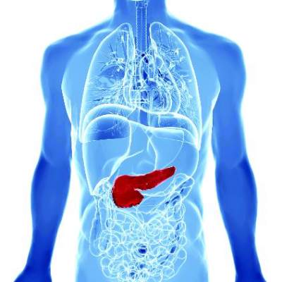
Furthermore, there were no malignant conversions among patients lacking American Gastroenterological Association high-risk features – that is, mural nodules, dilated pancreatic ducts, or cysts measuring more than 3 cm, said Dr. Wilson Kwong at the University of California San Diego Health Sciences in La Jolla. “There is a very low risk of malignant transformation of asymptomatic neoplastic pancreatic cysts after 5 years,” he and his associates wrote.
Up to 20% of cross-sectional imaging studies reveal incidental pancreatic cysts, the researchers noted. Cysts with neoplastic features are recommended for indefinite surveillance, even though there is little or no data on their natural history and malignant potential beyond 5- 10 years, they added. Therefore, they studied 310 patients who underwent endoscopic ultrasound of pancreatic cysts at an academic medical center, a Veterans’ Affairs hospital, and two community health care systems in California between 2002 and 2010. The most common age at enrollment was 66 years, 60% of patients were women, and the median follow-up period was 87 months (range, 60 to 189 months). A total of 90% of patients were followed for 5-10 years, while 10% were followed for more than 10 years (Clin Gastroenterol Hepatol. 2016 Feb 10. doi: 10.1016/j.cgh.2015.11.013).
Source: American Gastroenterological Association
In all, three patients developed invasive pancreatic malignancies after 6, 8, and 11 years of follow-up, for an overall conversion rate of 1%. Conversion rates by subgroup were 0% for patients with no high-risk AGA features, 1% (one case) for patients with one high-risk feature, and 15% (two cases) for patients with two high-risk features. “Because the risk of malignant transformation beyond 5 years is lower than the 1.4% mortality risk of pancreatic resection at high-volume centers, the argument can be made that discontinuing surveillance after 5 years is justified,” the researchers said. Specifically, surveillance could be discontinued after 5 years for neoplastic pancreatic cysts with up to one high-risk feature, particularly if patients have significant comorbidities that increase their risk of imminent death from other causes, they added. In contrast, healthy patients in their 60s and 70s might benefit from long-term surveillance given their longer life expectancy, they said. “Among patients with two high-risk features who remain surgically fit, discussion of surgery or surveillance beyond 5 years should be considered,” they emphasized.
A total of two patients developed high-grade dysplasia – a risk factor for invasive pancreatic cancer – but even so, the aggregate rate of cancer and high-grade dysplasia was 1.6%, only slightly higher than the fatality rate associated with pancreatic resection, the researchers noted. By excluding patients with recent acute pancreatitis (because of the likelihood of pseudocysts), they might have inadvertently excluded “a small number” of patients with pancreatic intraductal papillary mucinous neoplasms, they added.
The University of California San Diego Health Care System supported the study. The investigators had no disclosures.
Kwong et al. present important data demonstrating a low risk of malignant transformation for pancreas cysts followed for more than 5 years, which is similar to the risk of surgical resection. Mortality from nonpancreatic causes was found to be eightfold higher than mortality from pancreatic cancer. The goal of pancreas cyst surveillance is to prevent death from pancreatic cancer, currently accomplished by identifying high-risk cysts for surgical resection. When evaluating the utility of surveillance, patient and cyst characteristics can be considered.
Elderly patients with multiple comorbidities are unlikely to benefit from long-term surveillance as they may be poor surgical candidates and are unlikely to die from the malignant progression of a pancreas cyst. Healthy patients with a family history of pancreatic cancer and/or identifiable genetic risk factors, however, may benefit from long-term surveillance. Although demonstrated to be infrequent, cysts that have been stable for 5-10 years rarely may progress to cancer. The presence of more than one high-risk cyst feature increased the risk of progression from approximately 1% to 15%. The study of larger groups of cysts with morphologic high-risk features is required. The addition of molecular and genetic cyst and patient features has the potential to assist in risk stratification.
Clarifying which cysts and patients are likely to benefit from surveillance and resection is of increasing importance as high-resolution, cross-sectional imaging identifies greater numbers of pancreas cysts.
Dr. Harry R. Aslanian, AGAF, is director, Advanced Endoscopy Fellowship, and associate professor, Yale University, New Haven, Conn. He is a consultant for Boston Scientific and Olympus.
Only 1% of adults with asymptomatic neoplastic pancreatic cysts developed invasive pancreatic adenocarcinoma after more than 5 years of follow-up, according to a multicenter retrospective study reported in the June issue of Clinical Gastroenterology and Hepatology.
Furthermore, there were no malignant conversions among patients lacking American Gastroenterological Association high-risk features – that is, mural nodules, dilated pancreatic ducts, or cysts measuring more than 3 cm, said Dr. Wilson Kwong at the University of California San Diego Health Sciences in La Jolla. “There is a very low risk of malignant transformation of asymptomatic neoplastic pancreatic cysts after 5 years,” he and his associates wrote.
Up to 20% of cross-sectional imaging studies reveal incidental pancreatic cysts, the researchers noted. Cysts with neoplastic features are recommended for indefinite surveillance, even though there is little or no data on their natural history and malignant potential beyond 5- 10 years, they added. Therefore, they studied 310 patients who underwent endoscopic ultrasound of pancreatic cysts at an academic medical center, a Veterans’ Affairs hospital, and two community health care systems in California between 2002 and 2010. The most common age at enrollment was 66 years, 60% of patients were women, and the median follow-up period was 87 months (range, 60 to 189 months). A total of 90% of patients were followed for 5-10 years, while 10% were followed for more than 10 years (Clin Gastroenterol Hepatol. 2016 Feb 10. doi: 10.1016/j.cgh.2015.11.013).
Source: American Gastroenterological Association
In all, three patients developed invasive pancreatic malignancies after 6, 8, and 11 years of follow-up, for an overall conversion rate of 1%. Conversion rates by subgroup were 0% for patients with no high-risk AGA features, 1% (one case) for patients with one high-risk feature, and 15% (two cases) for patients with two high-risk features. “Because the risk of malignant transformation beyond 5 years is lower than the 1.4% mortality risk of pancreatic resection at high-volume centers, the argument can be made that discontinuing surveillance after 5 years is justified,” the researchers said. Specifically, surveillance could be discontinued after 5 years for neoplastic pancreatic cysts with up to one high-risk feature, particularly if patients have significant comorbidities that increase their risk of imminent death from other causes, they added. In contrast, healthy patients in their 60s and 70s might benefit from long-term surveillance given their longer life expectancy, they said. “Among patients with two high-risk features who remain surgically fit, discussion of surgery or surveillance beyond 5 years should be considered,” they emphasized.
A total of two patients developed high-grade dysplasia – a risk factor for invasive pancreatic cancer – but even so, the aggregate rate of cancer and high-grade dysplasia was 1.6%, only slightly higher than the fatality rate associated with pancreatic resection, the researchers noted. By excluding patients with recent acute pancreatitis (because of the likelihood of pseudocysts), they might have inadvertently excluded “a small number” of patients with pancreatic intraductal papillary mucinous neoplasms, they added.
The University of California San Diego Health Care System supported the study. The investigators had no disclosures.
FROM CLINICAL GASTROENTEROLOGY AND HEPATOLOGY
Key clinical point: Asymptomatic neoplastic pancreatic cysts rarely become malignant, especially in the absence of multiple American Gastroenterological Association high-risk features.
Major finding: Only 1% of patients developed invasive pancreatic adenocarcinoma after more than 5 years of surveillance.
Data source: A multicenter retrospective study of 310 patients who underwent endoscopic ultrasound evaluations of pancreatic cysts.
Disclosures: The University of California San Diego Health Care System supported the study. The investigators had no disclosures.

VIDEO: High sensitivity–CRP, IL-6 predicted inflammatory bowel disease
Women with high circulating levels of interleukin-6 and high-sensitivity C-reactive protein were at significantly greater risk of inflammatory bowel disease (IBD) compared with those testing in the lowest quintiles, according to a prospective nested case-control study.
The findings point to a preclinical state in IBD, in which patients are not yet symptomatic but have positive serologic markers, as occurs in rheumatoid arthritis and systemic lupus erythematosus, said Dr. Paul Lochhead at Massachusetts General Hospital in Boston, and his associates. “To our knowledge, no previous study has examined prediagnostic inflammatory markers in relation to IBD risk,” the investigators added. “Characterizing preclinical inflammation in IBD could give insights into the natural history of [Crohn’s disease] and [ulcerative colitis], and might help identify potential windows for early therapeutic or preventive interventions in high-risk individuals.”
SOURCE: American Gastroenterological Society
The study included 83 patients with Crohn’s disease, 90 patients with ulcerative colitis, and 344 matched controls. Patients were from two national prospective cohort studies – the Nurses’ Health Study, which includes female nurses aged 35-55 years at enrollment, and the Nurses’ Health Study II, which includes female nurses aged 24-42 years at enrollment. Both studies are ongoing, with follow-up rates exceeding 90%. To assemble the cohort, the researchers extracted questionnaire data and then obtained medical records for blinded review. They confirmed diagnoses of Crohn’s disease and ulcerative colitis using standard case definitions, they said (Clin Gastroenterol Hepatol. 2016 Feb 13. doi: 10.1016/j.cgh.2016.01.016).
Participants testing in the highest quintiles for circulating hs-CRP and IL-6 were at greater risk of Crohn’s disease and ulcerative colitis than were those in the lowest quintiles, even after accounting for age, smoking status, body mass index, oral contraceptive use, and cumulative physical activity. For IL-6, odds ratios were 4.7 for Crohn’s disease (95% confidence interval; 1.9-11.5), and 3.4 for ulcerative colitis (95% CI; 1.4-8.2). For hs-CRP, odds ratios were 2.8 for Crohn’s disease (95% CI; 1.15-6.9) and 1.8 for ulcerative colitis (95% CI; 0.8-4.0). The longest interval between testing and diagnosis of IBD was 20 years, Crohn’s disease patients were diagnosed within 10 years, and patients testing in the upper quintile for the inflammatory markers were diagnosed an average of 10.6 years later, the researchers said.
Study participants tended to be in their early 50s when first tested, which exceeds the typical age of Crohn’s disease and ulcerative colitis onset and might limit the generalizability of the findings, the investigators said. They tried to eliminate confounding from undiagnosed baseline IBD by excluding participants diagnosed within 2 years of blood collection, they added. “The differences in overall median inflammatory marker levels between cases and control subjects in our study were small; however, differences of similar magnitude have been reported between groups with disparate outcomes in studies of cardiovascular disease,” they noted. “Moreover, when comparing extreme quintiles of median inflammatory marker levels, where risk of [Crohn’s disease] or [ulcerative colitis] was most evident, the differences were more substantial.”
The study was funded by the National Institutes of Health, National Institute of Diabetes and Digestive and Kidney Diseases, Crohn’s and Colitis Foundation of America, and the American Gastroenterological Association. Dr. Lochhead had no disclosures. Two coinvestigators disclosed relationships with Exact Sciences, AbbVie, Cubist Pharmaceuticals, Bayer Healthcare, Pfizer, and Pozen.
FROM CLINICAL GASTROENTEROLOGY AND HEPATOLOGY
Key clinical point: Patients might have subclinical inflammation for several years before developing inflammatory bowel disease.
Major finding: Participants testing in the highest quintiles for circulating high-sensitivity C-reactive protein and interleukin-6 were at greater risk of Crohn’s disease and ulcerative colitis, compared with individuals testing in the lowest quintiles for each marker, with estimated odds ratios of 1.8, 2.8, 3.4, and 4.7.
Data source: A prospective nested case-control study of female nurses, including 83 with Crohn’s disease, 90 with ulcerative colitis, and 344 matched controls.
Disclosures: The study was funded by the National Institutes of Health, National Institute of Diabetes and Digestive and Kidney Diseases, Crohn’s and Colitis Foundation of America, and the American Gastroenterological Association. Dr. Lochhead had no disclosures. Two coinvestigators disclosed relationships with Exact Sciences, AbbVie, Cubist Pharmaceuticals, Bayer Healthcare, Pfizer, and Pozen.

Combination OCs tied to increased risk of surgery for Crohn’s
Women with Crohn’s disease who were prescribed combination oral contraceptive pills for more than 3 years were 68% more likely to need gastrointestinal surgery than patients who did not use oral contraceptives, according to a national prospective cohort study reported in the June issue of Gastroenterology.
“Our data suggest the importance of carefully evaluating contraceptive options among women with established Crohn’s disease. Future studies should focus on mechanisms by which oral contraceptive use alters risk and progression,” said Dr. Hamed Khalili of Harvard Medical School in Boston and his associates at Harvard and Karolinska Institutet, Solna, Sweden.

Several studies have linked OC exposure to Crohn’s disease itself. But past studies of OCs and Crohn’s disease progression were small, retrospective, or did not adequately ascertain OC exposure, Dr. Khalili and his associates said. To help fill this gap, they identified 4,036 women with Crohn’s disease aged 16-51 years through the Swedish National Patient Register, and ascertained OC exposure by analyzing Sweden’s national prescription database (Gastroenterology. 2016 Feb 23. doi: 10.1053/j.gastro.2016.02.041).
During a median follow-up period of 58 months, 482 patients (12%) underwent surgery related to Crohn’s disease, the researchers said. Use of OCs was associated with surgery, but the link only reached statistical significance among women prescribed combination (estrogen-containing) regimens for more than 3 years (adjusted hazard ratio, 1.68; 95% confidence interval, 1.06-2.67) or for more than 900 doses (aHR, 1.60; 95% CI, 1.1-2.34). For each additional year that combination OCs were prescribed, surgery risk rose by nearly 30% (aHR, 1.29; 95% CI, 1.05-1.57). Thus, one extra surgery was needed for every 83 patients who received combination OCs for at least 1 year, said the investigators. Progestin-only prescriptions did not increase the likelihood of needing surgery, and there was no link between current or prior OC exposure and the chances of being prescribed steroids, they noted.
Only one other study has linked OC exposure with Crohn’s disease progression, and it included only 158 patients followed for just a year, Dr. Khalili and his associates said. Exactly how estrogen exposure might trigger Crohn’s disease progression is unclear, but OCs have been linked to changes in intestinal barrier function, increased humoral immunity, and modulation of testosterone levels, which in turn affects cytokine function, they added. “Regardless of the potential mechanism, the effect of OCs on Crohn’s disease progression appears to be related to consistent and long-term use of these medications. Similar patterns of associations have also been reported with other chronic illnesses, such as breast cancer and cardiovascular diseases,” said the researchers. Current OC use itself might not have predicted surgery in the study because about one in four women in Sweden stop taking OCs or switch to a nonhormonal form within 6 months of being prescribed them, they added.
The work was funded by the Crohn’s and Colitis Foundation of America, the National Institute of Diabetes and Digestive and Kidney Diseases, the American Gastroenterological Association, and the American College of Gastroenterology. Dr. Khalili reported receiving consulting fees from Abbvie. One coinvestigator reported consulting relationships with Bayer Healthcare, Pfizer, and Pozen. The other investigators had no disclosures.
Women with Crohn’s disease who were prescribed combination oral contraceptive pills for more than 3 years were 68% more likely to need gastrointestinal surgery than patients who did not use oral contraceptives, according to a national prospective cohort study reported in the June issue of Gastroenterology.
“Our data suggest the importance of carefully evaluating contraceptive options among women with established Crohn’s disease. Future studies should focus on mechanisms by which oral contraceptive use alters risk and progression,” said Dr. Hamed Khalili of Harvard Medical School in Boston and his associates at Harvard and Karolinska Institutet, Solna, Sweden.
Several studies have linked OC exposure to Crohn’s disease itself. But past studies of OCs and Crohn’s disease progression were small, retrospective, or did not adequately ascertain OC exposure, Dr. Khalili and his associates said. To help fill this gap, they identified 4,036 women with Crohn’s disease aged 16-51 years through the Swedish National Patient Register, and ascertained OC exposure by analyzing Sweden’s national prescription database (Gastroenterology. 2016 Feb 23. doi: 10.1053/j.gastro.2016.02.041).
During a median follow-up period of 58 months, 482 patients (12%) underwent surgery related to Crohn’s disease, the researchers said. Use of OCs was associated with surgery, but the link only reached statistical significance among women prescribed combination (estrogen-containing) regimens for more than 3 years (adjusted hazard ratio, 1.68; 95% confidence interval, 1.06-2.67) or for more than 900 doses (aHR, 1.60; 95% CI, 1.1-2.34). For each additional year that combination OCs were prescribed, surgery risk rose by nearly 30% (aHR, 1.29; 95% CI, 1.05-1.57). Thus, one extra surgery was needed for every 83 patients who received combination OCs for at least 1 year, said the investigators. Progestin-only prescriptions did not increase the likelihood of needing surgery, and there was no link between current or prior OC exposure and the chances of being prescribed steroids, they noted.
Only one other study has linked OC exposure with Crohn’s disease progression, and it included only 158 patients followed for just a year, Dr. Khalili and his associates said. Exactly how estrogen exposure might trigger Crohn’s disease progression is unclear, but OCs have been linked to changes in intestinal barrier function, increased humoral immunity, and modulation of testosterone levels, which in turn affects cytokine function, they added. “Regardless of the potential mechanism, the effect of OCs on Crohn’s disease progression appears to be related to consistent and long-term use of these medications. Similar patterns of associations have also been reported with other chronic illnesses, such as breast cancer and cardiovascular diseases,” said the researchers. Current OC use itself might not have predicted surgery in the study because about one in four women in Sweden stop taking OCs or switch to a nonhormonal form within 6 months of being prescribed them, they added.
The work was funded by the Crohn’s and Colitis Foundation of America, the National Institute of Diabetes and Digestive and Kidney Diseases, the American Gastroenterological Association, and the American College of Gastroenterology. Dr. Khalili reported receiving consulting fees from Abbvie. One coinvestigator reported consulting relationships with Bayer Healthcare, Pfizer, and Pozen. The other investigators had no disclosures.
FROM GASTROENTEROLOGY
Key clinical point: Long-term use of combination oral contraceptives significantly increased the risk of surgery among women with Crohn’s disease.
Major finding: Women who used combination OCs for more than 3 years were 68% more likely to need surgery than were nonusers.
Data source: A prospective national registry study of 4,036 women with Crohn’s disease.
Disclosures: The study was funded by the Crohn’s and Colitis Foundation of America, the National Institute of Diabetes and Digestive and Kidney Diseases, the American Gastroenterological Association, and the American College of Gastroenterology. Dr. Khalili reported receiving consulting fees from Abbvie. One coinvestigator reported consulting relationships with Bayer Healthcare, Pfizer, and Pozen. The other investigators had no disclosures.

Cell-based strategy curbs constipation
Treatment at the nanomolecular level may become an alternative for various types of constipation, in particular for the opioid-induced constipation that is common after surgery, based on data from a proof of concept study involving mice and a small-molecule activator.
“Activation of the cystic fibrosis transmembrane conductance regulator (CFTR) chloride channel is the primary pathway that drives fluid secretion in the intestine, which maintains lubrication of luminal contents,” wrote Dr. Onur Cil of the University of California, San Francisco, and colleagues. The researchers examined whether direct activation of the CFTR would prompt fluid secretion and reverse stool dehydration when applied to constipated mice. The findings were published online in the May issue of the journal Cellular and Molecular Gastroenterology and Hepatology (2016. doi: 10.1016/j.jcmgh.2015.12.010).
The researchers identified a promising activator, the phenylquinoxalinone CFTRact-J027. Mice received up to 10 mg/kg CFTRact-J027 either orally or intraperitoneally (IP), with doses including 0.1 mg/kg, 0.3 mg/kg, 1 mg/kg, 3 mg/kg, and 10 mg/kg.
Overall, IP doses of CFTRact-J027 at 10 mg/kg normalized stool in the constipated mice, and dose-response studies showed a 50% effective dose of 2 mg/kg in these mice.
When given orally, CFTRact-J027 “normalized stool output and water content in a loperamide-induced mouse model of constipation with a 50% effective dose of approximately 0.5 mg/kg,” that was significantly lower than the IP administration, the researchers noted. An oral dose of 10 mg/kg CFTRact-J027 1 hour before inducing constipation also was effective in normalizing stool output and water content in loperamide-treated mice, with no effect in control nonconstipated mice.
The activator was not effective against constipation in cystic fibrosis mice that were missing a functional CFTR, they added.
The researchers used an in vivo closed loop model to specifically test the effects of CFTRact-J027 on intestinal fluid secretion and absorption and found that CFTRact-J027 caused “a 140% increase in loop weight/length ratio, indicating fluid secretion into the intestinal lumen in wild-type mice.” However, there was no effect in cystic fibrosis mice, further supporting the CFTR-selective mechanism of action, the researchers said.
As for potential toxic effects of the treatment, CFTRact-J027 showed no impact on the major serum chemistry and blood parameters of the mice after 7 days, and had no apparent impact on body weight. No accumulation of fluid (the most significant potential adverse effect) was noted in the airway or lungs of the treated mice.
Additional toxicity data are needed to continue preclinical development, the researchers said. However, “our data provide evidence for the prosecretory action of a CFTR activator in mouse intestine and proof of concept for its use in the treatment of various types of constipation, which could include opioid-induced constipation, chronic idiopathic constipation, and irritable bowel syndrome with constipation predominance,” they wrote. In addition, a CFTR activator similar to that used in this study may have clinical applications for other conditions including asthma, dry eye, cholestatic liver disease, chronic obstructive pulmonary disease and bronchitis, and cigarette smoke–induced lung dysfunction, they added.
Dr. Cil and two coauthors are inventors on a provisional patent filing, with rights owned by the University of California, San Francisco. The study was funded in part by several grants from organizations including the National Institutes of Health and the Cystic Fibrosis Foundation.
FROM CELLULAR AND MOLECULAR GASTROENTEROLOGY AND HEPATOLOGY
Key clinical point: Treatment at the nanomolecular level may become an alternative for various types of constipation.
Major finding: Oral doses of CFTRact-J027 normalized stool output and water content in a loperamide-induced mouse model of constipation with a 50% effective dose of approximately 0.5 mg/kg.
Data source: A proof-of-concept study in which a cell-based screen was performed for 120,000 druglike, synthetic small molecules that were then tested in constipation-induced mice and control mice.
Disclosures: Dr. Cil and two coauthors are inventors on a provisional patent filing, with rights owned by the University of California, San Francisco. The study was funded in part by several grants from organizations including the National Institutes of Health and the Cystic Fibrosis Foundation.
Cirrhosis 30-day readmissions down 40% with quality improvement initiative
Using checklists and electronic decision support in an inpatient liver unit, quality improvement (QI) care protocols reduced 30-day readmissions of patients with cirrhosis by 40%, due mostly to a drop in readmissions for hepatic encephalopathy (HE) according to a report published in the May issue of Clinical Gastroenterology and Hepatology.
For patients initially admitted for overt HE, the 30-day readmission rate was 26.0% (27 of 104), compared with 48.9% (66 of 135) before implementation of QI. The proportion of total readmissions due to HE after QI was 9.6% (14 of 146), compared with 40.7% (79 of 194) before QI. In addition, length of stay for HE patients was significantly reduced (–1.34 days; 95% confidence interval, –2.38 to –0.32; P = .01). There were no significant changes in 90-day mortality.
Source: American Gastroenterological Association
“Our study advances the current literature on QI for patients with cirrhosis by presenting an inexpensive, easy to implement, and generalizable approach,” wrote Dr. Elliot Tapper of Beth Israel Deaconess Medical Center, Boston, and his colleagues. Previous studies have addressed readmission interventions among patients with cirrhosis, but the protocols required costly infrastructure, expertise, and institutional commitments. The current study supports the value of standard checklists and education, according to the investigators, “showing that outcomes improve further when checklist items are hard-wired into the ordering system.” (Clin Gastroenterol Hepatol. 2016 Apr 7. doi: 10.1016/j.cgh.2015.08.041).
The QI initiative encompassed several aspects of care. All HE patients were designated to receive rifaximin, and their lactulose dosing was adjusted to mental status using the Richmond Agitation and Sedation Scale. For patients with spontaneous bacterial peritonitis (SBP), timely administration of the correct dose of antibiotics and albumin was promoted, as were prophylactic measures for all patients, such as variceal hemorrhage prophylaxis and subcutaneous heparin for the prevention of venous thrombosis.
The three-part program entailed a run-in phase for preliminary checklist troubleshooting, a hand-held checklist phase, including the HE protocol, SBP treatment, and prophylactic measures, and a final electronic phase in which checklist items were incorporated into the hospital’s electronic provider order entry system using mandatory preset doses and linked medications.
Individual protocol items were demonstrated to affect the readmission rate. Rifaximin use for HE patients rose from 78.1% to 96.3%, and use of rifaximin was associated with lower adjusted odds of 30-day readmission (OR, 0.39; 95% CI, 0.16-0.87; P = .02). The dose/frequency of lactulose for HE patients increased, and patients who had 6 or more cups of lactulose on the day of their readmission had significantly lower adjusted length of stay (–2.36 days; 95% CI, –3.40 to –1.31; P less than .0001). Patients taking SBP prophylaxis had lower readmission rates (OR, 0.51; 95% CI, 0.31-0.83; P = .007).
The prospective study from 2011 to 2013 evaluated patients with cirrhosis who were admitted to the liver unit of Beth Israel Deaconess Medical Center, Boston. Patients were diagnosed with cirrhosis caused by hepatitis C (44.9%), alcoholic liver disease (34%), hepatitis B (5.4%), and biliary cirrhosis (1.8%). In total, 824 unique patients were admitted 1,720 times; 485 (58.9%) were admitted once, 268 (32.5%) were admitted 2-4 times, and 71 (8.6%) were admitted 5 or more times. The median length of stay for all patients was 4.0 days (interquartile range, 2.0-8.0).
Dr. Tapper and his coauthors reported having no disclosures.
Key clinical point: Care protocols implemented by electronic decision support reduced 30-day readmissions of patients with cirrhosis by 40% in an inpatient liver unit.
Major finding: The drop was likely driven by fewer readmissions for hepatic encephalopathy (HE): the 30-day HE readmission rate was 26.0% (27 of 104), compared with 48.9% (66 of 135) before implementation of quality improvement.
Data sources: The prospective study evaluated 824 patients who were admitted 1,720 times to the liver unit of Beth Israel Deaconess Medical Center, Boston.
Disclosures: Dr. Tapper and his coauthors reported having no disclosures.