User login
AML risk is doubled in low-risk thyroid cancer patients unnecessarily given radioactive iodine therapy
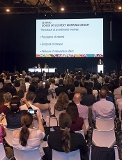
MADRID – Radioactive iodine treatment is associated with nearly twice the risk of developing acute myeloid leukemia (AML) in patients with well-differentiated thyroid cancer, based on data from the Surveillance Epidemiology and End Results (SEER) registry.
Up to 40% of patients in Europe and North America with well-differentiated thyroid cancer still receive radioactive iodine treatment “even though RAI has no proven benefit in this population,” Remco Molenaar, MD, PhD, of the University of Amsterdam reported at the European Society of Medical Oncology Congress.
Of 148,215 patients treated for well-differentiated thyroid cancer between 1973 and 2014, 55% had surgery only and 45% received surgery plus radioactive iodine treatment. After a median 4.3 years of follow-up, 44 patients developed AML. When cases in those exposed to RAI were cross-referenced to those who were not, the relative risk was increased more than fivefold. When the analysis controlled for an extensive list of potentially confounding variables, the hazard ratio of 1.79 remained statistically significant (P = .03).
“There is a nearly twofold increased risk even though radioactive iodine treatment is not indicated in this population,” Dr. Molenaar said. Moreover, AML following treatment for well-differentiated thyroid cancer was associated with a substantial reduction in expected overall survival, falling from a median 24.4 years to 7.5 years.
Compared with other AML patients, “those who develop AML after RAI also have a worse prognosis,” added Dr. Molenaar, noting the difference in overall survival is highly statistically significant (1.2 vs. 3.5 years; P = .004).
The ESMO-invited discussant, Tim Somervaille, MD, senior group leader of the Leukemia Biology Laboratory at the Cancer Research UK Manchester Institute, called this analysis “a more thorough and detailed study” than previous retrospective analyses, but he added a note of caution: Despite the almost twofold increase in risk, AML remains a rare iatrogenic event in thyroid cancer patients even if it is avoidable by withholding RAI therapy.
“These data do suggest that the risk is measurable and will further provide some downward pressure on the numbers of patients having unnecessary RAI therapy for well-differentiated thyroid cancer,” he said.
In the discussion that followed the presentation, one audience member suggested that telling patients they have a twofold increased risk of AML after RAI therapy is misleading. It was emphasized that a twofold increase of a very small number is still a very small number, but Dr. Molenaar suggested that this misses the point.
“I don’t think this is something that you need to discuss with patients, because you should not be giving RAI therapy to thyroid cancer patients with low- or intermediate-risk disease,” he said. Any AML case caused “by a therapy with no proven benefit is one too many,” especially since unnecessary RAI adds inconvenience and cost to treatment.
AT ESMO 2017
Key clinical point:
Major finding: The hazard ratio for AML after RAI therapy in well-differentiated thyroid cancer patients is almost doubled (HR = 1.79).
Data source: Population-based, retrospective study of 148,215 patients treated for well-differentiated thyroid cancer between 1973 and 2014.
Disclosures: Dr. Molenaar reported that he had no relevant financial relationships to disclose.
Report details progress, obstacles in cancer research and care
Deaths from cancer are on the decline in the US, but new cases of cancer are on the rise, according to the 7th annual American Association for Cancer Research (AACR) Cancer Progress Report.
The data suggest the cancer death rate declined by 35% from 1991 to 2014 for children and by 25% for adults, a reduction that translates to 2.1 million cancer deaths avoided.
However, 600,920 people in the US are projected to die from cancer in 2017.
And the number of new cancer cases is predicted to rise from 1.7 million in 2017 to 2.3 million in 2030.
The report also estimates there will be 62,130 new cases of leukemia in 2017 and 24,500 leukemia deaths this year.
This includes:
- 5970 cases of acute lymphocytic leukemia and 1440 deaths
- 20,110 cases of chronic lymphocytic leukemia and 4660 deaths
- 21,380 cases of acute myeloid leukemia (AML) and 10,590 deaths
- 8950 cases of chronic myeloid leukemia and 1080 deaths.
The estimate for lymphomas is 80,500 new cases and 21,210 deaths.
This includes:
- 8260 cases of Hodgkin lymphoma (HL) and 1070 deaths
- 72,240 cases of non-Hodgkin lymphoma and 20,140 deaths.
The estimate for myeloma is 30,280 new cases and 12,590 deaths.
The report says the estimated new cases of cancer are based on cancer incidence rates from 49 states and the District of Columbia from 1995 through 2013, as reported by the North American Association of Central Cancer Registries. This represents about 98% of the US population.
The estimated deaths are based on US mortality data from 1997 through 2013, taken from the National Center for Health Statistics of the Centers for Disease Control and Prevention.
Drug approvals
The AACR report notes that, between August 1, 2016, and July 31, 2017, the US Food and Drug Administration (FDA) approved new uses for 15 anticancer agents, 9 of which had no previous FDA approval.
Five of the agents are immunotherapies, which the report dubs “revolutionary treatments that are increasing survival and improving quality of life for patients.”
Among the recently approved therapies are 3 used for hematology indications:
- Ibrutinib (Imbruvica), approved to treat patients with relapsed/refractory marginal zone lymphoma who require systemic therapy and have received at least 1 prior anti-CD20-based therapy
- Midostaurin (Rydapt), approved as monotherapy for adults with advanced systemic mastocytosis and for use in combination with standard cytarabine and daunorubicin induction, followed by cytarabine consolidation, in adults with newly diagnosed AML who are FLT3 mutation-positive, as detected by an FDA-approved test.
- Pembrolizumab (Keytruda), approved to treat adult and pediatric patients with refractory classical HL or those with classical HL who have relapsed after 3 or more prior lines of therapy.
Disparities and costs
The AACR report points out that advances against cancer have not benefited everyone equally, and cancer health disparities are some of the most pressing challenges.
Among the disparities listed is the fact that adolescents and young adults (ages 15 to 39) with AML have a 5-year relative survival rate that is 22% lower than that of children (ages 1 to 14) with AML.
And Hispanic children are 24% more likely to develop leukemia than non-Hispanic children.
Another concern mentioned in the report is the cost of cancer care. The direct medical costs of cancer care in 2014 were estimated to be nearly $87.6 billion. This number does not include the indirect costs of lost productivity due to cancer-related morbidity and mortality.
With this in mind, the AACR is calling for a $2 billion increase in funding for the National Institutes of Health in fiscal year 2018, for a total funding level of $36.2 billion.
The AACR also recommends an $80 million increase in the FDA budget, bringing it to $2.8 billion for fiscal year 2018. ![]()
Treatment for WDTC tied to increased risk of AML
MADRID—New research suggests patients with well-differentiated thyroid cancer (WDTC) are more likely to develop acute myeloid leukemia (AML) if they receive radioactive iodine (RAI) after surgery.
Of the WDTC patients studied, those who only underwent surgery were less likely to develop AML.
WDTC patients who did develop AML had a far worse prognosis than WDTC patients without AML and patients with de novo AML.
These results were presented at the ESMO 2017 Congress (abstract 996O).
Researchers already knew that the risk of developing leukemia is increased after radiation and RAI treatment for WDTC.
However, the risk of AML following RAI treatment in WDTC survivors had not been fully characterized, according to Remco J. Molenaar, an MD/PhD candidate at Academic Medical Centre in Amsterdam, Netherlands.
Therefore, Molenaar and colleagues reviewed data from all 18 registries in the Surveillance Epidemiology and End Results database for WDTC cases treated solely with surgery or by surgery and RAI.
The researchers identified 148,215 patients who were diagnosed with WDTC from 1973 to 2014. Fifty-five percent were treated with surgery alone, and 45% were treated with surgery and RAI.
The median follow-up was 4.3 person-years. AML occurred in 44 patients in the surgery-only arm and 56 patients in the RAI arm.
A comparison to the background rates in the general population showed that patients receiving surgery plus RAI had an increased risk of developing AML (relative risk=5.6; 95% confidence interval [CI] 3.8, 8.1; P<0.0001), after correcting for age, sex, and year of WDTC diagnosis.
This risk peaked within the first 3 years following RAI and subsequently regressed to baseline rates.
In multivariate analysis that corrected for sex and WDTC tumor size, 3 variables emerged as independent predictors of AML development. There was an increased risk of AML associated with:
- Patient age (hazard ratio [HR]=1.03; 95% CI 1.02, 1.05; P<0.001)
- WDTC tumor stage (HR=1.36; 95% CI 1.04, 1.79; P=0.03)
- Receiving RAI after thyroidectomy vs thyroidectomy alone (HR=1.38; 95% CI 1.09, 1.75; P=0.007).
The researchers also found prognosis is significantly poorer in WDTC patients who develop AML following RAI and in patients with spontaneous AML development.
Case-control analyses revealed that WDTC patients who developed AML after surgery and RAI survived one-third as long as matched control patients who were successfully treated for WDTC but did not develop AML. The median overall survival was 7.4 years and 24.4 years, respectively (P<0.0001).
Patients who were diagnosed with AML after RAI treatment also had a significantly worse prognosis than patients with de novo AML. The median overall survival was 1.2 years and 3.5 years, respectively (P=0.004).
The researchers noted that rates of AML in WDTC survivors will likely continue to rise due to the increasing incidence of WDTC, the young ages at which most WDTC diagnoses are made, and the otherwise high survival rates of patients with WDTC. ![]()
CCSs have higher burden of chronic conditions
Adult survivors of childhood cancer have a greater cumulative burden of chronic health conditions than the general public, according to research published in The Lancet.
The study showed that, by age 50, childhood cancer survivors (CCSs) had experienced, on average, 17.1 chronic health conditions, and matched control subjects had experienced 9.2.
“The cumulative burden of chronic disease revealed in this analysis, along with the complexity and severity of chronic conditions some survivors experience, found childhood cancer survivors to be a vulnerable, medically complex population,” said study author Nickhill Bhakta, MD, of St. Jude Children’s Research Hospital in Memphis, Tennessee.
For this study, Dr Bhakta and his colleagues assessed the lifelong impact of 168 chronic health conditions—such as hepatic, thyroid, ocular, and reproductive disorders—on CCSs and control subjects.
The 3010 evaluable CCSs had survived 10 years or longer from their initial cancer diagnosis and were 18 years or older as of June 30, 2015. The 272 controls had no history of pediatric cancer and were matched to CCSs by age and sex.
At age 50, the cumulative incidence of chronic health conditions (of any grade) was 99.9% in CCSs and 96.0% in controls (P<0.0001). The cumulative incidence of grade 3 to 5 chronic health conditions was 96.0% and 84.9%, respectively (P<0.0001).
The cumulative burden for CCSs was 17.1 chronic health conditions, including 4.7 that were grade 3 to 5. For controls, the cumulative burden was 9.2 chronic health conditions, including 2.3 that were grade 3 to 5 (P<0.0001 for both comparisons).
The researchers said second neoplasms, spinal disorders, and pulmonary disease were major contributors to the excess total cumulative burden observed in CCSs. However, there was “notable heterogeneity” in burden according to the patients’ primary cancer diagnosis.
For instance, growth hormone deficiency was in the top 10th percentile of chronic health conditions for survivors of acute lymphoblastic leukemia but not for controls.
And pulmonary function deficits were in the top 10th percentile for survivors of acute myeloid leukemia and Hodgkin lymphoma but not for controls or survivors of acute lymphoblastic leukemia or non-Hodgkin lymphoma.
“This study found that the average childhood cancer survivor has a cumulative burden of chronic disease that requires a significant time investment by healthcare providers to disentangle and manage—time that community providers are unlikely to have,” Dr Bhakta said.
“The results suggest that childhood cancer survivors may benefit from the integrated, specialized healthcare delivery that is being tried for individuals infected with HIV or those with other complex, chronic health problems.” ![]()
New AML approvals changing the treatment landscape
With a recent flurry of new drug approvals, the treatment landscape for acute myeloid leukemia has expanded, raising new questions about how to incorporate those drugs into patient care.
Until about a decade ago, advances in AML therapy centered mainly around iterations of daunorubicin and cytarabine. Now, novel and targeted agents, many specifically going after mutational byproducts, are yielding some great results and raising hopes for better survival outcomes, Jeffrey Lancet, MD, said in an interview.
“When I go to sleep at night, I often dream about ... 10-year survival rates in the 80% range. And then I wake up ... and I realize this is actually [the survival curve for chronic myeloid leukemia]. This is where we’d like to be [with AML].” Those outcomes are a long way off, but appreciable incremental gains may lie ahead with the recent advances in AML therapy, said Dr. Lancet, chair of the department of malignant hematology at Moffitt Cancer Center in Tampa.
In addition to the new approvals, 16 drugs are in late stage clinical development and will likely contribute to an AML market that is expected to surpass $1.5 billion by 2026, according to projections by the market intelligence company GlobalData.
Vyxeos
The liposome-encapsulated combination of daunorubicin and cytarabine (Vyxeos) was approved in August by the Food and Drug Administration for the treatment of therapy-related AML and AML with myelodysplasia-related changes.
In a phase 3 randomized trial, the fixed-dose combination product was associated with median overall survival of 9.6 months, compared with 5.9 months with a standard combination of cytarabine and daunorubicin (7+3).
“I would envision that Vyxeos will hold and become the primary standard of care for fit chemotherapy-suitable older patients, or any patients for that matter, who are dealing with secondary-like AML or high-risk AML, based on the phase 3 results that we demonstrated,” Dr. Lancet, the principal investigator for the trial, said in an interview.
Asked whether the improved survival with Vyxeos is primarily related to more patients becoming transplant eligible or to significant reductions in disease burden, Dr. Lancet remarked that it’s likely a mixture of both.
The high remission rate with Vyxeos vs. standard 7+3 therapy means Vyxeos has the ability to stand on its own, and “the potential to send more patients to transplant and to get better results.”
“Transplant is part of the continuum of care of AML, including in older patients, and Vyxeos is going to become a standard part of that care,” he remarked. But transplant outcomes were not a predesignated component of the phase 3 trial, and further study will be needed to determine Vyxeos’ role as a bridge to transplant. “At this stage I can reasonably state that it has a role in the upfront therapy of secondary and high-risk AML, regardless of whether the patient is being considered for transplant.”
The early stages of working Vyxeos into the therapeutic mix come with some challenges, however, according to Donna Capozzi, PharmD.
Vyxeos is a fixed-dose combination that comes in vials containing 44 mg daunorubicin and 100 mg cytarabine encapsulated in liposomes. Patient dosing is based on the daunorubicin component and calculated based on body surface area (mg/m2), meaning the cytarabine dose does not need to be calculated. There are both pros and cons to this approach, she explained.
Benefits include a longer half-life with Vyxeos vs. standard 7+3, and the fact that during induction the drug is delivered on days 1, 3, and 5 for 90 minutes rather than continuously for 7 days as with 7+3, Dr. Capozzi said.
The main concern relates to ensuring that the dosing is calculated based on the proper component, she said.
“We had our first patient last week. It was very time consuming, with double and triple checking to make sure everything was correct,” she said. Preparing the drug is also time-consuming, as it involves multiple steps, such as warming, which is not required with standard 7+3; the additional labor factors will have to be built into workflow, she noted.
“The other piece not fully in place right now is building [the use of Vyxeos] into electronic health records,” she said, adding that safeguards put into place through EHRs will also help to streamline the administration process.
For example, cardiac toxicity is a known effect of daunorubicin; the EHR will help track lifetime cumulative dosing of that component, which is otherwise challenging, especially when using a combination product, she said.
The process will get easier over time, as use of Vyxeos becomes more prevalent in practice, she added. “None of these are insurmountable issues.”
Cost is another matter. Based on average wholesale prices, the cost per cycle is approximately $40,000 with Vyxeos vs. about $4,300 for conventional 7+3 therapy, Dr. Capozzi said. Given the differential, there will be a great deal of debate as to which patients will derive the most benefit from Vyxeos, she said.
Also, it will take time to figure out the extent of adverse events. “For liposomal products in general, rash-type side effects can be really significant. Hand-foot syndrome was not reported in the initial trials, but we’ll keep our eyes open to see how that plays out,” she said noting that the one patient treated so far at the University of Pennsylvania is doing very well. “We will learn more with real world experience.”
Oral targeted therapies
Enasidenib (Idhifa) was approved under priority review in August in conjunction with a companion diagnostic IDH2 assay for patients with relapsed or refractory disease and specific mutations in the IDH2 gene. Midostaurin (Rydapt) was approved in April for use in conjunction with standard daunorubicin and cytarabine induction and cytarabine consolidation in adults with FLT3 mutation-positive AML.
In a phase 1 dose escalation study reported at the annual meeting of the European Hematology Association, enasidenib was associated with an overall response rate of 37% in patients with relapsed/refractory AML, including 20.1% complete responses and 7.9% complete responses with incomplete recovery of platelets or incomplete hematologic recovery, 3.7% with partial responses, and 5.1% with a morphologic leukemia-free state. Patients who had a CR had a median overall survival of 22.9 months. For patients with responses other than CR, the median OS was 15.1 months. For patients with no response to the drug, the median OS was 5.6 months, Dr. Eytan M. Stein, of Memorial Sloan Kettering Cancer Center in New York, reported.
Additionally, need for transfusions was reduced in 34% of 157 patients who required transfusions at study entry.
“In a relapsed or refractory group of patients where there’s no true standard of care, this drug definitely represents a major breakthrough and has a lot of utility as a single agent, as a potential bridge to a transplant, and in combination with new or even old drugs – including regular old induction chemotherapy as a way to improve responses and outcomes in the future,” Dr. Lancet said, adding that as an oral agent it has potential for development as a maintenance strategy.
This agent could have a large impact, he said, adding: “I think this sets the paradigm for novel targeted therapies.”
Midostaurin has also emerged as a new standard of care, particularly for younger patients, Dr. Lancet said.
The approval of the multitargeted kinase inhibitor was based on the results of the randomized, placebo-controlled phase 3 RATIFY trial, which demonstrated significantly longer overall and event-free survival vs. placebo and standard chemotherapy in newly diagnosed AML patients with FLT3 gene mutations.
“I think this will be the new comparator for future studies, whatever they may be, for this patient population,” he said.
Dr. Capozzi noted that she has had some difficulty obtaining prior authorization for enasidenib due to its high cost (about $1,000/day).
The drug is taken orally on days 8-21 of a 28-day treatment cycle. In RATIFY, patients who achieved complete remission after induction therapy received four 28-day cycles of consolidation therapy.
Dr. Capozzi noted that the dosing regimen can be confusing, as it changes depending on whether it is used for induction or consolidation. It remains to be seen how these agents will fit into the treatment setting, she said.
Targeted therapies in development
Other targeted therapies in development for AML include an IDH1 inhibitor, the BCL2 inhibitor venetoclax, and several second-generation FLT3 inhibitors such as gilteritinib, Dr. Lancet said.
Venetoclax, which is currently approved for chronic lymphocytic leukemia, has shown single agent activity, but is even more promising in combination with low-dose cytarabine or aza-nucleosides, he noted.
For example, in one recent study reported at the annual congress of the European Hematology Association, response rates in older, newly diagnosed AML patients were as high as 72% for azacitidine plus venetoclax, and 76% for decitabine plus venetoclax.
“So there’s a lot of interest and promise,” Dr. Lancet said, adding that venetoclax may have broad application in AML. “We’ll be seeing a lot more data in the next year or two.”
An unusual aspect of venetoclax, which is used often for CLL, is the need for observation during dose escalation, Dr. Capozzi noted. Patients tend to question the need for admission for observation with the use of an oral agent, thus efforts are underway to develop criteria for outpatient observation.
Otherwise, venetoclax is fairly easy to access and use, and is well tolerated, she said.
“I expect as we learn more about where (venetoclax) fits in, it will be a much more commonplace agent” as part of AML therapy, she said.
Gilteritinib, as well as the second generation FLT3 inhibitors quizartinib and crenolanib, are also of interest in AML. With midostaurin already on the market, however, different strategies are being pursued, Dr. Lancet said.
“I believe gilteritinib is entering the fray in relapsed/refractory disease, and crenolanib is being looked at in the upfront FLT3 AML-positive setting and ultimately will be compared to midostaurin in combination with chemotherapy in that setting,” he added, noting that these drugs have the advantage of being more potent and selective inhibitors of FLT3, and some appear to have the ability to target resistance-conferring mutations.
“It still remains to be determined what the ultimate role will be, especially now that midostaurin is approved as frontline therapy and, in my opinion, will likely be entrenched there for awhile,” he said. “It’s a fairly competitive field right now, but certainly one where there’s a lot of excitement. The encouraging part is the second generation inhibitors, especially crenolanib and gilteritinib, are able to rescue some patients who may have failed primary therapy with an FLT3 inhibitor.”
Future direction and outcomes
So how should one go about selecting therapies, in the absence of data on combining therapies, for patients with multiple mutations?
Ideally, that means teasing out which of the AML patient’s mutations is clonal and the driver of their disease, and which one is subclonal. There are no guarantees, but that seems like a rational way to begin and move the field forward to studies of combination therapies, Dr. Lancet said.
“I think with the right combinations that target leukemias that are mutationally driven, there is potential to treat subsets of patient with very targeted therapies that will lead to prolonged survival. Right now, for the most part, we don’t have drugs for many of the targets that are very important in AML, and we don’t always know which target is driving the disease ... these are considerations that remain to be discovered,” he said. “But I do think that in 10 years we will have the ability with novel drugs and increased understanding of the clinical relevance of these targets to really personalize the approach more so than we are today, and to increase response rates significantly and improve survival as a result.”
Dr. Lancet is a consultant for Jazz Pharmaceuticals, Daiichi Sankyo, and Celgene. Dr. Capozzi reported having no disclosures.
Studies of donor CAR T cells placed on hold
The US Food and Drug Administration (FDA) has placed a clinical hold on both phase 1 studies of UCART123, a universal (allogeneic) chimeric antigen receptor (CAR) T-cell therapy targeting CD123.
One study was designed for patients with acute myeloid leukemia (AML), and the other was designed for patients with blastic plasmacytoid dendritic cell neoplasm (BPDCN).
The clinical hold is due to the death of the first patient treated in the BPDCN trial.
The hold means no new subjects can be enrolled in either trial, and there can be no further dosing of subjects who are already enrolled.
BPDCN patient
The first patient treated in the BPDCN trial was a 78-year-old male who had received 1 prior therapy. He presented with relapsed/refractory BPDCN with 30% blasts in his bone marrow and cutaneous lesions (biopsy-proven BPDCN) at baseline.
The patient first received pre-conditioning with fludarabine (30 mg/m²/day for 4 days) and cyclophosphamide (1 g/m²/day for 3 days).
On August 16, 2017 (Day 0), the patient received UCART123 at 6.25 x 10⁵ cells/kg, the first dose level explored in the protocol, without complication.
By Day 5, the patient had developed grade 2 cytokine release syndrome (CRS) and a grade 3 lung infection, which improved after a first dose of tocilizumab and the administration of broad-spectrum, intravenous antibiotics.
On Day 8, the patient was found to have more severe CRS (ultimately grade 5) and grade 4 capillary leak syndrome. Despite receiving treatment in keeping with CRS management, including the administration of corticosteroids and tociluzumab as well as intensive care unit support, the patient died on Day 9.
AML patient
The first patient treated in the AML study experienced similar adverse effects as the BPDCN patient but is still alive.
The AML patient was a 58-year-old woman with 84% blasts in her bone marrow at baseline.
On June 27, 2017 (Day 0), the patient received the same pre-conditioning regimen and the same dose of UCART123 as the BPDCN patient, without complication.
By Day 8, the AML patient had developed grade 2 CRS. This worsened to grade 3 on Day 9 and resolved on Day 11 with treatment in the intensive care unit.
The patient also experienced grade 4 capillary leak syndrome on Day 9 that resolved on Day 12.
Next steps
The data safety monitoring board for these trials met on August 28 and recommended lowering the dose of UCART123 to 6.25 x 10⁴ cells/kg in both studies and capping cyclophosphamide to a total dose of 4 g over 3 days.
Cellectis, the company developing UCART123, said it is working with study investigators and the FDA to resume both trials with an amended protocol that includes these dose adjustments. ![]()
Withdrawn AML drug back on market in US
The US Food and Drug Administration (FDA) has approved use of gemtuzumab ozogamicin (GO, Mylotarg), a treatment that was initially approved by the agency in 2000 but later pulled from the US market.
GO is an antibody-drug conjugate that consists of the cytotoxic agent calicheamicin attached to a monoclonal antibody targeting CD33.
GO is now approved to treat adults with newly diagnosed, CD33-positive acute myeloid leukemia (AML) and patients age 2 and older with CD33-positive, relapsed or refractory AML.
GO can be given alone or in combination with daunorubicin and cytarabine.
The prescribing information for GO includes a boxed warning detailing the risk of hepatotoxicity, including veno-occlusive disease or sinusoidal obstruction syndrome, associated with GO.
GO originates from a collaboration between Pfizer and Celltech, now UCB. Pfizer has sole responsibility for all manufacturing, clinical development, and commercialization activities for this molecule.
Market withdrawal and subsequent trials
GO was originally approved under the FDA’s accelerated approval program in 2000 for use as a single agent in patients with CD33-positive AML who had experienced their first relapse and were 60 years of age or older.
In 2010, Pfizer voluntarily withdrew GO from the US market due to the results of a confirmatory phase 3 trial, SWOG S0106.
This trial showed there was no clinical benefit for patients who received GO plus daunorubicin and cytarabine over patients who received only daunorubicin and cytarabine.
In addition, the rate of fatal, treatment-related toxicity was significantly higher in the GO arm of the study.
Because of the unmet need for effective treatments in AML, investigators expressed an interest in evaluating different doses and schedules of GO.
These independent investigators, with Pfizer’s support, conducted clinical trials that yielded more information on the efficacy and safety of GO.
The trials—ALFA-0701, AML-19, and MyloFrance-1—supported the new approval of GO. Updated data from these trials are included in the prescribing information, which is available for download at www.mylotarg.com. ![]()
FDA reapproves gemtuzumab ozogamicin for CD33-positive AML treatment
The Food and Drug Administration has approved gemtuzumab ozogamicin (Mylotarg) for the treatment of newly diagnosed CD33-positive acute myeloid leukemia in adults, according to a press release.
Approval was based on results from three clinical trials. In the first, newly diagnosed AML patients who received gemtuzumab ozogamicin plus chemotherapy had significantly longer event-free survival than did patients who received chemotherapy alone. In a second trial, patients who received gemtuzumab ozogamicin alone had better overall survival compared to those who received only best supportive care. In the third clinical trial, 26% of patients who had experienced a relapse and received gemtuzumab ozogamicin experienced a remission.
Common side effects of gemtuzumab ozogamicin include fever, nausea, infection, vomiting, bleeding, thrombocytopenia, stomatitis, constipation, rash, headache, elevated liver function tests, and neutropenia; it is not recommended for women who are pregnant or breastfeeding.
Gemtuzumab ozogamicin was also approved to treat patients older than 2 years old who have experienced a relapse or have not responded to initial treatment.
“Mylotarg’s history underscores the importance of examining alternative dosing, scheduling, and administration of therapies for patients with cancer, especially in those who may be most vulnerable to the side effects of treatment,” Richard Pazdur, MD, director of the FDA’s Oncology Center of Excellence, said in the press release.
Enasidenib data in IDH2-mutated AML are basis for combination therapy trials
Enasidenib was a well-tolerated, efficacious alternative to cytotoxic chemotherapy for patients with IDH2-mutated relapsed or refractory acute myeloid leukemia (AML), the results of a phase 1/2 study showed.
The Food and Drug Administration recently approved enasidenib (Idhifa, formerly AG-221; Celgene) for the treatment of adult patients with relapsed or refractory AML having an IDH2 mutation as detected by a companion diagnostic test (RealTime IDH2 Assay; Abbott Molecular Inc.). The drug was generally safe and well tolerated, according to reported trial results (Blood. 2017 Aug. 10;130[6]:722-31). The main grade 3-4 enasidenib-related adverse events were hyperbilirubinemia (12%) and IDH inhibitor–associated differentiation syndrome (6%).
The drug is an oral selective inhibitor of mutant isocitrate dehydrogenase 2 (IDH2) enzymes that is not myeloablative. Although enasidenib appears to work by inducing myeloblast differentiation, predictors of response to the drug generally remain elusive, according to Eytan M. Stein, MD, a hematologic oncologist at Memorial Sloan Kettering Cancer Center, New York, and his coauthors.
About 12% of patients with AML have an IDH2 mutation. This genetic defect leads to accumulation of the metabolite 2-hydroxyglutarate (2-HG), causing DNA and histone hypermethylation. Hypermethylation, in turn, blocks hematopoietic cellular differentiation.
In a phase 1/2 trial sponsored by Celgene and Agios Pharmaceuticals, the investigators tested daily enasidenib monotherapy among 239 patients with IDH2-mutated advanced myeloid malignancies.
Among the 176 patients with relapsed or refractory AML (more than half of whom had already received at least two regimens directed against the disease), the overall response rate was 40.3%, and the complete response/remission rate was 19.3%. The median duration of response was 5.8 months.
Evaluation of serial bone marrow aspirates showed that responses were associated with myeloid cellular differentiation and maturation, generally without evidence of aplasia or hypoplasia.
With a median follow-up of 7.7 months, median overall survival was 9.3 months in the entire AML cohort and 19.7 months in the subset achieving complete response/remission. Ten percent of patients went on to allogeneic stem cell transplantation.
Although nearly all patients had a marked reduction in plasma levels of 2-HG, the extent of reduction did not predict response, suggesting that this metabolite is not a biomarker for benefit, and additional mechanisms are at play.
“Continuous daily enasidenib treatment was generally well tolerated and induced hematologic responses in patients for whom prior AML therapy had failed,” Dr. Stein and his coinvestigators wrote. “Inducing differentiation of myeloblasts, not cytotoxicity, seems to drive the clinical efficacy of enasidenib.”
They noted that an ongoing multicenter, randomized phase 3 trial, called IDHENTIFY (NCT02577406), is testing enasidenib against conventional regimens in older adults with late-stage AML harboring an IDH2 mutation.
In a companion translational study, investigators led by Michael D. Amatangelo, PhD, a scientist with Celgene in Summit, N.J., further explored mechanisms of enasidenib response and sought to identify alternative biomarkers for benefit in the AML cohort. They measured 2-HG levels, mutant IDH2 allele burden, and co-occurring somatic mutations in samples serially collected during the trial, and used flow cytometry to assess inhibition of mutant IDH2 on hematopoietic differentiation.
Enasidenib therapy induced 2-HG suppression regardless of whether patients’ IDH2 mutation affected the R140 or the R172 residue, according to reported results (Blood. 2017 Aug. 10;130[6]:732-41). However, analyses confirmed that 2-HG suppression alone did not predict response, as most nonresponders also had suppression.
In some patients with complete remission, mutant IDH2 persisted, but there was normalization of hematopoietic stem and progenitor compartments, and emergence of functional neutrophils having the IDH2 mutation. And some patients saw a reduction in mutant IDH2 allele burden, with levels remaining undetectable during response.
Analyses failed to identify any single mutation predictive of response to enasidenib. However, patients who did not achieve a response more commonly had mutations in NRAS and other MAPK pathway effectors, suggesting that RAS signaling plays a role in primary resistance to the drug.
“Although this is only a subgroup analysis of a large single-arm experience, taken together, the clinical response and translational data demonstrate that single-agent [mutant IDH2] inhibition by enasidenib in [relapsed or refractory AML] represents a critical and novel differentiation therapy,” Dr. Amatangelo and his coinvestigators wrote.
“Although enasidenib responses are clinically durable, the genetic heterogeneity observed in our patients suggests that combination with other therapies may be required to achieve long-term disease remission in more patients,” they added. “Current clinical studies combining enasidenib with combination chemotherapy or azacitidine (NCT02677922 and NCT02632708) and future orthogonal targeted therapies will address this question.”
Dr. Stein disclosed that he received grants and personal fees from Celgene and Agios Pharmaceuticals. Dr. Amatangelo disclosed that he is employed by and owns equity in Celgene.
Stein et al. provide for the first time answers to the questions of whether IDH inhibitors are tolerated in patients and whether these compounds induce clinical responses and, excitingly, both answers are positive.
Data from the phase 1/2 trial are encouraging, given that the population studied has a notoriously dismal prognosis when treated with conventional modalities.
Findings from the companion translational study suggest heterogeneity in the dynamics of response to enasidenib, with some responding patients retaining mutant alleles in mature cells, and a smaller group of responding patients clearing the mutation.
These findings raise the important question of whether enasidenib can target leukemic stem cells, the holy grail of AML therapy. Additional questions arising from the research pertain to the long-term effects of the drug, whether it will induce clonal selection, and how it affects leukemic cells that lack an IDH2 mutation.
Together, the results of these studies argue for the further clinical exploration of IDH inhibitors. It is expected that for more powerful responses, differentiation-based IDH2 inhibition will need to be combined with orthogonal treatment modalities, such as standard chemotherapy or other types of mechanism-based targeted therapy. Obviously, in the next phase, enasidenib-based regimens should be compared head to head to standard regimens in a randomized controlled fashion. Such studies, and studies with other IDH2 and IDH1 inhibitors, will address the full role of IDH inhibition in AML treatment.
Bas J. Wouters, MD, PhD, a hematologist at Erasmus University Medical Center, Rotterdam, the Netherlands, made his remarks in a related commentary ( Blood. 2017;130:693-4 ). Dr. Wouters reported no competing financial interests.
Enasidenib was a well-tolerated, efficacious alternative to cytotoxic chemotherapy for patients with IDH2-mutated relapsed or refractory acute myeloid leukemia (AML), the results of a phase 1/2 study showed.
The Food and Drug Administration recently approved enasidenib (Idhifa, formerly AG-221; Celgene) for the treatment of adult patients with relapsed or refractory AML having an IDH2 mutation as detected by a companion diagnostic test (RealTime IDH2 Assay; Abbott Molecular Inc.). The drug was generally safe and well tolerated, according to reported trial results (Blood. 2017 Aug. 10;130[6]:722-31). The main grade 3-4 enasidenib-related adverse events were hyperbilirubinemia (12%) and IDH inhibitor–associated differentiation syndrome (6%).
The drug is an oral selective inhibitor of mutant isocitrate dehydrogenase 2 (IDH2) enzymes that is not myeloablative. Although enasidenib appears to work by inducing myeloblast differentiation, predictors of response to the drug generally remain elusive, according to Eytan M. Stein, MD, a hematologic oncologist at Memorial Sloan Kettering Cancer Center, New York, and his coauthors.
About 12% of patients with AML have an IDH2 mutation. This genetic defect leads to accumulation of the metabolite 2-hydroxyglutarate (2-HG), causing DNA and histone hypermethylation. Hypermethylation, in turn, blocks hematopoietic cellular differentiation.
In a phase 1/2 trial sponsored by Celgene and Agios Pharmaceuticals, the investigators tested daily enasidenib monotherapy among 239 patients with IDH2-mutated advanced myeloid malignancies.
Among the 176 patients with relapsed or refractory AML (more than half of whom had already received at least two regimens directed against the disease), the overall response rate was 40.3%, and the complete response/remission rate was 19.3%. The median duration of response was 5.8 months.
Evaluation of serial bone marrow aspirates showed that responses were associated with myeloid cellular differentiation and maturation, generally without evidence of aplasia or hypoplasia.
With a median follow-up of 7.7 months, median overall survival was 9.3 months in the entire AML cohort and 19.7 months in the subset achieving complete response/remission. Ten percent of patients went on to allogeneic stem cell transplantation.
Although nearly all patients had a marked reduction in plasma levels of 2-HG, the extent of reduction did not predict response, suggesting that this metabolite is not a biomarker for benefit, and additional mechanisms are at play.
“Continuous daily enasidenib treatment was generally well tolerated and induced hematologic responses in patients for whom prior AML therapy had failed,” Dr. Stein and his coinvestigators wrote. “Inducing differentiation of myeloblasts, not cytotoxicity, seems to drive the clinical efficacy of enasidenib.”
They noted that an ongoing multicenter, randomized phase 3 trial, called IDHENTIFY (NCT02577406), is testing enasidenib against conventional regimens in older adults with late-stage AML harboring an IDH2 mutation.
In a companion translational study, investigators led by Michael D. Amatangelo, PhD, a scientist with Celgene in Summit, N.J., further explored mechanisms of enasidenib response and sought to identify alternative biomarkers for benefit in the AML cohort. They measured 2-HG levels, mutant IDH2 allele burden, and co-occurring somatic mutations in samples serially collected during the trial, and used flow cytometry to assess inhibition of mutant IDH2 on hematopoietic differentiation.
Enasidenib therapy induced 2-HG suppression regardless of whether patients’ IDH2 mutation affected the R140 or the R172 residue, according to reported results (Blood. 2017 Aug. 10;130[6]:732-41). However, analyses confirmed that 2-HG suppression alone did not predict response, as most nonresponders also had suppression.
In some patients with complete remission, mutant IDH2 persisted, but there was normalization of hematopoietic stem and progenitor compartments, and emergence of functional neutrophils having the IDH2 mutation. And some patients saw a reduction in mutant IDH2 allele burden, with levels remaining undetectable during response.
Analyses failed to identify any single mutation predictive of response to enasidenib. However, patients who did not achieve a response more commonly had mutations in NRAS and other MAPK pathway effectors, suggesting that RAS signaling plays a role in primary resistance to the drug.
“Although this is only a subgroup analysis of a large single-arm experience, taken together, the clinical response and translational data demonstrate that single-agent [mutant IDH2] inhibition by enasidenib in [relapsed or refractory AML] represents a critical and novel differentiation therapy,” Dr. Amatangelo and his coinvestigators wrote.
“Although enasidenib responses are clinically durable, the genetic heterogeneity observed in our patients suggests that combination with other therapies may be required to achieve long-term disease remission in more patients,” they added. “Current clinical studies combining enasidenib with combination chemotherapy or azacitidine (NCT02677922 and NCT02632708) and future orthogonal targeted therapies will address this question.”
Dr. Stein disclosed that he received grants and personal fees from Celgene and Agios Pharmaceuticals. Dr. Amatangelo disclosed that he is employed by and owns equity in Celgene.
FROM BLOOD
Key clinical point:
Major finding: The overall response rate was 40.3%; 2-HG levels and co-occurring mutations were not reliable predictors of response.
Data source: A phase 1/2 trial of enasidenib monotherapy including 176 patients with relapsed or refractory IDH2-mutated AML, and a companion translational study of mechanisms and biomarkers of response in the same patients.
Disclosures: Dr. Stein disclosed that he received grants and personal fees from Celgene and Agios Pharmaceuticals, the trial sponsors. Dr. Amatangelo disclosed that he is employed by and owns equity in Celgene. The study was supported by the National Institutes of Health, National Cancer Institute.
Vitamin C regulates HSCs, curbs AML development
Researchers have described a molecular mechanism that could help explain the link between low vitamin C (ascorbate) levels and acute myeloid leukemia (AML).
The team found that mice with low levels of ascorbate in their blood experience a notable increase in hematopoietic stem cell (HSC) frequency and function.
This, in turn, accelerates AML development, partly by inhibiting the tumor suppressor Tet2.
Sean Morrison, PhD, of the University of Texas Southwestern Medical Center in Dallas, and his colleagues reported these findings in Nature.
“We have known for a while that people with lower levels of ascorbate (vitamin C) are at increased cancer risk, but we haven’t fully understood why,” Dr Morrison said. “Our research provides part of the explanation, at least for the blood-forming system.”
Dr Morrison and his colleagues developed a technique for analyzing the metabolic profiles of rare cell populations and used it to compare HSCs to restricted hematopoietic progenitors.
The researchers found that each hematopoietic cell type had a “distinct metabolic signature,” and both human and mouse HSCs had “unusually high” levels of ascorbate, which decreased as the cells differentiated.
To determine if ascorbate is important for HSC function, the team studied mice that lacked gulonolactone oxidase (Gulo), an enzyme mice use to synthesize their own ascorbate. Loss of the enzyme requires Gulo-deficient mice to obtain ascorbate exclusively through their diet like humans do.
So when the researchers fed the mice a standard diet, which contains little ascorbate, the animals’ ascorbate levels were depleted.
The team expected ascorbate depletion might lead to loss of HSC function, but they found the opposite was true. HSCs actually gained function, and this increased the incidence of AML in the mice.
This increase is partly tied to how ascorbate affects Tet2. The researchers found that ascorbate depletion can limit Tet2 function in tissues in a way that increases the risk of AML.
In addition, ascorbate depletion cooperated with Flt3 internal tandem duplication mutations to accelerate leukemogenesis. But the researchers were able to suppress leukemogenesis by feeding animals higher levels of ascorbate.
“Stem cells use ascorbate to regulate the abundance of certain chemical modifications on DNA, which are part of the epigenome,” said study author Michalis Agathocleous, PhD, of the University of Texas Southwestern Medical Center.
“So when stem cells don’t receive enough vitamin C, the epigenome can become damaged in a way that increases stem cell function but also increases the risk of leukemia.”
The researchers said further studies are needed to better understand the potential clinical implications of these findings.
However, the findings may have implications for older patients with clonal hematopoiesis. This condition increases a person’s risk of developing leukemia, but it is not well understood why certain patients develop leukemia and others do not. The results of this study might offer an explanation.
“One of the most common mutations in patients with clonal hematopoiesis is a loss of one copy of TET2,” Dr Morrison said. “Our results suggest patients with clonal hematopoiesis and a TET2 mutation should be particularly careful to get 100% of their daily vitamin C requirement. Because these patients only have one good copy of TET2 left, they need to maximize the residual TET2 tumor-suppressor activity to protect themselves from cancer.” ![]()


