User login
RBC storage duration doesn’t affect outcomes in kids

Photo by Elise Amendola
ORLANDO, FL—The storage duration of red blood cells (RBCs) doesn’t affect transfusion outcomes in children with lactic acidosis due to severe anemia, according to a new study.
Investigators found no significant differences in lactate levels, 30-day recovery, survival, or adverse events between children who received RBCs stored for 25 to 35 days and children who received RBCs stored for 1 to 10 days.
These results were published in JAMA and presented at the 2015 ASH Annual Meeting as abstract 769.
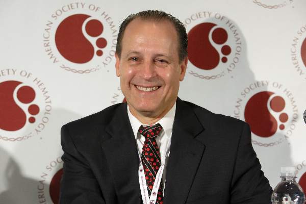
Christine Cserti-Gazdewich, MD, of University Health Network in Toronto, Canada, provided insights on the data during a press conference at ASH.
A concern of the investigators, according to Dr Cserti-Gazdewich, was that previous studies on blood storage were conducted in “high-income countries in high-technology care settings with blood inventory wealth, and findings may not be generalizable to the other half of the world.”
She pointed out that, in less developed countries, the shortfall in blood availability compared to the need is up to 3-fold.
So the investigators examined whether longer-stored red blood cells actually deliver oxygen in a manner not worse than shorter-stored or fresh blood and examined it at the extremes of storage duration in the context of a very high-dose need.
The team evaluated 290 children (aged 6 months to 60 months) with elevated blood lactate levels due to severe anemia who presented at a university-affiliated national referral hospital in Kampala, Uganda.
The children were randomized to receive RBC units stored for 25 to 35 days (longer-storage group, n=145) or RBCs stored for 1 to 10 days (shorter-storage group, n=145).
All units were leukoreduced prior to storage. All patients received 10 mL/kg of RBCs during hours 0 through 2 and, if indicated per protocol, an additional 10 mL/kg during hours 4 through 6.
In the entire population, the mean presenting hemoglobin level was 3.7 g/dL, and the mean lactate level was 9.3 mmol/L. The median RBC unit storage duration was 8 days (range, 7-9) for the shorter-storage group and 32 days (range, 30-34) for the longer-storage group.
Results
The investigators found that RBC units maintained under standard storage conditions for 25 to 35 days were not inferior to RBC units stored for up to 10 days.
The study’s primary endpoint was the proportion of patients with a lactate level of 3 mmol/L or lower at 8 hours, using a margin of noninferiority equal to an absolute difference of 25%.
The proportion of patients meeting this endpoint was 0.61 in the longer-storage group and 0.58 in the shorter-storage group (P<0.001 for noninferiority).
Average lactate levels were not statistically different between the 2 groups at 0, 2, 4, 6, 8, or 24 hours. And there was no statistical difference in the median time to achieve a blood lactate of 3 mmol/L or lower at 4 hours (hazard ratio=0.99, P=0.92).
Cerebral tissue oxygen saturation rose significantly during transfusion, but there was no significant difference between the 2 storage groups. The median area under the curve of cerebral tissue oxygen saturation during transfusion was 679 (range, 334-1156) for the longer-storage group and 521 (range, 303-835) for the shorter-storage group (P=0.25).
There was no significant difference between the longer-storage group and the shorter-storage group in the persistence of stupor or coma 8 hours after transfusion (12.6% and 19.6%, respectively, P=0.11) or the persistence of respiratory distress at 8 hours (28.7% and 30%, respectively, P=0.79).
The median length of hospital stay was 4 days (range, 2-6) in the longer-storage group and 4 days (range, 3-7) in the shorter-storage group.
There were 8 deaths, 3 in the longer-storage group and 5 in the shorter-storage group, during the 24 hours from the start of transfusion. Four additional patients, 2 in each group, died in the hospital after the initial 24-hour observation period.
The proportion of patients who had returned to good health by 30 days was 86% of the longer-storage group and 93% of the shorter-storage group (P=0.13).
“By every single measure we explored, and we explored many, we found that long-stored blood was not inferior . . . ,” Dr Csert-Gazdewich said. “We truly found no justification to shorten the current storage duration of red cells as judged by the fundamental role to deliver oxygen.” ![]()
Photo by Elise Amendola
ORLANDO, FL—The storage duration of red blood cells (RBCs) doesn’t affect transfusion outcomes in children with lactic acidosis due to severe anemia, according to a new study.
Investigators found no significant differences in lactate levels, 30-day recovery, survival, or adverse events between children who received RBCs stored for 25 to 35 days and children who received RBCs stored for 1 to 10 days.
These results were published in JAMA and presented at the 2015 ASH Annual Meeting as abstract 769.
Christine Cserti-Gazdewich, MD, of University Health Network in Toronto, Canada, provided insights on the data during a press conference at ASH.
A concern of the investigators, according to Dr Cserti-Gazdewich, was that previous studies on blood storage were conducted in “high-income countries in high-technology care settings with blood inventory wealth, and findings may not be generalizable to the other half of the world.”
She pointed out that, in less developed countries, the shortfall in blood availability compared to the need is up to 3-fold.
So the investigators examined whether longer-stored red blood cells actually deliver oxygen in a manner not worse than shorter-stored or fresh blood and examined it at the extremes of storage duration in the context of a very high-dose need.
The team evaluated 290 children (aged 6 months to 60 months) with elevated blood lactate levels due to severe anemia who presented at a university-affiliated national referral hospital in Kampala, Uganda.
The children were randomized to receive RBC units stored for 25 to 35 days (longer-storage group, n=145) or RBCs stored for 1 to 10 days (shorter-storage group, n=145).
All units were leukoreduced prior to storage. All patients received 10 mL/kg of RBCs during hours 0 through 2 and, if indicated per protocol, an additional 10 mL/kg during hours 4 through 6.
In the entire population, the mean presenting hemoglobin level was 3.7 g/dL, and the mean lactate level was 9.3 mmol/L. The median RBC unit storage duration was 8 days (range, 7-9) for the shorter-storage group and 32 days (range, 30-34) for the longer-storage group.
Results
The investigators found that RBC units maintained under standard storage conditions for 25 to 35 days were not inferior to RBC units stored for up to 10 days.
The study’s primary endpoint was the proportion of patients with a lactate level of 3 mmol/L or lower at 8 hours, using a margin of noninferiority equal to an absolute difference of 25%.
The proportion of patients meeting this endpoint was 0.61 in the longer-storage group and 0.58 in the shorter-storage group (P<0.001 for noninferiority).
Average lactate levels were not statistically different between the 2 groups at 0, 2, 4, 6, 8, or 24 hours. And there was no statistical difference in the median time to achieve a blood lactate of 3 mmol/L or lower at 4 hours (hazard ratio=0.99, P=0.92).
Cerebral tissue oxygen saturation rose significantly during transfusion, but there was no significant difference between the 2 storage groups. The median area under the curve of cerebral tissue oxygen saturation during transfusion was 679 (range, 334-1156) for the longer-storage group and 521 (range, 303-835) for the shorter-storage group (P=0.25).
There was no significant difference between the longer-storage group and the shorter-storage group in the persistence of stupor or coma 8 hours after transfusion (12.6% and 19.6%, respectively, P=0.11) or the persistence of respiratory distress at 8 hours (28.7% and 30%, respectively, P=0.79).
The median length of hospital stay was 4 days (range, 2-6) in the longer-storage group and 4 days (range, 3-7) in the shorter-storage group.
There were 8 deaths, 3 in the longer-storage group and 5 in the shorter-storage group, during the 24 hours from the start of transfusion. Four additional patients, 2 in each group, died in the hospital after the initial 24-hour observation period.
The proportion of patients who had returned to good health by 30 days was 86% of the longer-storage group and 93% of the shorter-storage group (P=0.13).
“By every single measure we explored, and we explored many, we found that long-stored blood was not inferior . . . ,” Dr Csert-Gazdewich said. “We truly found no justification to shorten the current storage duration of red cells as judged by the fundamental role to deliver oxygen.” ![]()
VIDEO: Novel GBT440 improves blood parameters in sickle cell disease
ORLANDO – A novel small molecule agent improved hematologic parameters and was associated with significant reduction in sickling of red blood cells in patients with sickle cell disease.
The drug was shown to increase hemoglobin in both healthy volunteers and patients, reduce reticulocytosis, and improve biomarkers of hemolysis and inflammation.
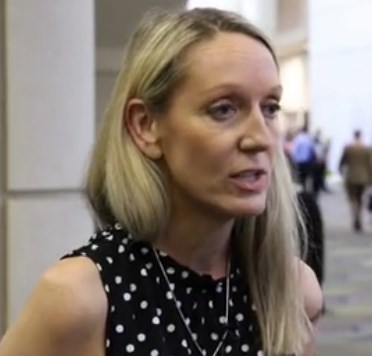
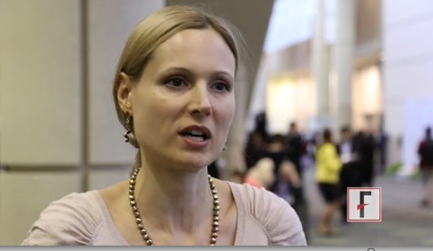
In an interview, Dr. Claire Hemmaway of Queens Hospital in Romford, England, discusses early results from a phase I/II randomized, double-blind, placebo-controlled, parallel-group trial.
The video associated with this article is no longer available on this site. Please view all of our videos on the MDedge YouTube channel
ORLANDO – A novel small molecule agent improved hematologic parameters and was associated with significant reduction in sickling of red blood cells in patients with sickle cell disease.
The drug was shown to increase hemoglobin in both healthy volunteers and patients, reduce reticulocytosis, and improve biomarkers of hemolysis and inflammation.
In an interview, Dr. Claire Hemmaway of Queens Hospital in Romford, England, discusses early results from a phase I/II randomized, double-blind, placebo-controlled, parallel-group trial.
The video associated with this article is no longer available on this site. Please view all of our videos on the MDedge YouTube channel
AT ASH 2015
ASH: Novel GBT440 reduces sickle cells, improves hematologic parameters
ORLANDO – An experimental agent that restores plasticity to red blood cells significantly improved hematologic parameters and reduced the deformation of red blood cells from sickle cell disease, suggest preliminary results from a phase I/II randomized clinical trial.
The drug, labeled GBT440, was well tolerated over one month, with no serious drug-related adverse events and no evidence of tissue hypoxia, said Dr. Claire Hemmaway from Queens Hospital in Essex, United Kingdom.
“We have emerging data with 28 days of dosing, and this supports the hypothesis that this drug inhibits polymerization of sickle hemoglobin, improves hemolysis, reduces red blood cell damage even in the micro-circulation, and improves oxygen delivery. Longer-term dosing is clearly required and this will define the optimal hematologic effects and the clinical benefit of this drug,” she said at the American Society of Hematology annual meeting.

Although its mechanism of action is not completely understood, GBT440 is a small-molecule hemoglobin modifier which is known to increase hemoglobin oxygen affinity. This first-in-class agent has been shown in both in vitro and in vivo studies to be a strong and direct anti-sickling agent. The drug has also been shown to inhibit polymerization of hemoglobin S, the central event in sickle cell disease, Dr. Hemmaway said.
“We hypothesized that patients on GBT440 will see a reduction in the red cell damage, see a reduction in hemolysis, and an improvement in the anemia, and with an inhibition of the formation of the sickle cells we’ll also see an improvement in blood flow, and this could potentially modify the course of sickle cell disease in our patients,” she said at a briefing prior to her presentation of the data in an oral abstract session.
To test the safety and efficacy of the drug, the investigators enrolled 64 healthy volunteers and 16 patients with homozygous HbSS sickle-cell disease into a phase I/II randomized, double-placebo controlled, parallel group trial.
The patients all had baseline hemoglobin levels from 6 to 10 g/dL, and had not had a vaso-occlusive crisis or transfusion with 30 days of screening.
The study was divided into parts, with part A testing single ascending doses, and part B testing multiple ascending doses compared with placebo on a 6:2 randomization basis.
As of July 24 2015, 54 healthy volunteers had completed the study, two were discontinued due to mild-to-moderate rash and/or headache, and eight were still on follow-up.
Eight patients with sickle cell disease completed part A, and eight were on follow-up in part B. No patients dropped out of the study, although one had a dose reduction from 700 mg to 400 mg because of abdominal discomfort.
Patients and volunteers generally tolerated the drug well, with generally mild adverse events, no deaths, and just one serious adverse event, an acute painful crisis in a patient on placebo.
The drug was shown to increase hemoglobin in both healthy volunteers and patients, reduce reticulocytosis, and improve biomarkers of hemolysis and inflammation. The hematologic effects correlate with GBT440 blood levels, Dr. Hemmaway said.
“You can see dramatic reductions in the reticulocyte counts, which maximally are reduced by more than 50%, and these are maintained over 28 days. The reduction in reticulocyte counts suggests an improvement in red cell life span,” she said.
One patient who received a 700 mg daily oral dose of GBT440 had evidence of a complete absence of sickled cells 28 days after starting on therapy, she noted.
The investigators have not been able to determine whether the hematologic improvements seen in the study correlate with symptomatic improvements, because they have only 28 days of dosing data and the results are still blinded, Dr. Hemmaway said.
A pediatric hematologist who was not involved in the study said that the early results offer hope for patients.
“I think many of us in the field continue to come back to the painful reality that as of today there is only one FDA-approved medication for sickle cell disease. That fundamentally is something that I think many of us are extremely troubled by, especially at a meeting like this where we see so many other opportunities in other conditions,” commented Dr. Alexis Thompson, a professor of hematology at the Robert H. Lurie Children’s Hospital of Chicago, Illinois.
Although the data are early, they are encouraging enough to justify moving forward with a phase II trial, which is planned to start in early 2016.
“I think the person who is going to be the ultimate beneficiary from many of these advances will be a child who can look forward to not only a longer lifespan but a lifespan that is less hampered by the complications of this disease,” she said.
Dr. Thompson moderated the briefing in which Dr. Hemmaway presented the data.
The study is supported by Global Blood Therapeutics. Dr. Hemmaway had no relevant disclosures. Several co-investigators are employees of the company. Dr, Thompson had no disclosures relevant to the study.
ORLANDO – An experimental agent that restores plasticity to red blood cells significantly improved hematologic parameters and reduced the deformation of red blood cells from sickle cell disease, suggest preliminary results from a phase I/II randomized clinical trial.
The drug, labeled GBT440, was well tolerated over one month, with no serious drug-related adverse events and no evidence of tissue hypoxia, said Dr. Claire Hemmaway from Queens Hospital in Essex, United Kingdom.
“We have emerging data with 28 days of dosing, and this supports the hypothesis that this drug inhibits polymerization of sickle hemoglobin, improves hemolysis, reduces red blood cell damage even in the micro-circulation, and improves oxygen delivery. Longer-term dosing is clearly required and this will define the optimal hematologic effects and the clinical benefit of this drug,” she said at the American Society of Hematology annual meeting.
Although its mechanism of action is not completely understood, GBT440 is a small-molecule hemoglobin modifier which is known to increase hemoglobin oxygen affinity. This first-in-class agent has been shown in both in vitro and in vivo studies to be a strong and direct anti-sickling agent. The drug has also been shown to inhibit polymerization of hemoglobin S, the central event in sickle cell disease, Dr. Hemmaway said.
“We hypothesized that patients on GBT440 will see a reduction in the red cell damage, see a reduction in hemolysis, and an improvement in the anemia, and with an inhibition of the formation of the sickle cells we’ll also see an improvement in blood flow, and this could potentially modify the course of sickle cell disease in our patients,” she said at a briefing prior to her presentation of the data in an oral abstract session.
To test the safety and efficacy of the drug, the investigators enrolled 64 healthy volunteers and 16 patients with homozygous HbSS sickle-cell disease into a phase I/II randomized, double-placebo controlled, parallel group trial.
The patients all had baseline hemoglobin levels from 6 to 10 g/dL, and had not had a vaso-occlusive crisis or transfusion with 30 days of screening.
The study was divided into parts, with part A testing single ascending doses, and part B testing multiple ascending doses compared with placebo on a 6:2 randomization basis.
As of July 24 2015, 54 healthy volunteers had completed the study, two were discontinued due to mild-to-moderate rash and/or headache, and eight were still on follow-up.
Eight patients with sickle cell disease completed part A, and eight were on follow-up in part B. No patients dropped out of the study, although one had a dose reduction from 700 mg to 400 mg because of abdominal discomfort.
Patients and volunteers generally tolerated the drug well, with generally mild adverse events, no deaths, and just one serious adverse event, an acute painful crisis in a patient on placebo.
The drug was shown to increase hemoglobin in both healthy volunteers and patients, reduce reticulocytosis, and improve biomarkers of hemolysis and inflammation. The hematologic effects correlate with GBT440 blood levels, Dr. Hemmaway said.
“You can see dramatic reductions in the reticulocyte counts, which maximally are reduced by more than 50%, and these are maintained over 28 days. The reduction in reticulocyte counts suggests an improvement in red cell life span,” she said.
One patient who received a 700 mg daily oral dose of GBT440 had evidence of a complete absence of sickled cells 28 days after starting on therapy, she noted.
The investigators have not been able to determine whether the hematologic improvements seen in the study correlate with symptomatic improvements, because they have only 28 days of dosing data and the results are still blinded, Dr. Hemmaway said.
A pediatric hematologist who was not involved in the study said that the early results offer hope for patients.
“I think many of us in the field continue to come back to the painful reality that as of today there is only one FDA-approved medication for sickle cell disease. That fundamentally is something that I think many of us are extremely troubled by, especially at a meeting like this where we see so many other opportunities in other conditions,” commented Dr. Alexis Thompson, a professor of hematology at the Robert H. Lurie Children’s Hospital of Chicago, Illinois.
Although the data are early, they are encouraging enough to justify moving forward with a phase II trial, which is planned to start in early 2016.
“I think the person who is going to be the ultimate beneficiary from many of these advances will be a child who can look forward to not only a longer lifespan but a lifespan that is less hampered by the complications of this disease,” she said.
Dr. Thompson moderated the briefing in which Dr. Hemmaway presented the data.
The study is supported by Global Blood Therapeutics. Dr. Hemmaway had no relevant disclosures. Several co-investigators are employees of the company. Dr, Thompson had no disclosures relevant to the study.
AT ASH 2015
Key clinical point: A first-in class small molecule drug improved hematologic parameters and reduced sickling of red blood cells.
Major finding: GBT440 inhibits polymerization of sickle hemoglobin, improves hemolysis, reduces red blood cell damage, and improves oxygen delivery.
Data source: Randomized, double blind, placebo-controlled phase I/II trial.
Disclosures: The study is supported by Global Blood Therapeutics. Dr. Hemmaway had no relevant disclosures. Several co-investigators are employees of the company. Dr, Thompson had no disclosures relevant to the study.
ASH: HLA-identical sibling transplants “excellent” in eligble SCD patients
ORLANDO – For patients with severe sickle cell disease, stem cell transplants from an HLA identical sibling are associated with “excellent” 3-year overall and event-free survival, results of a retrospective analysis show.
Data on 1,000 hematopoietic stem cell transplants (HSCT) in children and adults with sickle-cell disease performed in 88 centers in 23 countries showed a 3-year event-free-survival (EFS) rate of 90%, and 3-year overall survival (OS) rate of 94%, reported Dr. Barbara Cappelli from the Eurocord International Registry in Paris, France.

“Early referral to transplant for patients with severe sickle-cell disease is warranted, as age is an independent predictor for both event-free survival and overall survival,” she said about the study at a briefing at the American Society of Hematology annual meeting.
Patients who received donor cord blood had the best 3-year OS, at 99%, compared with 94% for patients who received bone marrow grafts, and 80% for those who received peripheral blood stem cells (PBSC).
“Transplants from peripheral blood are not recommended, because the result that there is higher mortality with transplants with peripheral blood,” she said.
The findings provide further evidence that for patients with severe sickle-disease who have an HLA-identical sibling available as a donor, HSCT can be curative with good safety, commented Dr. Alexis Thompson, a professor of hematology at the Robert H. Lurie Children’s Hospital of Chicago, Illinois.
Dr. Thompson was not involved in the study, but moderated a briefing where the data were presented.
International Registries
HSCT is the only therapy currently known to be curative for sickle-cell disease, but due to the risk for early and late complications it is generally reserved for patients with the most severe anemia or most disabling symptoms.
To evaluate the efficacy and safety of HSCT for sickle-cell disease, the investigators took a retrospective look at data from bone marrow transplant registries covering Europe, Brazil, the United States, and other reporting centers.
They searched for data on all patients with sickle-cell disease, children and adults, who received a transplant from an HLA identical sibling from 1986 through 2013.
The majority of patients (85%) were younger than 16, with the median age at HSCT of 9 years (range 1 to 54 years). Stroke was the most common indication for HSCT.
In all, 87% of patients received a myeloablative conditioning regimen based on busulfan combined with either cyclophosphamide or fludarabine. The remaining 13% of patients underwent a reduced-intensity conditioning regimen, primarily with fludarabine and cyclophosphamide.
In vivo T-cell depletion was performed in 70% of patients, either with anti-thymocyte globulin (630 patients) or with alemtumumab (76).
Prophylaxis for graft vs. host disease (GVHD) was predominantly cyclosporine as monotherapy or in combination with methotrexate.
A large majority of patients (84%) received bone marrow grafts, 7% got PBSCs, and 9% received umbilical cord blood.
By 60 days, the cumulative incidence of engraftment was 98%, with a median cumulative incidence of platelet engraftment of 25 days.
The cumulative incidence of acute GVHD (within 100 days of transplant) 14.3%, and the cumulative incidence of chronic GVHD (out to 3 years) was 13.3%
In all, 71 patients (7%) required autologous reconstitution, 45 because of late graft failure, 31 patients (3%) underwent a second transplant and 67 patients (7%) died. Of the patients who died, 6% had received bone marrow, 1% had received cord blood, and 21% had received PBSC.
As noted before, respective EFS and OS 3 years after transplant were 90% and 94%.
In multivariate analysis controlled for transplant and demographic characteristics, the authors found that younger age at transplant and use of bone marrow or cord blood were independently associated with better EFS and OS. In addition, 3-year overall survival was significantly better among patients who received transplants from the year 2000 on.
The risk for acute GVHD was significantly associated with increasing age, but none of the variables tested were associated with chronic GVHD.
Dr. Cappelli said that ways to improve outcomes include performing pre-or post-natal diagnosis in at-risk families, performing HLA typing of all family members at the time of diagnosis, and going to transplant as soon as the criteria for severe sickle-cell disease are met.
The study was supported by Eurocord-Monacord, EBMT Paediatric Disease Working Party and CIBMTR. Dr. Cappelli and Dr. Johnson reported having no conflicts of interest.
ORLANDO – For patients with severe sickle cell disease, stem cell transplants from an HLA identical sibling are associated with “excellent” 3-year overall and event-free survival, results of a retrospective analysis show.
Data on 1,000 hematopoietic stem cell transplants (HSCT) in children and adults with sickle-cell disease performed in 88 centers in 23 countries showed a 3-year event-free-survival (EFS) rate of 90%, and 3-year overall survival (OS) rate of 94%, reported Dr. Barbara Cappelli from the Eurocord International Registry in Paris, France.
“Early referral to transplant for patients with severe sickle-cell disease is warranted, as age is an independent predictor for both event-free survival and overall survival,” she said about the study at a briefing at the American Society of Hematology annual meeting.
Patients who received donor cord blood had the best 3-year OS, at 99%, compared with 94% for patients who received bone marrow grafts, and 80% for those who received peripheral blood stem cells (PBSC).
“Transplants from peripheral blood are not recommended, because the result that there is higher mortality with transplants with peripheral blood,” she said.
The findings provide further evidence that for patients with severe sickle-disease who have an HLA-identical sibling available as a donor, HSCT can be curative with good safety, commented Dr. Alexis Thompson, a professor of hematology at the Robert H. Lurie Children’s Hospital of Chicago, Illinois.
Dr. Thompson was not involved in the study, but moderated a briefing where the data were presented.
International Registries
HSCT is the only therapy currently known to be curative for sickle-cell disease, but due to the risk for early and late complications it is generally reserved for patients with the most severe anemia or most disabling symptoms.
To evaluate the efficacy and safety of HSCT for sickle-cell disease, the investigators took a retrospective look at data from bone marrow transplant registries covering Europe, Brazil, the United States, and other reporting centers.
They searched for data on all patients with sickle-cell disease, children and adults, who received a transplant from an HLA identical sibling from 1986 through 2013.
The majority of patients (85%) were younger than 16, with the median age at HSCT of 9 years (range 1 to 54 years). Stroke was the most common indication for HSCT.
In all, 87% of patients received a myeloablative conditioning regimen based on busulfan combined with either cyclophosphamide or fludarabine. The remaining 13% of patients underwent a reduced-intensity conditioning regimen, primarily with fludarabine and cyclophosphamide.
In vivo T-cell depletion was performed in 70% of patients, either with anti-thymocyte globulin (630 patients) or with alemtumumab (76).
Prophylaxis for graft vs. host disease (GVHD) was predominantly cyclosporine as monotherapy or in combination with methotrexate.
A large majority of patients (84%) received bone marrow grafts, 7% got PBSCs, and 9% received umbilical cord blood.
By 60 days, the cumulative incidence of engraftment was 98%, with a median cumulative incidence of platelet engraftment of 25 days.
The cumulative incidence of acute GVHD (within 100 days of transplant) 14.3%, and the cumulative incidence of chronic GVHD (out to 3 years) was 13.3%
In all, 71 patients (7%) required autologous reconstitution, 45 because of late graft failure, 31 patients (3%) underwent a second transplant and 67 patients (7%) died. Of the patients who died, 6% had received bone marrow, 1% had received cord blood, and 21% had received PBSC.
As noted before, respective EFS and OS 3 years after transplant were 90% and 94%.
In multivariate analysis controlled for transplant and demographic characteristics, the authors found that younger age at transplant and use of bone marrow or cord blood were independently associated with better EFS and OS. In addition, 3-year overall survival was significantly better among patients who received transplants from the year 2000 on.
The risk for acute GVHD was significantly associated with increasing age, but none of the variables tested were associated with chronic GVHD.
Dr. Cappelli said that ways to improve outcomes include performing pre-or post-natal diagnosis in at-risk families, performing HLA typing of all family members at the time of diagnosis, and going to transplant as soon as the criteria for severe sickle-cell disease are met.
The study was supported by Eurocord-Monacord, EBMT Paediatric Disease Working Party and CIBMTR. Dr. Cappelli and Dr. Johnson reported having no conflicts of interest.
AT ASH 2015
Key clinical point: Bone marrow transplants from HLA-identical siblings are frequently curative of sickle-cell disease.
Major finding: Three-year event-free survival among 1000 patients was 90%, and 3-year overall survival was 94%.
Data source: Retrospective review of registry data on 1000 patients who underwent hematopoietic stem cell transplants with HLA-identical sibling donors.
Disclosures: The study was supported by Eurocord-Monacord, EBMT Paediatric Disease Working Party and CIBMTR. Dr. Cappelli and Dr. Johnson reported having no conflicts of interest.
VIDEO: RBCs have extended shelf life, randomized trial shows
ORLANDO – Stored red blood cells kept for longer than a few weeks do not impair outcomes or harm the patients who receive them, Dr. Christine Cserti-Gazdewich reported at the annual meeting of the American Society of Hematology.
In a randomized clinical trial conducted in Kanpala, Uganda, where severe anemia with lactic acidosis is common, children who received RBCs that had been stored from 25-35 days had outcomes that were not inferior to those of children who received RBCs delivered within 10 days of collection. The findings have significant, positive implications for countries and geographic regions where there are chronic shortages of blood products, said Dr. Cserti-Gazdewich, of Toronto General Hospital.
The video associated with this article is no longer available on this site. Please view all of our videos on the MDedge YouTube channel
AT ASH 2015
ASH: Oral drug offers alternative to lifelong transfusions in sickle cell
ORLANDO – Oral hydroxyurea is as good as chronic red blood cell transfusions for prevention of primary stroke in children at high-risk for this devastating complication of sickle cell disease, results of the TWiTCH study show.
No child suffered a stroke with either hydroxyurea or monthly transfusions and transcranial doppler (TCD) velocities were maintained in both arms.
The study was stopped early, however, after noninferiority was shown for the primary end point of TCD mean velocities on the index side at 24 months, with a post-hoc analysis suggesting hydroxyurea may even be superior, study author Dr. Russell E. Ware, director of hematology at Cincinnati (Ohio) Children’s Hospital Medical Center, reported during the plenary session at the annual meeting of the American Society of Hematology.
“Hydroxyurea therapy can substitute for chronic transfusions to maintain TCD velocities and help prevent primary stroke,” he said during a press briefing.
The final mean TCD velocities were 143 cm/second in the transfusion arm and 138 cm/sec in the hydroxyurea arm, resulting in P values of 8.82 x 10⁻¹⁶ for non-inferiority by intention-to-treat analysis and 0.023 for superiority in a post-hoc analysis.
Hydroxyurea also had the added benefit of improving iron overload status more than monthly transfusions based on a greater average change in serum ferritin (-1,085 ng/mL vs. -38 ng/mL; P less than .001) and liver iron concentrations (-1.9 mg/g vs. +2.4 mg/g; P = .001), according to their report.
Press briefing moderator Dr. Alexis Thompson, of the Ann & Robert H. Lurie Children’s Hospital of Chicago, commented, “This truly is one of the abstracts that are being presenting at the meeting today that can be defined as practice changing. There are many families who have great difficulty accepting the reality, prior to the TWiTCH study, of their children having to be transfused lifelong.”
Strokes occur in up to 10% of children with sickle cell disease (SCD). Transfusions are effective for stroke prophylaxis in this setting, but have to be continued lifelong and can lead to iron overload and other complications.
Based on the participating sites, at least 80% of children with abnormal TCD velocities currently on blood transfusions to prevent stroke would be eligible for treatment with hydroxyurea, Dr. Ware said.
Hydroxyurea increases the amount of fetal hemoglobin and fetal red blood cells and was approved more than a decade ago to ameliorate the acute and chronic complications of SCD. Its use could provide dramatic cost savings for families since a transfusion costs about $1,000 to $2,000 every month, whereas hydroxyurea costs less than a dollar a day, Dr. Ware said in an interview.
TWiTCH (TCD with Transfusions Changing to Hydroxyurea) was conducted at 26 pediatric programs and used TCD to identify 121 children with SCD who were at elevated risk of stroke based on abnormally high cerebral artery flow velocities of at least 200 cm/sec. The children were evenly randomized to 24 months of treatment. TCD velocities were obtained every 12 weeks and reviewed centrally, with local investigators blinded to the results. All children had received transfusions for at least 12 months, but had not developed severe vasculopathy.
The transfusion arm was maintained at a target hemoglobin S level of less than 30% and chelation used to manage elevated liver concentrations. Transfusions were allowed in the hydroxyurea arm until a stable maximum tolerated dose (MTD) of hydroxyurea was reached, and were then replaced by serial phlebotomy to reduce iron overload. The MTD was reached after 6 months at an average dose of about 25 mg/kg/day.
The transfusion overlap with hydroxyurea was designed as a safety measure to avoid strokes if monthly transfusions were abruptly discontinued in the hydroxyurea arm before the children had time to achieve MTD.
“The fact that that overlap was about 6 months and the fact we had about 24 months of follow-up tracking the TCD velocities over time, we feel that doesn’t affect the end statistical analysis,” Dr. Ware said.
As for whether hydroxyurea is superior to monthly transfusions, he noted that the trial was not designed for superiority and superiority was seen in a post-hoc analysis. “What we can say with certainty is that it’s non-inferior to the standard treatment,” Dr. Ware said.
The final analysis was based on 42 patients randomized to the transfusion arm who completed all 24 months of treatment, 11 with truncated treatment, and 8 withdrawals, and 41 patients assigned to the hydroxyurea arm who completed all treatment, 13 with truncated treatment and 6 withdrawals.
Sickle cell-related serious adverse events were more common in the hydroxyurea arm than the transfusion arm (23 vs. 15), but none were related to the study drug or procedures.
There were 29 new centrally adjudicated neurological events including 3 transient ischemic attacks in each arm. In the transfusion arm, one child was withdrawn per protocol after developing TCD velocities exceeding 240 cm/sec and a second developed new vasculopathy. The safety of long-term hydroxyurea has been established in multiple pediatric studies, Dr. Ware said.
TWiTCH was funded by the National Heart, Lung and Blood Institute and sponsored by Cincinnati Children’s Hospital Medical Center. Dr. Ware reported serving as a data safety monitoring board member for Eli Lilly, consultancy for Bayer, and research funding from Bristol Myers Squibb. Dr. Thompson disclosed research funding from Amgen, Baxalta, bluebird bio, Celgene, Eli Lilly, GlaxoSmithKline, Mast, and Novartis, and consultancy for ApoPharma, bluebird bio, and Novartis.
Abnormal transcranial Doppler findings in sickle cell disease indicates that the pediatric patient is at high risk of a primary stroke. To date, the standard of care has been lifelong transfusion for these patients.
This study indicates there may be a viable alternative to transfusions with less invasive therapy and reduced risk of iron loading. What is not addressed, however, is whether we use hydroxyurea as front line therapy for children with abnormal findings on transcranial Doppler. The study patients had been on transfusions for at least 12 months when they were enrolled in the study and had normalized velocities on transcranial Doppler.
Do we still need to screen sickle cell disease patient patients with transcranial Doppler for stroke risk? The answer is 'yes.' What if they are already on hydroxyurea? Still the answer is 'yes.'
Should transcranial Doppler screening also be linked with MRI/MRA to look at vessels? This study refers to a select group of patients with no significant vasculopathy noted on brain imaging.
So how does the community provider interpret these finding for the real-world care of a patient? How do we not over interpret or under interpret the data? In partnership with a hematologist each child should continue to get transcranial Doppler screening annually and transfusions initiated for abnormal transcranial Doppler and study methods should be followed with crossing over to hydroxyurea for patients who normalize their transcranial Doppler results at 12 months and have no evidence of significant vasculopathy on brain imaging.
Transcranial Doppler screening should continue annually per guidelines even if the patient is already on hydroxyurea for other indications or was switched to hydroxyurea from transfusions. This is a situation that is more likely to occur in the real world clinical setting as hydroxyurea use is advocated for children as early as 9 months of age.
What happens then if transcranial Doppler findings become abnormal while on hydroxyurea? If compliance is assured and the hydroxyurea dose is optimized, then we are back to square one. Primary stroke prevention with lifelong transfusion is currently the standard of care. At least until another randomized study is done to prove non inferiority.
Dr. Ifeyinwa Osunkwo is the medical director for the sickle cell program at the Levine Cancer Institute, Carolinas Healthcare Systems, Charlotte, NC. and a member of the editorial board for Hematology News.
ORLANDO – Oral hydroxyurea is as good as chronic red blood cell transfusions for prevention of primary stroke in children at high-risk for this devastating complication of sickle cell disease, results of the TWiTCH study show.
No child suffered a stroke with either hydroxyurea or monthly transfusions and transcranial doppler (TCD) velocities were maintained in both arms.
The study was stopped early, however, after noninferiority was shown for the primary end point of TCD mean velocities on the index side at 24 months, with a post-hoc analysis suggesting hydroxyurea may even be superior, study author Dr. Russell E. Ware, director of hematology at Cincinnati (Ohio) Children’s Hospital Medical Center, reported during the plenary session at the annual meeting of the American Society of Hematology.
“Hydroxyurea therapy can substitute for chronic transfusions to maintain TCD velocities and help prevent primary stroke,” he said during a press briefing.
The final mean TCD velocities were 143 cm/second in the transfusion arm and 138 cm/sec in the hydroxyurea arm, resulting in P values of 8.82 x 10⁻¹⁶ for non-inferiority by intention-to-treat analysis and 0.023 for superiority in a post-hoc analysis.
Hydroxyurea also had the added benefit of improving iron overload status more than monthly transfusions based on a greater average change in serum ferritin (-1,085 ng/mL vs. -38 ng/mL; P less than .001) and liver iron concentrations (-1.9 mg/g vs. +2.4 mg/g; P = .001), according to their report.
Press briefing moderator Dr. Alexis Thompson, of the Ann & Robert H. Lurie Children’s Hospital of Chicago, commented, “This truly is one of the abstracts that are being presenting at the meeting today that can be defined as practice changing. There are many families who have great difficulty accepting the reality, prior to the TWiTCH study, of their children having to be transfused lifelong.”
Strokes occur in up to 10% of children with sickle cell disease (SCD). Transfusions are effective for stroke prophylaxis in this setting, but have to be continued lifelong and can lead to iron overload and other complications.
Based on the participating sites, at least 80% of children with abnormal TCD velocities currently on blood transfusions to prevent stroke would be eligible for treatment with hydroxyurea, Dr. Ware said.
Hydroxyurea increases the amount of fetal hemoglobin and fetal red blood cells and was approved more than a decade ago to ameliorate the acute and chronic complications of SCD. Its use could provide dramatic cost savings for families since a transfusion costs about $1,000 to $2,000 every month, whereas hydroxyurea costs less than a dollar a day, Dr. Ware said in an interview.
TWiTCH (TCD with Transfusions Changing to Hydroxyurea) was conducted at 26 pediatric programs and used TCD to identify 121 children with SCD who were at elevated risk of stroke based on abnormally high cerebral artery flow velocities of at least 200 cm/sec. The children were evenly randomized to 24 months of treatment. TCD velocities were obtained every 12 weeks and reviewed centrally, with local investigators blinded to the results. All children had received transfusions for at least 12 months, but had not developed severe vasculopathy.
The transfusion arm was maintained at a target hemoglobin S level of less than 30% and chelation used to manage elevated liver concentrations. Transfusions were allowed in the hydroxyurea arm until a stable maximum tolerated dose (MTD) of hydroxyurea was reached, and were then replaced by serial phlebotomy to reduce iron overload. The MTD was reached after 6 months at an average dose of about 25 mg/kg/day.
The transfusion overlap with hydroxyurea was designed as a safety measure to avoid strokes if monthly transfusions were abruptly discontinued in the hydroxyurea arm before the children had time to achieve MTD.
“The fact that that overlap was about 6 months and the fact we had about 24 months of follow-up tracking the TCD velocities over time, we feel that doesn’t affect the end statistical analysis,” Dr. Ware said.
As for whether hydroxyurea is superior to monthly transfusions, he noted that the trial was not designed for superiority and superiority was seen in a post-hoc analysis. “What we can say with certainty is that it’s non-inferior to the standard treatment,” Dr. Ware said.
The final analysis was based on 42 patients randomized to the transfusion arm who completed all 24 months of treatment, 11 with truncated treatment, and 8 withdrawals, and 41 patients assigned to the hydroxyurea arm who completed all treatment, 13 with truncated treatment and 6 withdrawals.
Sickle cell-related serious adverse events were more common in the hydroxyurea arm than the transfusion arm (23 vs. 15), but none were related to the study drug or procedures.
There were 29 new centrally adjudicated neurological events including 3 transient ischemic attacks in each arm. In the transfusion arm, one child was withdrawn per protocol after developing TCD velocities exceeding 240 cm/sec and a second developed new vasculopathy. The safety of long-term hydroxyurea has been established in multiple pediatric studies, Dr. Ware said.
TWiTCH was funded by the National Heart, Lung and Blood Institute and sponsored by Cincinnati Children’s Hospital Medical Center. Dr. Ware reported serving as a data safety monitoring board member for Eli Lilly, consultancy for Bayer, and research funding from Bristol Myers Squibb. Dr. Thompson disclosed research funding from Amgen, Baxalta, bluebird bio, Celgene, Eli Lilly, GlaxoSmithKline, Mast, and Novartis, and consultancy for ApoPharma, bluebird bio, and Novartis.
AT ASH 2015
Key clinical point: Hydroxyurea can substitute for chronic transfusions to prevent primary stroke in children with sickle cell disease and abnormal transcranial doppler velocities.
Major finding: Mean TCD velocities were 143 cm/sec with transfusions vs. 138 cm/sec with hydroxyurea (P less than 8.82 x 10⁻¹⁶ for noninferiority; P = .023 for superiority).
Data source: Phase III, noninferiority study in 121 children with sickle cell disease.
Disclosures: TWiTCH was funded by the National Heart, Lung and Blood Institute and sponsored by Cincinnati Children’s Hospital Medical Center. Dr. Ware reported serving as a data safety monitoring board member for Eli Lilly, consultancy for Bayer, and research funding from Bristol Myers Squibb. Dr. Thompson disclosed research funding from Amgen, Baxalta, bluebird bio, Celgene, Eli Lilly, GlaxoSmithKline, Mast, and Novartis, and consultancy for ApoPharma, bluebird bio, and Novartis.

FDA approves generic imatinib

Photo by Steven Harbour
The US Food and Drug Administration (FDA) has approved the use of imatinib mesylate, a generic version of Novartis’s Gleevec being developed by a subsidiary of Sun Pharmaceuticals Limited.
Under the terms of a settlement agreement with Novartis, the Sun Pharma subsidiary is allowed to launch its generic imatinib in the US on February 1, 2016.
The drug will be available in 100 mg and 400 mg tablets.
The Sun Pharma subsidiary was the first company to file an abbreviated new drug application for generic imatinib with a para IV certification and is therefore eligible for 180 days of marketing exclusivity in the US.
Sun Pharma’s imatinib mesylate is approved for the following indications:
- Newly diagnosed adult and pediatric patients with Philadelphia chromosome positive chronic myeloid leukemia (Ph+ CML) in chronic phase
- Patients with (Ph+ CML) in blast crisis, accelerated phase, or in chronic phase after failure of interferon-alpha therapy
- Adult patients with relapsed or refractory Ph+ acute lymphoblastic leukemia
- Adult patients with myelodysplastic/myeloproliferative diseases associated with PDGFR gene re-arrangements
- Adult patients with aggressive systemic mastocytosis without the D816V c-Kit mutation or with c-Kit mutational status unknown
- Adult patients with hypereosinophilic syndrome (HES) and/or chronic eosinophilic leukemia (CEL) who have the FIP1L1-PDGFRα fusion kinase and for patients with HES and/or CEL who are FIP1L1- PDGFRα fusion kinase negative or unknown
- Adult patients with unresectable, recurrent and/or metastatic dermatofibrosarcoma protuberans.
The drug is not approved to treat patients with KIT (CD117)-positive unresectable and/or metastatic malignant gastrointestinal stromal tumors. ![]()
ASH: Gene therapy reduces transfusion needs in beta-thalassemia major
ORLANDO – Lentiviral gene therapy with LentiGlobin BB305 boosts beta-globin production in patients with beta-thalassemia, but frees only some from lifelong dependence on blood transfusions, updated results of the Northstar study show.
Five patients with non-Beta-0/Beta-0 genotypes were able to stop transfusions shortly after their infusion, and remain transfusion independent for up to 16.4 months.
In four patients with the more severe form of beta-thalassemia, the Beta-0/Beta-0 genotype, red blood cell transfusion volume was reduced by 33% to 100%, with one patient stopping transfusions entirely, Dr. Mark C. Walters of the University of California-San Francisco Benioff Children’s Hospital in Oakland reported at the annual meeting of the American Society of Hematology.
Preliminary findings reported over the last two years have raised hopes that the experimental lentiviral-based therapy could be a functional cure for beta-thalassemia major and severe sickle cell disease.
Patients with beta-thalassemia major, also called Cooley’s anemia, rely on frequent blood transfusions to correct the anemia, with less than a quarter undergoing curative treatment with an allogeneic hematopoietic transplant.
In the ongoing Northstar study, 13 patients with transfusion-dependent beta-thalassemia major have been infused as of Oct. 28, 2015 with autologous CD34-positive cells transduced ex-vivo with LentiGlobin BB305 (Bluebird bio, Cambridge, Mass.), a self-inactivating, second-generation lentiviral vector containing a functioning, engineered beta-globin gene (A-T87Q). Their median age was 21 years and 11 were women.Vector-derived hemoglobin AT87Q was detectable at 6 months in 8 of 9 evaluable patients with at least six months follow-up and in 100% at 9 months, Dr. Walters reported. The median HbAT87Q level was 4.9 g/dL at 6 months, 6.5 g/dL at 9 months, and 4.2 g/dL at 12 months.
The difference in transfusion independence between genotypes is explained by endogenous non-HbAT87Q production, he said during a press briefing. While lentiglobin production was the same in patients with Beta-0/Beta-0 and non-Beta-0/Beta-0 genotypes, the Beta-0/Beta-0 patients made much smaller amounts of native hemoglobin.
Three serious post-infusion events occurred: grade 2 thrombosis, grade 3 skin infection and grade 3 veno-occlusive liver disease.
Importantly, there was no evidence of clonal dominance or replication competent lentivirus with up to 19 months follow-up.
“This is a significant advancement in the treatment of thalassemia for several reasons,” Dr. Walters told reporters. “First, compared to a bone marrow transplant, which is the only curative therapy that’s been approved, this appears to be a safer treatment in that none of these patients had a life-threatening complication. Second, because the treatment uses a thalassemia patient’s own stem cells, this bypasses the need to find a healthy bone marrow donor and thus should be more broadly available to patients affected by this disease.”
Dr. George Daley of Harvard Medical School in Boston and moderator of the press briefing, agreed that the results are an welcome advancement after decades of disappointments in the field of gene therapy including the development of insertional mutigenesis.
The video associated with this article is no longer available on this site. Please view all of our videos on the MDedge YouTube channel
In addition to the Northstar study (Ab. 201), results will be presented at the meeting from a second study examining LentiGlobin BB305 gene therapy for severe sickle cell disease and beta-thalassemia major (Ab. 202) and from the recently expanded phase I HGB-206 study in severe sickle cell disease (Ab. 3233).
Sickle cell disease represents a much larger potential market for LentiGlobin BB305, with an estimated 90,000 to 100,000 Americans affected compared with about 15,000 patients in America and Europe living with beta-thalassemia major.
AT ASH 2015
Key clinical point: Lentiviral-based gene therapy with LentiGlobin BB305 restarts hemoglobin production and leads to transfusion independence in some patients with beta-thalassemia major.
Major finding: Five patients with the non-Beta-0/Beta-0 genotype were transfusion independent post-infusion.
Data source: Phase I/II study in 13 patients with transfusion-dependent beta-thalassemia major.
Disclosures: Dr. Walters reported financial relationships with ViaCord and AllCells Inc. Several co-authors have financial relationships including employment with Bluebird bio, the study sponsor. Dr. Daley disclosed consultancy with True North Therapeutics and serving as an advisory committee member for Raze Therapeutics, Ocata Therapeutics, MPM Capital, and Solasia.
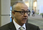
Device linked to anemia in blood donors
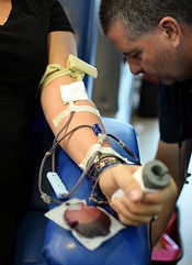
A device used to measure hemoglobin can deliver inaccurate results and may have prompted an increase in anemia among blood donors in Ireland, according to the Irish Blood Transfusion Service (IBTS).
The IBTS began using the device—called haemospect—in July 2014 but recently discovered that it cannot detect anemia accurately in all cases.
This may have resulted in anemic individuals donating blood or caused individuals to become anemic by donating.
The IBTS said this issue likely affected female donors more than males, so the organization has decided to temporarily stop taking blood donations from women who have donated in the last 18 months.
The IBTS also said it will perform a full blood count on all male and female donors going forward, until this issue has been resolved.
“Until we have a resolution to the problem that has arisen, we are asking male donors to attend our clinics and give blood,” said IBTS Medical and Scientific Director William Murphy, MD.
“We need male donors to make an extra effort to donate and maintain the blood supply. We will be double-checking all hemoglobin results on these donors.”
Discovering the problem
Haemospect is a noninvasive device that uses white light to detect hemoglobin levels. The device is made by the German company MBR Optical Systems.
IBTS learned of the potential for inaccuracies with haemospect after a blood donor contacted the organization. She had been diagnosed as severely anemic by her doctor but had given blood the week before.
“We have now discovered that the device gives inaccurate results in some individuals with anemia down to, and probably below, 8.4 g/dL,” Dr Murphy said. “As a result of the issue . . . , some women, and probably a much smaller number of men, could have been rendered iron-deficient and anemic from blood donation in the past 18 months.”
Steps to resolution
Since the issue was discovered, more than 30 women with anemia have been identified and contacted. The IBTS said it will contact all donors who are found to be anemic.
“Over the next few weeks, we will introduce new software to reanalyze all the electronic results from all donors who have been tested and accepted for donation since we introduced this device,” Dr Murphy said. “Any discrepant results will be notified to the donors involved.”
Dr Murphy also said donors who think they might be anemic or iron-deficient should visit their doctors for testing, and the IBTS will foot the bill. Concerned donors can contact the IBTS at 1850 731137.
The IBTS plans to replace haemospect with an alternative device as soon as possible. The organization has contacted the Health Products Regularity Authority about the issue with haemospect, which is used in Austria and Germany as well.
“We are determined to have this matter resolved as soon as possible,” Dr Murphy said. “We are confident that this issue . . . has not had any impact on blood received by patients.” ![]()
Team characterizes EMH niche

Image by John Perry
Previous studies have shown that hematopoietic stresses—such as myelofibrosis, anemia, and myeloablation—can induce extramedullary hematopoiesis (EMH), in which hematopoietic stem cells (HSCs) are mobilized to sites outside the bone marrow.
The splenic red pulp is known to be a prominent site of EMH in both mice and humans, but not much is known about the EMH niche.
Now, investigators say they have characterized this niche.
They detailed their findings in Nature.
The team used mouse models to examine the expression patterns of 2 known niche cell factors, SCF and CXCL12.
They discovered that the hematopoietic microenvironment in the spleen is found near sinusoidal blood vessels and is created by endothelial cells and perivascular stromal cells, just like the microenvironment in the bone marrow.
“Under emergency conditions, the endothelial cells and perivascular stromal cells that reside in the spleen are induced to proliferate so they can sustain all the new blood-forming stem cells that migrate into the spleen,” explained study author Sean Morrison, PhD, of the University of Texas Southwestern Medical Center, Dallas.
“We determined that this process in the spleen is physiologically important for responding to hematopoietic stress. Without it, the mice we studied could not maintain normal blood cell counts during pregnancy or quickly regenerate blood cell counts after bleeding or chemotherapy.”
The investigators believe these findings could aid the development of therapeutic interventions to enhance blood formation following chemotherapy or HSC transplant and thus accelerate the recovery of blood cell counts. ![]()