User login
Risk of anaphylaxis with IV iron products
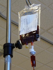
Researchers have compared the risk of anaphylaxis with different intravenous (IV) iron products and found evidence to suggest that iron dextran poses the greatest risk.
Compared with nondextran formulations, iron dextran was associated with a higher cumulative risk of anaphylaxis and an increased risk of anaphylaxis at first administration.
Iron sucrose was associated with the lowest risk of anaphylaxis, both cumulative and at first administration.
Cunlin Wang, MD, PhD, of the US Food and Drug Administration in Silver Spring, Maryland, and his colleagues conducted this research and reported the results in JAMA.
The researchers studied 688,183 recipients of IV iron enrolled in the fee-for-service Medicare program from January 2003 to December 2013.
The team examined administrations of IV iron dextran, gluconate, sucrose, or ferumoxytol. They identified 247,500 iron dextran and 440,683 nondextran users during the study period.
Overall, there were 274 cases of anaphylaxis at first exposure to IV iron and an additional 170 cases during subsequent iron administrations.
At first administration, iron dextran was associated with a higher anaphylaxis risk than nondextran formulations. The risk of anaphylaxis was 68 per 100,000 persons for iron dextran and 24 per 100,000 persons for all nondextran products combined. The odds ratio—adjusted for age, indication, history of coronary heart disease, and hypertension—was 2.6 (P<0.001).
Among the nondextran products, the risk of anaphylaxis at first administration was higher with both iron gluconate and ferumoxytol than with iron sucrose. When compared with iron sucrose, the adjusted odds ratio of anaphylaxis was 3.6 for iron dextran, 2.0 for iron gluconate, and 2.2 for ferumoxytol.
Because each IV iron product has a specific recommended dose and schedule of administration, the researchers also calculated the cumulative risk of anaphylaxis based on both the number of administrations and the clinically relevant repletion level of iron (1000 mg) achieved within 12 weeks.
The cumulative risk of anaphylaxis over multiple administrations was highest for iron dextran, followed by ferumoxytol, iron gluconate, and iron sucrose.
The estimated cumulative anaphylaxis risk following total iron repletion of 1000 mg administered within a 12-week period was highest with iron dextran (82 per 100,000 persons) and lowest with iron sucrose (21 per 100,000 persons).
The researchers noted that the mechanism of anaphylactic reaction after IV iron remains unknown. ![]()
What’s on tap at ASH 2015
FROM A TELECONFERENCE – The American Society of Hematology’s (ASH) 57th annual meeting in Orlando is chock-full of much-anticipated results in cancer immunotherapies such as CAR T cell therapies and checkpoint inhibitors, advances in sickle cell disease, and practical advice on managing the latest drugs in the clinic, ASH officials said in a teleconference. Here are some of the day-by-day picks selected by ASH president Dr. David Williams and ASH secretary Dr. Stephanie J. Lee, who gave their recommendations during a conference call for the press. Meeting abstracts are now available online.
Saturday, Dec. 5
Clinical applications of newly approved drugs
The popular special education session on clinical applications of newly approved drugs returns on Saturday, Dec. 5 at 9:30 a.m., with didactic presentations that address issues clinicians may face such as drug-drug interactions, side effects, and adverse events. The three drugs to be discussed this year are: idarucizumab (Praxbind), the first specific reversal agent approved for dabigatran reversal; blinatumomab (Blincyto), approved for second-line treatment of Philadelphia chromosomenegative acute lymphoblastic leukemia; and the histone deacetylase (HDAC) inhibitor panobinostat (Farydak), approved for the treatment of multiple myeloma.
Adoptive immunotherapy
One presentation to look out for next month is abstract 99at 12:30 p.m. on Saturday, Dec. 5 in the adoptive immunotherapy session, Dr. Williams told reporters. The chimeric antigen receptor (CAR)-T-cell approach has relied on genetically engineering the patient’s own T cells to rev up the immune system. This group’s approach is to treat B-cell malignancies after allogeneic hematopoietic stem cell transplantation using a single infusion of anti-CD19 CAR-T cells from the patient’s transplant donor.
Eight of 20 patients treated with this strategy achieved remission, including six complete remissions and two partial remissions. Importantly, none of these patients developed acute graft-versus-host disease, a potential consequence of using allogeneic rather than autologous T cells, he said. The authors also noted that patients who responded and went into remission were marked by higher numbers of these infused CAR-T cells in their circulation, suggesting a biomarker of response.
Checkpoint, please?
Immunotherapy is a “very hot area,” so ASH has put together a special session at 4 p.m. Saturday called “Checkpoint, Please?” Dr. Williams said. Topics include the role of programmed death (PD)-1 and PD-ligand 1 in acute and chronic graft-versus-host disease, checkpoint blockade with neoantigen cancer vaccines, and insights into the mechanisms of action of anti-CTLA-4 (cytotoxic T-lymphocyte–associated protein 4) antibody therapy.
Sunday, Dec. 6
Precision medicine
Sunday’s plenary scientific session will include several noteworthy personalized medicine abstracts featuring emerging therapies targeted to specific genetic subtypes, Dr. Lee, from the University of Washington, Seattle, said.
Plenary abstract 6 is a large, multinational study looking at whether adding the multikinase inhibitor midostaurin to standard induction therapy and carried through 1 year of maintenance would improve outcomes in newly diagnosed acute myeloid leukemia with FLT3 mutations. Patients with these deleterious mutations do enter remission with chemotherapy, but often relapse.
Overall and event-free survival were better at 5 years by about 7% to 8% in the experimental arm using midostaurin, she said. Caveats are that complete response rates were similar in both arms and lower than reported in other trials.
“Because we know that patients with this FLT3 mutation have a very poor prognosis with standard chemotherapy, more than half of the patients in this trial received an allogeneic transplant,” Dr. Lee noted. “But the abstract does say that the results are similar if you censor at the time of the transplant.”
In this same vein of precision medicine is plenary abstract 1, testing whether adding rituximab to standard chemotherapy improves outcomes in adults with CD-20–positive, Philadelphia chromosome–negative, B-cell precursor acute lymphoblastic leukemia (ALL). Rituximab (Rituxan) binds to CD-20, which is found in about 30% to 50% of adult B-cell ALL, she said.
At 2 years, patients treated with rituximab had longer event-free survival than controls (65% vs. 52%; P = .038), but similar overall survival (71% vs. 64%; P = .09), according to the abstract. The rituximab arm also received more allogeneic transplants, but again, after censoring the data, the abstract states that both event-free and overall survival were longer with rituximab, Dr. Lee said.
Sickle cell anemia
Sunday’s plenary session will also feature the very important TWiTCH (TCD with Transfusions Changing to Hydroxyurea) study evaluating hydroxyurea therapy as an alternative to chronic blood transfusions to prevent stroke. Stroke is one of the most dreaded complications of sickle cell disease, occurring in up to 10% of children, Dr. Williams said. Though transfusions are effective, they have to be continued indefinitely and lead to iron overload. Hydroxyurea increases the amount of fetal hemoglobin and fetal red blood cells and has become a standard therapy to attenuate the complications of sickle cell.
The phase III noninferiority study, which used Transcranial Doppler (TCD) screening to identify children at elevated risk for stroke, showed that hydroxyurea “was as good as current therapy with red cell transfusions and there was some indication, although not significant, that it might even be superior in lowering the TCD levels,” Dr. Williams said. An added benefit of the hydroxyurea was that it improved the patients’ iron overload status. There were no strokes in either group.
Sunday’s abstract 202 is another presentation “that I’m sure will get a lot of attention,” Dr. Williams said. It offers updated details on outcomes from patients with sickle cell disease (SCD) treated with a novel gene therapy transduced with the LentiGlobin BB305 (Bluebird Bio) lentiviral vector. Patients with beta thalassemia major have remained transfusion-independent for more than a year after this treatment, with results now available from four patients with SCD. One patient with a severe phenotype has had no sickle cell complications and has been able to stop his transfusion therapy, while two of the other four patients are also transfusion-independent.
“This is an early study showing what appears to be efficacy of the gene therapy approach not in thalassemia, but in sickle cell disease,” Dr. Williams said, noting that abstract 3233 will also feature results using LentiGlobin gene therapy in severe SCD.
ASH/EHA joint symposium
Also noteworthy is a special joint ASH/European Hematology Association symposium looking at how well genomic data are being incorporated into practice in the U.S. and Europe.
Monday, Dec. 7
ASH/FDA joint symposium
A joint ASH/FDA symposium on late-breaking drug approvals is new this year and features drugs that gained approval in November 2015. FDA product-reviewers will discuss safety and efficacy issues in the clinical approval trials and toxicity studies, while clinicians will share their experiences in the real-world use of these drugs.
“This is information that is really going to be very hot off the press and presented in conjunction with the FDA,” Dr. Lee said.
Dr. Williams reported research funding from Bluebird Bio. Dr. Lee reported having no conflicts of interest.
Study raises questions about iron dosing

A new study suggests it may be difficult for the body to absorb iron in the necessary or desired quantities when iron supplements are administered in 24-hour intervals.
Investigators believe this is due to hepcidin. They found that giving subjects iron supplements at doses of 60 mg or higher increased hepcidin levels for up to 24 hours and was associated with lower iron absorption on the following day.
The team postulated that administering low-dose iron on alternate days may overcome this problem, but they said more research is needed to confirm this.
The investigators reported their findings in Blood.
Diego Moretti, PhD, of the Swiss Federal Institute of Technology Zürich, and his colleagues conducted this study in 54 young women.
The women had depleted iron reserves but were not yet anemic (plasma ferritin ≤20 mcg/L). They received a daily dose of at least 40 mg of iron, as is commonly prescribed in cases of iron deficiency.
Afterward, the investigators measured how the hepcidin concentration developed and quantified its effect on the absorption of subsequent doses of iron.
To analyze iron reabsorption, the team used stable iron isotopes as indicator substances. These substances have a modified ratio of stable iron isotopes. Iron-56 is the most frequent naturally occurring stable iron isotope (91.7%), followed by iron-54 (5.8%), and iron-57 (2.1%). Iron-58 occurs only in trace amounts.
The investigators used tablets with increased quantities of iron-57, iron-54, and iron-58. And they were able to measure endogenous iron absorption by observing isotope ratio changes within the body.
The team found that hepcidin reached its peak concentration after 6 to 8 hours, but even 24 hours after the first dose of iron, it was still present in high enough quantities to markedly reduce absorption of the second dose.
The body was only partly able to absorb a second dose of iron, which was given either on the same day or 24 hours after the first dose.
“To improve the percentage of iron absorbed, it would likely be more efficient to wait longer between doses,” Dr Moretti said.
However, he noted that more research is needed to confirm this, particularly because this study had 2 limitations. The first is that participants were all healthy young women without anemia, and the second is that iron absorption was observed over 2 days only.
The investigators are now preparing to conduct a follow-up study to analyze hepcidin concentration over the course of an iron supplementation regimen lasting several weeks. ![]()
Experiment reveals new method of RBC production
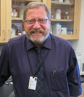
Photo courtesy of
Josh Barney/University of
Virginia Health System
An unexpected result of a lab experiment has led researchers to a new way to trigger red blood cell (RBC) production.
They found that engaging a stress receptor on type 1 conventional dendritic cells can induce stress erythropoiesis in mice.
The team believes that, eventually, this method could be used to turn on RBC production in humans when necessary, perhaps to replace a blood transfusion or as a stop-gap measure when a transfusion is delayed.
Thomas J. Braciale, MD, PhD, of the University of Virginia in Charlottesville, and his colleagues conducted this research and reported the results in The Journal of Clinical Investigation.
The team was not investigating RBC production when they made their discovery. They were looking into the role of dendritic cells in the lungs.
Dendritic cells have traditionally been thought to be sensors of infection and inflammation, but a lab test involving the flu virus produced an unexpected effect in mice that ultimately revealed a new aspect to the cells’ function.
The researchers injected mice with the flu virus and an αCD24 monoclonal antibody, which resulted in splenomegaly. The team was baffled at this outcome, so they repeated the experiment, only to get the same results.
“We did it again, and I didn’t believe it, and we did it again, and I didn’t believe it,” Dr Braciale recalled. “I asked whether you needed flu to infect the mice when you injected this antibody. So the postdoc did the experiment, and he just injected the antibody without flu-injecting the mice—giant spleens. After much consultation, after talking with my colleagues in pathology, we decided we were inducing stress erythropoiesis.”
Specifically, the researchers found that engaging CD24 on type 1 conventional dendritic cells upregulates expression of the Kit ligand stem cell factor, which results in Kit-mediated proliferative expansion of early erythroid progenitors and transient reticulocytosis.
“In a very basic way, what we’ve discovered is that the process of regulating stress in the body is mediated—certainly in part, at least—by these dendritic cells,” Dr Braciale explained. “And stress can be a variety of different stresses.”
“It doesn’t have to be infection. It doesn’t have to be inflammation. It can be anemia. It can be hemorrhage. And these cells act to initiate this response that, until this report, there’s been really no evidence that [dendritic] cells ever participate in making red blood cells.”
More work is needed before this approach to RBC production can be tested in humans. However, Dr Braciale is optimistic, based on the findings so far.
“We’re very excited to see where this goes,” he said. “We know that the same things can be done in humans in the following sense. There are mice called humanized mice. These are mice that are engineered so they have a human blood system. And if you inject these mice with this antibody, they’ll make red blood cells.” ![]()
A new category of cytopenias

New research suggests that clonality may underlie the cytopenias observed in patients with idiopathic cytopenias of undetermined significance (ICUS).
So researchers say these patients should be classified as having clonal cytopenias of undetermined significance (CCUS).
The team believes that recognition of CCUS as a clinical entity would help identify it as a formally defined diagnostic group and could aid future studies.
“We don’t know to what extent patients who have low blood counts and mutations are at increased risk of developing an overt malignancy,” said Rafael Bejar, MD, PhD, of the UCSD Moores Cancer Center in La Jolla, California.
“We hope that by defining CCUS, future studies will follow these patients to learn what these mutations mean for their future, as their genetically abnormal cells may represent early stages of subsequent blood cancers.”
Dr Bejar and his colleagues defined CCUS in Blood. Their research was funded, in part, by Genoptix Medical Laboratory, and several study authors are employees of Genoptix.
The researchers prospectively examined bone marrow samples from 144 patients with unexplained cytopenias. Seventeen percent of these patients were diagnosed with myelodysplastic syndromes (MDS), 15% with ICUS and some evidence of dysplasia, and 69% with ICUS and no dysplasia.
The team then sequenced patient samples looking for mutations in 22 frequently mutated myeloid malignancy genes. They identified somatic mutations in 71% of MDS patients, 62% of patients with ICUS and some dysplasia, and 20% of ICUS patients with no dysplasia.
Overall, 35% of ICUS patients carried a somatic mutation or chromosomal abnormality indicative of clonal hematopoiesis.
To build upon these findings, the researchers performed a retrospective analysis of data from 91 patients with lower-risk MDS and 249 patients with ICUS.
The team identified mutations in 79% of patients with MDS, 45% of patients with ICUS and dysplasia, and 17% of patients with ICUS and no dysplasia.
The researchers said the spectrum of mutated genes was similar among the 3 groups. The exception was SF3B1, which was rarely mutated in patients without dysplasia.
The team added that variant allele fractions were comparable between clonal ICUS (CCUS) and MDS, and the same was true for the patients’ mean age and blood counts. But CCUS was a more frequent diagnosis than MDS.
Dr Bejar and his colleagues said larger, carefully controlled studies will be needed to confirm the findings of this research. ![]()
Iron chelator tablets may now be crushed

Photo courtesy of the CDC
The US Food and Drug Administration (FDA) has approved a label change for Jadenu, an oral formulation of the iron chelator Exjade (deferasirox).
Jadenu comes in tablet form, and the previous label stated that Jadenu tablets must be swallowed whole.
Now, the medication can also be crushed to help simplify administration for patients who have difficulty swallowing whole tablets.
Jadenu tablets may be crushed and mixed with soft foods, such as yogurt or applesauce, immediately prior to use.
The label notes that commercial crushers with serrated surfaces should be avoided for crushing a single 90 mg tablet. The dose should be consumed immediately and not stored.
Jadenu was granted accelerated approval from the FDA earlier this year.
It is approved to treat patients 2 years of age and older who have chronic iron overload resulting from blood transfusions, as well as to treat chronic iron overload in patients 10 years of age and older who have non-transfusion-dependent thalassemia.
The full prescribing information for Jadenu can be found at http://www.pharma.us.novartis.com/product/pi/pdf/jadenu.pdf. ![]()
Technique enables SCD detection with a smartphone

Photo by Daniel Sone
Researchers say they’ve developed a simple technique for diagnosing and monitoring sickle cell disease (SCD) that could be used in regions where advanced medical technology and training are scarce.
The team created a 3D-printed box that can be attached to an Android smartphone and used to test a small blood sample.
The testing method involves magnetic levitation, which allows the user to differentiate sickle cells from normal red blood cells with the naked eye.
Savas Tasoglu, PhD, of the University of Connecticut in Storrs, and his colleagues described this technique in Nature Scientific Reports.
First, a clinician takes a blood sample from a patient and mixes it with a common, salt-based solution that draws oxygen out of sickle cells, making them denser and easier to detect via magnetic levitation. The denser sickle cells will float at a lower height than healthy red blood cells, which are not affected by the solution.
The sample is then loaded into a disposable micro-capillary that is inserted into the tester attached to the smartphone. Inside the testing apparatus, the micro-capillary passes between 2 magnets that are aligned so that the same poles face each other, creating a magnetic field.
The capillary is then illuminated with an LED that is filtered through a ground glass diffuser and magnified by an internal lens.
The smartphone’s built-in camera captures the resulting image and presents it digitally on the phone’s external display. The blood cells floating inside the capillary—whether higher-floating healthy red blood cells or lower-floating sickle cells—can be easily observed.
The device also provides clinicians with a digital readout that assigns a numerical value to the sample density to assist with the diagnosis. The entire process takes less than 15 minutes.
“With this device, you’re getting much more specific information about your cells than some other tests,” said Stephanie Knowlton, a graduate student at the University of Connecticut.
“Rather than sending a sample to a lab and waiting 3 days to find out if you have this disease, with this device, you get on-site and portable results right away. We believe a device like this could be very helpful in third-world countries where laboratory resources may be limited.”
Dr Tasoglu’s lab has filed a provisional patent for the device and is working on expanding its capabilities so it can be applied to other diseases. ![]()
Immunosuppressant can treat autoimmune cytopenias
New research suggests the immunosuppressant sirolimus may be a promising treatment option for patients with refractory autoimmune cytopenias.
The drug proved particularly effective in children with autoimmune lymphoproliferative syndrome (ALPS), producing complete responses in all of the ALPS patients studied.
On the other hand, patients with single-lineage autoimmune cytopenias, such as immune thrombocytopenia (ITP), did not fare as well.
David T. Teachey, MD, of The Children’s Hospital of Philadelphia in Pennsylvania, and his colleagues reported these results in Blood.
The group studied sirolimus in 30 patients with refractory autoimmune cytopenias who were 5 to 19 years of age. All of the patients were refractory to or could not tolerate corticosteroids.
Twelve patients had ALPS, 6 had single-lineage autoimmune cytopenias (4 with ITP and 2 with autoimmune hemolytic anemia [AIHA]), and 12 patients had multi-lineage cytopenias secondary to common variable immune deficiency (n=2), Evans syndrome (n=8), or systemic lupus erythematosus (n=2).
The patients received 2 mg/m2 to 2.5 mg/m2 per day of sirolimus in liquid or tablet form for 6 months. After 6 months, those who benefited from the drug were allowed to continue treatment with follow-up appointments to monitor toxicities.
Of the 12 children with ALPS, 11 had complete responses—normalization of blood cell counts—from 1 to 3 months after receiving sirolimus. The remaining patient achieved a complete response after 18 months.
All ALPS patients were successfully weaned off steroids and discontinued all other medications within 1 week to 1 month after starting sirolimus.
The patients with multi-lineage cytopenias also responded well to sirolimus. Eight of the 12 patients had complete responses, although these occurred later than for most ALPS patients (after 3 months).
The 6 patients with single-lineage cytopenias had less robust results—1 complete response and 2 partial responses. One child with ITP achieved a partial response but had to discontinue therapy.
One of the patients with AIHA had a complete response by 6 months and was able to stop taking other medications within a month. The other child with AIHA achieved a partial response.
For all patients, the median time on sirolimus was 2 years (range, 1–4.5 years).
The most common adverse event observed in this study was grade 1-2 mucositis (n=10). Other toxicities included elevated triglycerides and elevated cholesterol (n=2), acne (n=1), sun sensitivity (n=1), and exacerbation of gastro-esophageal reflux disease (n=1).
One patient developed hypertension 2 years after starting sirolimus, but this was temporally related to starting a new psychiatric medication.
Another patient (with Evans syndrome) developed a headache with associated white matter changes (4 different lesions). The changes were attributed to disease-associated vasculitis, and the lesions resolved over a few months with the addition of steroids. The patient was eventually diagnosed with a primary T-cell immune deficiency and underwent hematopoietic stem cell transplant.
“This study demonstrates that sirolimus is an effective and safe alternative to steroids, providing children with an improved quality of life as they continue treatment into adulthood,” Dr Teachey said. “While further studies are needed, sirolimus should be considered an early therapy option for patients with autoimmune blood disorders requiring ongoing therapy.” ![]()
New research suggests the immunosuppressant sirolimus may be a promising treatment option for patients with refractory autoimmune cytopenias.
The drug proved particularly effective in children with autoimmune lymphoproliferative syndrome (ALPS), producing complete responses in all of the ALPS patients studied.
On the other hand, patients with single-lineage autoimmune cytopenias, such as immune thrombocytopenia (ITP), did not fare as well.
David T. Teachey, MD, of The Children’s Hospital of Philadelphia in Pennsylvania, and his colleagues reported these results in Blood.
The group studied sirolimus in 30 patients with refractory autoimmune cytopenias who were 5 to 19 years of age. All of the patients were refractory to or could not tolerate corticosteroids.
Twelve patients had ALPS, 6 had single-lineage autoimmune cytopenias (4 with ITP and 2 with autoimmune hemolytic anemia [AIHA]), and 12 patients had multi-lineage cytopenias secondary to common variable immune deficiency (n=2), Evans syndrome (n=8), or systemic lupus erythematosus (n=2).
The patients received 2 mg/m2 to 2.5 mg/m2 per day of sirolimus in liquid or tablet form for 6 months. After 6 months, those who benefited from the drug were allowed to continue treatment with follow-up appointments to monitor toxicities.
Of the 12 children with ALPS, 11 had complete responses—normalization of blood cell counts—from 1 to 3 months after receiving sirolimus. The remaining patient achieved a complete response after 18 months.
All ALPS patients were successfully weaned off steroids and discontinued all other medications within 1 week to 1 month after starting sirolimus.
The patients with multi-lineage cytopenias also responded well to sirolimus. Eight of the 12 patients had complete responses, although these occurred later than for most ALPS patients (after 3 months).
The 6 patients with single-lineage cytopenias had less robust results—1 complete response and 2 partial responses. One child with ITP achieved a partial response but had to discontinue therapy.
One of the patients with AIHA had a complete response by 6 months and was able to stop taking other medications within a month. The other child with AIHA achieved a partial response.
For all patients, the median time on sirolimus was 2 years (range, 1–4.5 years).
The most common adverse event observed in this study was grade 1-2 mucositis (n=10). Other toxicities included elevated triglycerides and elevated cholesterol (n=2), acne (n=1), sun sensitivity (n=1), and exacerbation of gastro-esophageal reflux disease (n=1).
One patient developed hypertension 2 years after starting sirolimus, but this was temporally related to starting a new psychiatric medication.
Another patient (with Evans syndrome) developed a headache with associated white matter changes (4 different lesions). The changes were attributed to disease-associated vasculitis, and the lesions resolved over a few months with the addition of steroids. The patient was eventually diagnosed with a primary T-cell immune deficiency and underwent hematopoietic stem cell transplant.
“This study demonstrates that sirolimus is an effective and safe alternative to steroids, providing children with an improved quality of life as they continue treatment into adulthood,” Dr Teachey said. “While further studies are needed, sirolimus should be considered an early therapy option for patients with autoimmune blood disorders requiring ongoing therapy.”
HSC finding may have range of implications
Image by Matthias Zepper
Murine research has provided new insight into the functionality of hematopoietic stem cells (HSCs).
Investigators found that a full complement of mini-chromosome maintenance (MCM) proteins is required to preserve HSC functionality and the proper differentiation and maturation of erythrocytes.
Downregulation of the gene MCM3 during embryonic development caused replication stress in hematopoietic progenitors and led to fetal anemia.
In adult mice, downregulation of MCM3 reduced life expectancy and promoted lymphomagenesis.
The investigators therefore believe that therapies designed to modulate replication stress could fight aging, anemia, and hematopoietic malignancies.
This research was published in Nature Communications.
A previous study revealed that replication stress drives the functional decline of HSCs that occurs with age. With the new study, investigators have managed to replicate this phenomenon in mouse embryos.
The team did this by reducing levels of MCM3, one of the components of the MCM complex that is responsible for separating the strands of the DNA double helix during replication. Cells need to maintain high levels of MCM during DNA replication or they experience replication stress.
“When we reduce the levels of the MCM3 gene in the entire organism, we observe that replication stress especially affects the stem cells that give rise to the other blood cells and, in particular, the red blood cell precursors,” explained study author Juan Méndez, PhD, of Centro Nacional de Investigaciones Oncologicas (CNIO) in Madrid, Spain.
“In adult organisms, the production and maturation of red blood cells takes place in the bone marrow, but, during embryonic development, it occurs mainly in the fetal liver. In animals with MCM3 deficiency, the stem cells of the fetal liver are deteriorated, and the embryos develop a severe form of anemia that prevents them from being born.”
“We could say that replication stress turns fetal stem cells, which should be in perfect working order, into very old cells. We have verified this finding in transplantation experiments, where fetal cells with replication stress were unable to adequately reconstitute the blood system in the recipient animals.”
However, the investigators managed to prevent embryonic lethality and reduce anemia by increasing the levels of another gene, CHK1.
“CHK1 is one of the genes responsible for protecting cells from replication stress,” Dr Méndez noted. “It supervises DNA replication. When something goes wrong, CHK1 slows down or halts cell division until the problem has been resolved.”
Embryos that were subjected to replication stress (due to loss of MCM3) but had higher levels of CHK1 showed less pronounced anemia. Four of every 10 embryos developed normally and completed their gestation.
The investigators said an interesting implication of this work is that the type of anemia caused by replication stress is very similar to the aplastic anemia that arises in patients receiving chemotherapy or radiation therapy.
Therefore, the team believes these results could aid the development of novel therapies for aplastic anemia.
NICE plans to recommend device for SCD patients
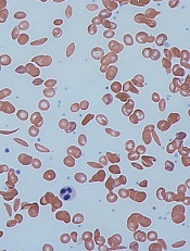
Image by Graham Beards
The UK’s National Institute for Health and Care Excellence (NICE) is asking for views on its plans to recommend a device that automatically replaces sickled red blood cells (RBCs) with healthy RBCs in patients with sickle cell disease (SCD).
NICE has issued a draft medical technology guidance supporting use of the Spectra Optia Apheresis System for automated RBC exchange in patients with SCD who need regular transfusions.
The guidance is open for comment until November 16.
The Spectra Optia system is made up of 3 components: the apheresis machine, embedded software, and a single-use disposable blood tubing set. The system is manufactured by Terumo.
NICE said the evidence examined indicates that the Spectra Optia system speeds up the process of RBC exchange compared to manual RBC exchange.
The evidence also suggests that patients require RBC exchange less frequently when using this system than they do with manual RBC exchange.
Furthermore, the Spectra Optia system is estimated to provide cost savings for most patients, when compared to manual RBC exchange or top-up transfusion.
Potential savings depend on the patient’s clinical circumstance and if devices already owned by the National Health Service can also be used to treat SCD.
However, NICE’s draft guidance also highlights a need for additional data on treatment outcomes, as there is limited clinical evidence for some outcomes.
“In particular, the independent Medical Technologies Advisory Committee would like to see long-term data on how manual and automated cell exchange affects the amount of iron in the body and the need to treat this complication,” said Carole Longson, director of the NICE Centre for Health Technology Evaluation. “We welcome comments on the draft guidance as part of this consultation.”