User login
Positive node risk defined for elderly breast cancer patients
LAS VEGAS – In women aged 70 years or older with hormone receptor–positive invasive breast cancer, their tumor size, grade, and histology – but not human epidermal growth factor receptor–2 status – predicted nodal positivity, according to a retrospective analysis.
Investigators at the Mayo Clinic, Rochester, Minn., reviewed 52,532 women in the National Cancer Database from 2010 to 2013 who were at least 70 years old with hormone receptor–positive invasive breast cancer and clinically node negative disease, who had axillary surgery performed. Two-thirds of the cohort was used to identify risk factors, and the remaining third to validate them. About 16% in both groups had cancer in their axillary lymph nodes.
On multivariate analysis, higher clinical T stage, higher grade, and invasive lobular and invasive mammary histology were all associated with positive nodes. Although significant on univariate analysis, age (P = .57) and HER2 status (P = .32) fell out on multivariate analysis.
Nodal positivity was more than five times as likely with clinical T2 tumors, compared with T1a tumors, and far less likely for patients with invasive mucinous carcinoma than for those with invasive ductal carcinoma.
The team expects to release a nomogram for general use in clinical practice to predict the risk of positive nodes for various combinations of tumor size, grade, and histology in older women. When the model predicted a less than 10% chance of node positive disease, the actual rate in the validation set was around 5.5%. When it predicted a 30%-39% chance, the actual rate was 32.6%. The area under the receiver operating characteristic curve was 0.7 in both the development and validation sets, indicating good discrimination.
The work grew out of an effort to implement the Society of Surgical Oncologists’ recommendation not to do routine sentinel node biopsies in clinically node negative, hormone receptor–positive invasive breast cancer in women over the age of 70 years, a recommendation the group made as part of its contribution to the Choosing Wisely campaign.
“After the guideline was released, we were sitting in the clinic thinking how to apply it to our patients,” lead investigator Jessemae Welsh, MD, a surgeon at the Mayo Clinic, said at the American Society of Breast Surgeons annual meeting.
The problem is that nodal positivity is important to know for other aspects of care, including regional nodal irradiation and duration of systemic hormone therapy, and axillary lymph node staging might still be indicated if older women are truly at high risk. “We [wanted] to develop a multivariate model that gives a precise estimate of nodal risk,” to help “patients and surgeons to decide together based on an individual risk” how best to proceed. Also, a prediction of low risk “can help reassure patients that they will do well without axillary surgery,” she said.
Mayo’s nomogram is unique in that it focuses specifically on women 70 years and older. Development of previous nomograms incorporated older women, but did not focus on them specifically, Dr. Welsh said.
Dr. Welsh said she had no relevant disclosures.
[email protected]
AT ASBS 2017
Key clinical point:
Major finding: On multivariate analysis, higher clinical T stage, higher grade, and invasive lobular and invasive mammary histology were all associated with positive nodes.
Data source: An analysis of data from 52,532 women in the National Cancer Database during 2010-2013.
Disclosures: The lead investigator had no disclosures.
Forgo axillary dissection for single suspicious node on ultrasound
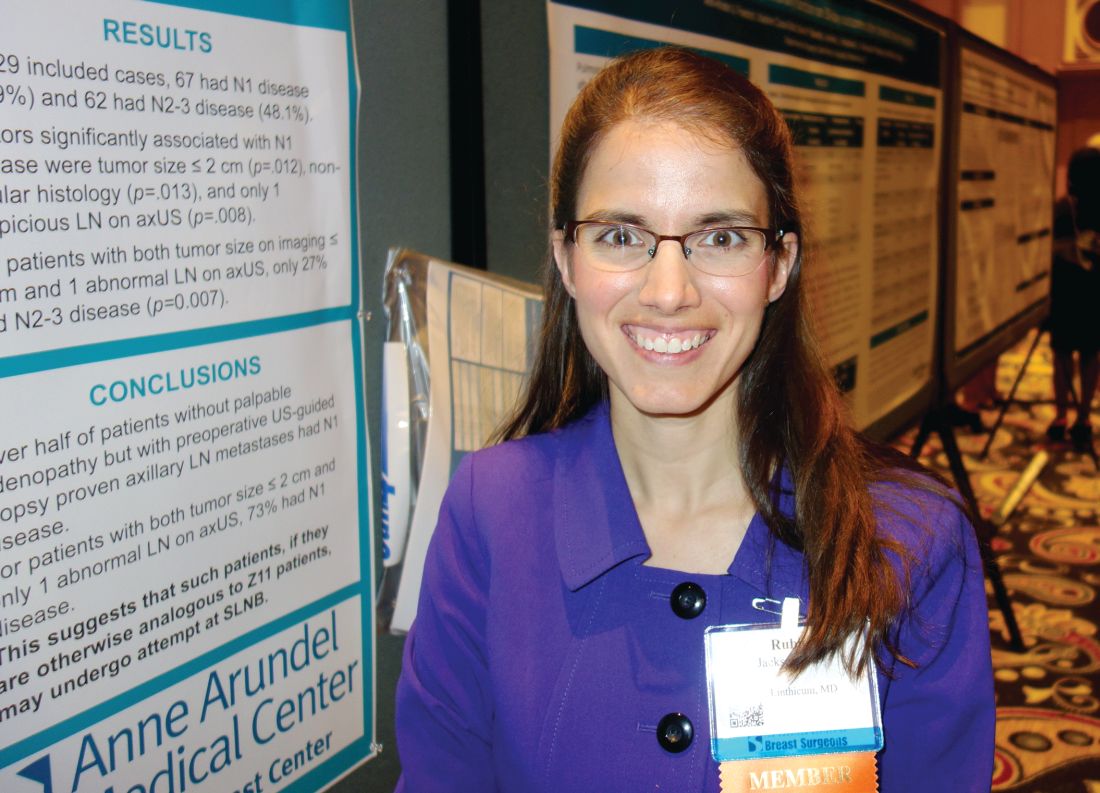
LAS VEGAS – About half of breast cancer patients without palpable lymphadenopathy but with preoperative ultrasound-guided, biopsy-proven axillary lymph node metastases have N1 disease, according to a review of 129 women.
Among the 30 women with a primary tumor 2 cm or smaller and only one abnormal lymph node on axillary ultrasound, 22 (73%) had metastases limited to the one lymph node, suggesting that such patients “may undergo ... sentinel lymph node biopsy” instead of a complete axillary lymph node dissection (ALND), the investigators concluded.
The problem that has come up in clinical practice is that axillary ultrasound is now a routine part of breast cancer workup, but the original trial didn’t address ultrasound, said senior investigator Rubie Sue Jackson, MD, at the annual meeting of the American Society of Breast Surgeons. As a result, “surgeons don’t know what to do with an ultrasound-detected suspicious node. I think a lot of surgeons, if they detect a positive lymph node by ultrasound, even if it’s nonpalpable, would not consider the patient a candidate for sentinel lymph node biopsy. Our data suggest that many of these patients are being overtreated if they have an upfront axillary lymph node dissection,” she said.
The 129 women had 1-3 suspicious, nonpalpable nodes on ultrasound that turned out to have metastatic disease on needle biopsy. They all had subsequent ALNDs.
On final pathology, 67 women (52%) had only one metastatic node. For those women, a sentinel lymph node biopsy was likely all that they required. “They probably did not benefit from having an ALND,” said Dr. Jackson, a breast surgeon at the Anne Arundel Medical Center in Annapolis, Md. The other 62 women (48%) had N2-3 disease.
A primary tumor sized 2 cm or smaller (P = .012); nonlobular histology (P = .013), and having only one suspicious nonpalpable node on ultrasound (P = .008) were all associated with NI disease. Of the women who met the criteria, only eight (27%) had N 2-3 disease (P = .007).
“Patients meeting the three criteria are particularly unlikely to have three or more positive sentinel lymph nodes” and require subsequent ALND. “You don’t need to do a complete axillary lymph node dissection upfront, as long as they are getting a lumpectomy and whole breast radiation,” Dr. Jackson said.
These days at Anne Arundel, “we do the axillary ultrasound, we biopsy the lymph node if it looks suspicious, but we don’t feel forced to do an ALND. If a patient has tumor biology that’s likely to be highly responsive, we do upfront chemotherapy. If they have luminal tumor biology that’s not going to be very responsive to neoadjuvant therapy,” with one or two suspicious nodes, “and they are planning to get breast conserving therapy, I would do a sentinel lymph node biopsy and x-ray the specimen to make sure that I’ve retrieved the clip,” she said.
Dr. Jackson didn’t have any disclosures, and there was no industry funding for the work.
[email protected]
AT ASBS 2017
Key clinical point:
Major finding: Among the 30 women with tumors 2 cm or smaller, and only one abnormal lymph node on axillary ultrasound, 22 (73%) had metastasis limited to the one lymph node.
Data source: A review of 129 women with breast cancer.
Disclosures: There was no industry funding, and the senior investigator had no disclosures.
Omitting ALND in some breast cancer patients may be the right choice
PHILADELPHIA – The safety of sentinel lymph node biopsy (SLNB) alone without axillary lymph node dissection (ALND) has been established for patients with cT1-2N0 cancer that are found to have one or two metastatic sentinel lymph nodes who undergo breast conservation therapy, but questions regarding the role of regional radiation have persisted.
This issue is addressed by the results of a large, prospective, 5+ year study at Memorial Sloan Kettering Cancer Center which confirmed the safety of omitting axillary lymph node dissection and suggested that regional radiation provides minimal benefit.

Dr. Morrow explained that, in August 2010, the breast surgery service at MSKCC adopted the guidelines that arose from the American College of Surgeons Oncology Group’s multicenter Z0011 trial and abandoned routine use of ALND in eligible patients. The goal of the study, she reported, was to determine how frequently axillary dissection was avoided in a consecutive, otherwise unselected, series of patients and to determine the incidence of local regional recurrence after SLNB alone in a population treated with known radiotherapy fields.
Eligible subjects had T1 or T2 node-negative breast cancer, were undergoing breast-conserving surgery with planned whole-breast irradiation, and were found to have hematoxylin-eosin-detected sentinel node metastases. Patients receiving neoadjuvant chemotherapy or requiring conversion to mastectomy, or those in whom partial breast irradiation or no radiotherapy was planned, were ineligible. Axillary imaging was not used in select patients. Criteria for axillary dissection were metastases in three or more sentinel nodes or the presence of matted nodes identified intraoperatively. The researchers did not use the MSKCC nomogram to predict the likelihood of non–sentinel node metastases.
Median patient age was 58 years and median tumor size 1.7 cm. With regard to tumor pathology, 87% had infiltrating ductal tumors, 94% had grade 2 or 3 disease, and the most common subtype was HR+, HER2– disease in 84%. “In this node-positive cohort of patients, 98% received adjuvant systemic therapy, most commonly both chemotherapy and endocrine therapy (received by 65%), and 93% completed radiotherapy,” Dr. Morrow said.
In the entire patient cohort, 84% (663) were treated with SLNB alone, Dr. Morrow said. Among the 130 patients requiring ALND, 68% (88) had metastases in three or more nodes, 26% (34) were found to have had matted nodes intraoperatively, and 6% (8) were eligible for SLNB alone but opted for ALND or had it recommended by their surgeon. “All of these occurred early in our experience, and this has not been repeated since,” Dr. Morrow said.
Among the SLNB-only patients, the 5-year event-free survival was 93%. “There were no isolated axillary recurrences,” Dr. Morrow said. The study reported four combined breast and axillary recurrences, three in nonradiated patients, and four combined nodal and distant recurrences, only one of which involved the axillary nodes. “The median time to any nodal recurrence was 25 months,” Dr. Morrow added. Among 484 patients who had 1 year or more of follow-up, 58% (280) received conventional supine breast tangents, 21% were treated prone – “meaning their axilla received essentially no radiotherapy,” Dr. Morrow said – and 21% had node field irradiation.
“If we compare patient characteristics based on radiotherapy fields treated, it’s clear that the patients who received nodal irradiation were a higher-risk group,” Dr. Morrow said. While all three groups had a median of one positive sentinel node, that “skewed towards two” in the nodal irradiation group, she said. This group also had higher rates of lymphovascular invasion (72% vs. 56% and 49% in the supine and prone groups, respectively) and extracapsular extension (41% vs. 31% and 25%).
The rates of nodal relapse were not statistically significant among the three groups: 1% in the prone group, 1.4% in the supine group, and 0% in the node irradiation group.
“Factors associated with a higher risk of distant metastases, such as young patient age, estrogen receptor negativity, or HER2 over-expression, were not associated with the need for axillary dissection and should not be used as priority selection criteria,” Dr. Morrow said. “Nodal recurrence was uncommon in the absence of routine nodal radiation therapy, and no isolated nodal failures were observed.
In his comments, Armando Giuliano, MD, of Cedars Sinai Medical Center in Los Angeles, principal investigator of the Z0011 trial, said the MSKCC study “extends and informs” the Z0011 findings. He noted that the prone treatment group in the MSKCC trial had a low rate of axillary recurrence. “Can you speculate how such excellent results are achieved without resection or irradiation?” he asked Dr. Morrow. “To me it appears that nodal irradiation provides very little benefit to this selected group of patients.”
The patients in the prone group were in the lowest-risk category of the study, Dr. Morrow said, but the fact that not all nodal disease becomes clinically evident, even in patients who do not receive radiotherapy or systemic therapy, along with the high use of systemic therapy in this group, may explain the low rates of axillary recurrence. “What I think we still need to find out, though, is whether or not failure to irradiate the nodes at all is in any way associated with decreased survival, as would be suggested in the MA.20 trial,” she said. “I think we will find that out from ongoing trials looking at no axillary dissection in mastectomy patients.”
Dr. Morrow and Dr. Giuliano reported no financial disclosures.
The complete manuscript of this study and its presentation at the American Surgical Association’s 137th Annual Meeting, April 2017, in Philadelphia, Pennsylvania, is to be published in Annals of Surgery pending editorial review.
PHILADELPHIA – The safety of sentinel lymph node biopsy (SLNB) alone without axillary lymph node dissection (ALND) has been established for patients with cT1-2N0 cancer that are found to have one or two metastatic sentinel lymph nodes who undergo breast conservation therapy, but questions regarding the role of regional radiation have persisted.
This issue is addressed by the results of a large, prospective, 5+ year study at Memorial Sloan Kettering Cancer Center which confirmed the safety of omitting axillary lymph node dissection and suggested that regional radiation provides minimal benefit.
Dr. Morrow explained that, in August 2010, the breast surgery service at MSKCC adopted the guidelines that arose from the American College of Surgeons Oncology Group’s multicenter Z0011 trial and abandoned routine use of ALND in eligible patients. The goal of the study, she reported, was to determine how frequently axillary dissection was avoided in a consecutive, otherwise unselected, series of patients and to determine the incidence of local regional recurrence after SLNB alone in a population treated with known radiotherapy fields.
Eligible subjects had T1 or T2 node-negative breast cancer, were undergoing breast-conserving surgery with planned whole-breast irradiation, and were found to have hematoxylin-eosin-detected sentinel node metastases. Patients receiving neoadjuvant chemotherapy or requiring conversion to mastectomy, or those in whom partial breast irradiation or no radiotherapy was planned, were ineligible. Axillary imaging was not used in select patients. Criteria for axillary dissection were metastases in three or more sentinel nodes or the presence of matted nodes identified intraoperatively. The researchers did not use the MSKCC nomogram to predict the likelihood of non–sentinel node metastases.
Median patient age was 58 years and median tumor size 1.7 cm. With regard to tumor pathology, 87% had infiltrating ductal tumors, 94% had grade 2 or 3 disease, and the most common subtype was HR+, HER2– disease in 84%. “In this node-positive cohort of patients, 98% received adjuvant systemic therapy, most commonly both chemotherapy and endocrine therapy (received by 65%), and 93% completed radiotherapy,” Dr. Morrow said.
In the entire patient cohort, 84% (663) were treated with SLNB alone, Dr. Morrow said. Among the 130 patients requiring ALND, 68% (88) had metastases in three or more nodes, 26% (34) were found to have had matted nodes intraoperatively, and 6% (8) were eligible for SLNB alone but opted for ALND or had it recommended by their surgeon. “All of these occurred early in our experience, and this has not been repeated since,” Dr. Morrow said.
Among the SLNB-only patients, the 5-year event-free survival was 93%. “There were no isolated axillary recurrences,” Dr. Morrow said. The study reported four combined breast and axillary recurrences, three in nonradiated patients, and four combined nodal and distant recurrences, only one of which involved the axillary nodes. “The median time to any nodal recurrence was 25 months,” Dr. Morrow added. Among 484 patients who had 1 year or more of follow-up, 58% (280) received conventional supine breast tangents, 21% were treated prone – “meaning their axilla received essentially no radiotherapy,” Dr. Morrow said – and 21% had node field irradiation.
“If we compare patient characteristics based on radiotherapy fields treated, it’s clear that the patients who received nodal irradiation were a higher-risk group,” Dr. Morrow said. While all three groups had a median of one positive sentinel node, that “skewed towards two” in the nodal irradiation group, she said. This group also had higher rates of lymphovascular invasion (72% vs. 56% and 49% in the supine and prone groups, respectively) and extracapsular extension (41% vs. 31% and 25%).
The rates of nodal relapse were not statistically significant among the three groups: 1% in the prone group, 1.4% in the supine group, and 0% in the node irradiation group.
“Factors associated with a higher risk of distant metastases, such as young patient age, estrogen receptor negativity, or HER2 over-expression, were not associated with the need for axillary dissection and should not be used as priority selection criteria,” Dr. Morrow said. “Nodal recurrence was uncommon in the absence of routine nodal radiation therapy, and no isolated nodal failures were observed.
In his comments, Armando Giuliano, MD, of Cedars Sinai Medical Center in Los Angeles, principal investigator of the Z0011 trial, said the MSKCC study “extends and informs” the Z0011 findings. He noted that the prone treatment group in the MSKCC trial had a low rate of axillary recurrence. “Can you speculate how such excellent results are achieved without resection or irradiation?” he asked Dr. Morrow. “To me it appears that nodal irradiation provides very little benefit to this selected group of patients.”
The patients in the prone group were in the lowest-risk category of the study, Dr. Morrow said, but the fact that not all nodal disease becomes clinically evident, even in patients who do not receive radiotherapy or systemic therapy, along with the high use of systemic therapy in this group, may explain the low rates of axillary recurrence. “What I think we still need to find out, though, is whether or not failure to irradiate the nodes at all is in any way associated with decreased survival, as would be suggested in the MA.20 trial,” she said. “I think we will find that out from ongoing trials looking at no axillary dissection in mastectomy patients.”
Dr. Morrow and Dr. Giuliano reported no financial disclosures.
The complete manuscript of this study and its presentation at the American Surgical Association’s 137th Annual Meeting, April 2017, in Philadelphia, Pennsylvania, is to be published in Annals of Surgery pending editorial review.
AT THE ANNUAL ASA MEETING
More early-stage cancer diagnosis since ACA implementation
Implementation of the Affordable Care Act (ACA) has been associated with a shift toward earlier stage at diagnosis for common screenable cancers, finds an analysis of nearly 273,000 patients reported in a presscast leading up to the annual meeting of the American Society of Clinical Oncology.
“Extensive evidence has shown that people without insurance are more likely to be diagnosed at later stage, especially for the cancers that can be detected earlier through screening or symptoms,” said lead study author Xuesong Han, PhD, strategic director of health policy and health care delivery research at the American Cancer Society in Atlanta. “In 2014, two major components of the Affordable Care Act – Medicaid expansion and marketplace exchange – were implemented. As a result, insurance coverage has substantially increased for nonelderly Americans.”
Study findings showed that, for four of five screenable cancers – breast and cervical cancer in women and lung and colorectal cancer in both sexes combined – the proportion of cancers that were stage I at diagnosis, and hence most curable, increased by an absolute 1% or so after the ACA was implemented. Prostate cancer was the outlier: the value for this malignancy decreased by 1%.
“The increases for the first four cancers were consistent with our hypothesis, with more people gaining insurance and access to screening services or access to physicians to detect early symptoms,” Dr. Han summarized. “But what about prostate cancer? We think [that pattern] may reflect the recent USPSTF recommendations against routine prostate cancer screening.”
“We think that this is an important study,” commented ASCO president-elect Bruce E. Johnson, MD, who is also chief clinical research officer and an institute physician at the Dana-Farber Cancer Institute in Boston. “Obviously, the changes are not enormous; they are not dramatic. But … because the uptake of screening is relatively slow, this is certainly consistent with the idea that, by doing additional screening, you can potentially find more stage I patients, and, the earlier the stage, the more likely one is to be cured.”
“The other important thing is that ASCO strongly supports the relative ease of access to screening capabilities, and that’s one of the characteristics of the Affordable Care Act, that most of the cancer screening is covered,” he further stated. “Whatever form our health care takes over the next several years, we advocate for patients to have early access to screening, which can identify cancers at an earlier stage in their more curable forms.”
Study details
For the study, the investigators used the National Cancer Database – which captures 70% of newly diagnosed cases in the United States – to identify patients younger than 65 who were eligible for cancer screening and who received a diagnosis of any of the five screenable cancers in 2013 or 2014. They compared stage distribution before ACA implementation (first nine months of 2013) and afterward (last nine months of 2014).
Analyses were based on data from 121,402 female breast cancer patients aged 40-64 years, 39,418 colorectal cancer patients aged 50-64 years, 11,190 cervical cancer patients aged 21-64 years, 59,210 prostate cancer patients aged 50-64 years, and 41,436 lung cancer patients aged 55-64 years.
Results showed that the proportion of cancers that were stage I at diagnosis increased after ACA implementation from 47.8% to 48.9% for breast cancer (adjusted prevalence ratio, 1.02) and from 47.3% to 48.8% for cervical cancer (APR, 1.02) in women, and from 16.6% to 17.7% for lung cancer (APR, 1.07) and from 22.8% to 23.7% for colorectal cancer (APR, 1.04) in men and women combined, Dr. Han reported.
Prostate cancer was the exception, with the proportion of cases that were stage I at diagnosis falling from 18.5% to 17.2% (APR, 0.93).
In a stratified analysis, the significant downshift in lung and colorectal cancer stage were seen only in states that had actually adopted the Medicaid expansion component of the ACA, which covers low-income individuals, according to Dr. Han. The downshift in female breast cancer stage and upshift in prostate cancer stage occurred regardless of whether states had done so.
FROM THE 2017 ASCO ANNUAL MEETING
Key clinical point:
Major finding: The proportion of cancers that were stage I when diagnosed increased by about 1% after ACA implementation for breast, cervical, lung, and colorectal cancer, while it decreased by 1% for prostate cancer.
Data source: A cohort study of 272,656 patients with these five cancers from the National Cancer Database.
Disclosures: Dr. Han reported that she had no disclosures.
Three drug combinations represent next level for high-risk breast cancer
BRUSSELS – Now that dual combinations of targeted breast cancer drugs are standard treatment, researchers see up-front triple drug combinations as the next treatment frontier.
“Doublets are simply not enough,” Dejan Juric, MD, said at a breast cancer conference sponsored by the European Society of Medical Oncology. Triple-drug combinations offer the possibility of substantially-reduced rates of resistance development and the possibility of reducing dosages to improve tolerability.

One way to leverage the potential of a triple regimen is to target all three drugs at the same oncogenic pathway. “If we target a pathway with multiple drugs do we still need the maximally-tolerated dose of each? Clearly no,” said René Bernards, PhD, a professor at the Netherlands Cancer Institute in Amsterdam.
Dr. Bernards said he has as-yet unpublished evidence that individual-drug dosages can be cut when using a combination regimen that joins treatments targeted to the sequential signaling steps of Raf, MEK, and ERK in a key signaling cascade tied to the epidermal growth factor receptor (BBA Mol Cell Res. 2007 Aug;1773[8]:1263-84). “We now have drugs for all these [targets]. You can use 10% of the effective dose of each of these drugs and get complete inhibition of the pathway and not make cells resistant to these low-doses,” he declared.
Having drugs that work well together is critical, agreed Dr. Juric. “We need to keep searching for drugs where you can achieve nice inhibition by the triplet.” The goal is to “completely shut down a pathway,” he said. While “upfront combination is always superior to a sequential strategy, we hope that a new combination is not just more hits at the goal but a new quality.” The conventional concept of first-line, second-line, and third-line treatments is an “obstacle” to development of the most rational combinations.
A challenge when testing a triple regimen as first-line treatment is deciding which patients to target, as some hormone-receptor positive patients can do well on just an aromatase inhibitor.
“I don’t know which patients will do well on a single agent and who will need a combination,” said Dr. Juric. Given that uncertainty, his priorities are testing combination regimens that are both tolerable and cost effective. He also stressed the need to “better understand the tumor we are dealing with, using blood and biopsies, and use an adaptive approach” based on each tumor’s combination of characteristics.
As long as tolerability is possible, preclinical models suggest that the biggest impact from combination regimens comes in treatment naive patients. That would mean targeting patients with high-risk, estrogen–receptor positive breast cancer “where we know the risk continues for 20, 30 years and the long-term prognosis for relapse is very poor,” said Nicholas Turner, MD, a molecular oncologist at the Institute of Cancer Research and the Royal Marsden Hospital, both in London. “Perhaps a triple combination, if tolerable, would have the most potential to change the prognosis of these patients.”
Dr. Juric has been a consultant to Eisai, EMD, Novartis, and Serono. Dr. Bernards owns a portion of Agendia, a company that markets a breast cancer genetic test he codeveloped. Dr. Turner has received honoraria from Lilly, Novartis, Pfizer, and Roche.
[email protected]
On Twitter @mitchelzoler
BRUSSELS – Now that dual combinations of targeted breast cancer drugs are standard treatment, researchers see up-front triple drug combinations as the next treatment frontier.
“Doublets are simply not enough,” Dejan Juric, MD, said at a breast cancer conference sponsored by the European Society of Medical Oncology. Triple-drug combinations offer the possibility of substantially-reduced rates of resistance development and the possibility of reducing dosages to improve tolerability.
One way to leverage the potential of a triple regimen is to target all three drugs at the same oncogenic pathway. “If we target a pathway with multiple drugs do we still need the maximally-tolerated dose of each? Clearly no,” said René Bernards, PhD, a professor at the Netherlands Cancer Institute in Amsterdam.
Dr. Bernards said he has as-yet unpublished evidence that individual-drug dosages can be cut when using a combination regimen that joins treatments targeted to the sequential signaling steps of Raf, MEK, and ERK in a key signaling cascade tied to the epidermal growth factor receptor (BBA Mol Cell Res. 2007 Aug;1773[8]:1263-84). “We now have drugs for all these [targets]. You can use 10% of the effective dose of each of these drugs and get complete inhibition of the pathway and not make cells resistant to these low-doses,” he declared.
Having drugs that work well together is critical, agreed Dr. Juric. “We need to keep searching for drugs where you can achieve nice inhibition by the triplet.” The goal is to “completely shut down a pathway,” he said. While “upfront combination is always superior to a sequential strategy, we hope that a new combination is not just more hits at the goal but a new quality.” The conventional concept of first-line, second-line, and third-line treatments is an “obstacle” to development of the most rational combinations.
A challenge when testing a triple regimen as first-line treatment is deciding which patients to target, as some hormone-receptor positive patients can do well on just an aromatase inhibitor.
“I don’t know which patients will do well on a single agent and who will need a combination,” said Dr. Juric. Given that uncertainty, his priorities are testing combination regimens that are both tolerable and cost effective. He also stressed the need to “better understand the tumor we are dealing with, using blood and biopsies, and use an adaptive approach” based on each tumor’s combination of characteristics.
As long as tolerability is possible, preclinical models suggest that the biggest impact from combination regimens comes in treatment naive patients. That would mean targeting patients with high-risk, estrogen–receptor positive breast cancer “where we know the risk continues for 20, 30 years and the long-term prognosis for relapse is very poor,” said Nicholas Turner, MD, a molecular oncologist at the Institute of Cancer Research and the Royal Marsden Hospital, both in London. “Perhaps a triple combination, if tolerable, would have the most potential to change the prognosis of these patients.”
Dr. Juric has been a consultant to Eisai, EMD, Novartis, and Serono. Dr. Bernards owns a portion of Agendia, a company that markets a breast cancer genetic test he codeveloped. Dr. Turner has received honoraria from Lilly, Novartis, Pfizer, and Roche.
[email protected]
On Twitter @mitchelzoler
EXPERT ANALYSIS FROM IMPAKT 2017
Educate patients about dense breasts and cancer risk
Monica Saini, MD, a radiologist in Santa Fe, New Mexico, and JoAnn Pushkin, executive director of the nonprofit educational website DenseBreast-info.org, engaged ObGyn attendees on “Breast density: Why it matters and what to do” at the American College of Obstetricians and Gynecologists (ACOG) 2017 Annual Clinical and Scientific Meeting (May 6–9, 2017) in San Diego, California. The program was sponsored by GE Healthcare.
DENSE BREASTS ARE A RISK FACTOR FOR CANCER
Breast density is the second largest risk factor for breast cancer after radiation treatment to the chest, so it is important to identify patients with dense breasts, according to Dr. Saini. The American College of Radiology’s Breast Imaging Reporting and Data System (BI-RADS) classifies breast density into 4 groups: 1) almost entirely fatty, 2) scattered fibroglandular densities, 3) heterogeneously dense, and 4) extremely dense. A woman whose mammograms show heterogeneously dense or extremely dense breasts is considered to have “dense breasts.”
Cancer is often difficult to identify with mammography in dense breasts because masses or lumps appear as white on a white (dense tissue) background; by contrast, a tumor in a nondense (fatty) breast would appear as white on a dark, fatty tissue background. Approximately one-third of cancers in dense breasts have a delayed diagnosis on mammography, and 70% of cancers occur in dense breasts, said Dr. Saini.
Having dense breasts is not an abnormal condition, however, and is actually common—about 40% of women aged 40 or older have dense breasts.
Supplement mammography with other screening modalities
While screening mammograms can save lives, mammography should not be viewed as a one-size-fits-all modality. Screening for breast cancer should be personalized, based on, among other factors, a woman’s personal and family history, age, genetic risk, lifestyle factors, and breast density.
Key point. Women with dense breasts should continue to have screening mammograms. In addition, mammography for these patients should be supplemented with other technologies, such as 3D mammography (digital tomosynthesis), handheld ultrasound, or automated breast ultrasound (ABUS). In women at higher risk (presence of BRCA1 or BRCA2 gene mutation, strong family history of breast cancer, or radiation treatment to the chest) magnetic resonance imaging (MRI) may be considered.
Data on adjunct screening modalities. Dr. Saini discussed the results of the ASTOUND trial, a prospective multicenter study that compared ultrasound and tomosynthesis for the detection of breast cancer in mammography-negative dense breasts.1 Among the 3,231 asymptomatic women included in the trial, 13 breast cancers were detected with tomosynthesis (incremental cancer detection rate [CDR], 4 per 1,000 screens; 95% confidence interval [CI], 1.8–6.2) and 23 were detected with ultrasound (incremental CDR, 7.1 per 1,000 screens; 95% CI, 4.4–10.0), P = .006. There were 107 false-positive results: 53 with tomosynthesis and 65 with ultrasound, a difference that was not statistically significant. The study authors noted that while ultrasound had better incremental breast cancer detection than tomosynthesis, and at a similar false-positive recall rate, tomosynthesis did detect more than half of the additional breast cancers in these women.1
Make screening easier for the patient
Dr. Saini noted that for women with dense breasts, performing mammography and adjunctive screening at the same visit is convenient for the patient. Physicians can also write prescriptions for follow-up based on density findings, for example, “3D mammography if available, if dense, order ultrasound.”
Read how to answer patient questions about breast density
ARE YOU READY TO ANSWER PATIENT QUESTIONS ABOUT BREAST DENSITY?
That is the question JoAnn Pushkin, executive director of DenseBreast-info.org, asked in her presentation. You should discuss with patients exactly what it means to have dense breasts, breast density as an independent risk factor for cancer, the breast imaging technologies available for screening (mammography, tomosynthesis, ultrasound, contrast-enhanced MRI), the risks and benefits of each screening modality, and surveillance intervals for women with dense breasts. Good communication with the patient’s radiology team assists in formulating an individualized screening strategy.
Patients may have concerns about the information provided—or not provided—in their state’s breast density notification letter after a mammogram. Currently, 31 states mandate some type of breast density notification, while 4 states have efforts for density reporting or education that do not require notification. The information given to patients and how they will be informed varies by state. Some states, for example, require that patients who have heterogeneously or extremely dense breasts be informed of this by letter, while other states require that all patients receive the same notification with information about dense breasts but does not tell them whether or not they have dense breasts.
A go-to resource for ObGyns and patients
The website of the nonprofit DenseBreast-Info.org (http://densebreast-info.org/), co-founded by Wendie Berg, MD, PhD, who serves as Chief Scientific Advisor to the organization and is Professor of Radiology at the University of Pittsburgh School of Medicine/Magee-Women’s Hospital of UPMC, provides an interactive US map that features state-by-state breast density reporting guidelines so you can stay up-to-date on notification legislation in your area.
Sections for patients offer comprehensive and clearly written information on categories of breast density, a patient risk checklist, screening test descriptions, frequently asked questions, educational videos, and a patient brochure in English and Spanish.
For health care providers, resources include:
- a screening decision support tool flowchart to help assess which patients need more screening
- a table summarizing the cancer detection rates for mammography alone and mammography plus another screening modality (tomosynthesis, ultrasound, MRI)
- a comparison of breast cancer screening guidelines from various medical societies, including the American College of Radiology/Society of Breast Imaging, the American Cancer Society, the American College of Obstetricians and Gynecologists, and the US Preventive Services Task Force.
A special section covers screening technology, and each page includes descriptions, benefits, and considerations for use. Photos of the equipment and images of breast scans with explanatory captions enhance understanding.
Screening for high-risk women
Ms. Pushkin noted that for high-risk patients with dense breasts, mammography plus MRI annually would be an appropriate option.
- Tagliafico AS, Calabrese M, Mariscotti G, et al. Adjunct screening with tomosynthesis or ultrasound in women with mammography-negative dense breasts: interim report of a prospective comparative trial [published online ahead of print March 9, 2015]. J Clin Oncol. doi:10.1200/JCO.2015.63.4147.
Monica Saini, MD, a radiologist in Santa Fe, New Mexico, and JoAnn Pushkin, executive director of the nonprofit educational website DenseBreast-info.org, engaged ObGyn attendees on “Breast density: Why it matters and what to do” at the American College of Obstetricians and Gynecologists (ACOG) 2017 Annual Clinical and Scientific Meeting (May 6–9, 2017) in San Diego, California. The program was sponsored by GE Healthcare.
DENSE BREASTS ARE A RISK FACTOR FOR CANCER
Breast density is the second largest risk factor for breast cancer after radiation treatment to the chest, so it is important to identify patients with dense breasts, according to Dr. Saini. The American College of Radiology’s Breast Imaging Reporting and Data System (BI-RADS) classifies breast density into 4 groups: 1) almost entirely fatty, 2) scattered fibroglandular densities, 3) heterogeneously dense, and 4) extremely dense. A woman whose mammograms show heterogeneously dense or extremely dense breasts is considered to have “dense breasts.”
Cancer is often difficult to identify with mammography in dense breasts because masses or lumps appear as white on a white (dense tissue) background; by contrast, a tumor in a nondense (fatty) breast would appear as white on a dark, fatty tissue background. Approximately one-third of cancers in dense breasts have a delayed diagnosis on mammography, and 70% of cancers occur in dense breasts, said Dr. Saini.
Having dense breasts is not an abnormal condition, however, and is actually common—about 40% of women aged 40 or older have dense breasts.
Supplement mammography with other screening modalities
While screening mammograms can save lives, mammography should not be viewed as a one-size-fits-all modality. Screening for breast cancer should be personalized, based on, among other factors, a woman’s personal and family history, age, genetic risk, lifestyle factors, and breast density.
Key point. Women with dense breasts should continue to have screening mammograms. In addition, mammography for these patients should be supplemented with other technologies, such as 3D mammography (digital tomosynthesis), handheld ultrasound, or automated breast ultrasound (ABUS). In women at higher risk (presence of BRCA1 or BRCA2 gene mutation, strong family history of breast cancer, or radiation treatment to the chest) magnetic resonance imaging (MRI) may be considered.
Data on adjunct screening modalities. Dr. Saini discussed the results of the ASTOUND trial, a prospective multicenter study that compared ultrasound and tomosynthesis for the detection of breast cancer in mammography-negative dense breasts.1 Among the 3,231 asymptomatic women included in the trial, 13 breast cancers were detected with tomosynthesis (incremental cancer detection rate [CDR], 4 per 1,000 screens; 95% confidence interval [CI], 1.8–6.2) and 23 were detected with ultrasound (incremental CDR, 7.1 per 1,000 screens; 95% CI, 4.4–10.0), P = .006. There were 107 false-positive results: 53 with tomosynthesis and 65 with ultrasound, a difference that was not statistically significant. The study authors noted that while ultrasound had better incremental breast cancer detection than tomosynthesis, and at a similar false-positive recall rate, tomosynthesis did detect more than half of the additional breast cancers in these women.1
Make screening easier for the patient
Dr. Saini noted that for women with dense breasts, performing mammography and adjunctive screening at the same visit is convenient for the patient. Physicians can also write prescriptions for follow-up based on density findings, for example, “3D mammography if available, if dense, order ultrasound.”
Read how to answer patient questions about breast density
ARE YOU READY TO ANSWER PATIENT QUESTIONS ABOUT BREAST DENSITY?
That is the question JoAnn Pushkin, executive director of DenseBreast-info.org, asked in her presentation. You should discuss with patients exactly what it means to have dense breasts, breast density as an independent risk factor for cancer, the breast imaging technologies available for screening (mammography, tomosynthesis, ultrasound, contrast-enhanced MRI), the risks and benefits of each screening modality, and surveillance intervals for women with dense breasts. Good communication with the patient’s radiology team assists in formulating an individualized screening strategy.
Patients may have concerns about the information provided—or not provided—in their state’s breast density notification letter after a mammogram. Currently, 31 states mandate some type of breast density notification, while 4 states have efforts for density reporting or education that do not require notification. The information given to patients and how they will be informed varies by state. Some states, for example, require that patients who have heterogeneously or extremely dense breasts be informed of this by letter, while other states require that all patients receive the same notification with information about dense breasts but does not tell them whether or not they have dense breasts.
A go-to resource for ObGyns and patients
The website of the nonprofit DenseBreast-Info.org (http://densebreast-info.org/), co-founded by Wendie Berg, MD, PhD, who serves as Chief Scientific Advisor to the organization and is Professor of Radiology at the University of Pittsburgh School of Medicine/Magee-Women’s Hospital of UPMC, provides an interactive US map that features state-by-state breast density reporting guidelines so you can stay up-to-date on notification legislation in your area.
Sections for patients offer comprehensive and clearly written information on categories of breast density, a patient risk checklist, screening test descriptions, frequently asked questions, educational videos, and a patient brochure in English and Spanish.
For health care providers, resources include:
- a screening decision support tool flowchart to help assess which patients need more screening
- a table summarizing the cancer detection rates for mammography alone and mammography plus another screening modality (tomosynthesis, ultrasound, MRI)
- a comparison of breast cancer screening guidelines from various medical societies, including the American College of Radiology/Society of Breast Imaging, the American Cancer Society, the American College of Obstetricians and Gynecologists, and the US Preventive Services Task Force.
A special section covers screening technology, and each page includes descriptions, benefits, and considerations for use. Photos of the equipment and images of breast scans with explanatory captions enhance understanding.
Screening for high-risk women
Ms. Pushkin noted that for high-risk patients with dense breasts, mammography plus MRI annually would be an appropriate option.
- Tagliafico AS, Calabrese M, Mariscotti G, et al. Adjunct screening with tomosynthesis or ultrasound in women with mammography-negative dense breasts: interim report of a prospective comparative trial [published online ahead of print March 9, 2015]. J Clin Oncol. doi:10.1200/JCO.2015.63.4147.
- Tagliafico AS, Calabrese M, Mariscotti G, et al. Adjunct screening with tomosynthesis or ultrasound in women with mammography-negative dense breasts: interim report of a prospective comparative trial [published online ahead of print March 9, 2015]. J Clin Oncol. doi:10.1200/JCO.2015.63.4147.
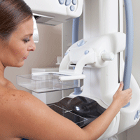
Genes may play role in breast cancer racial disparities
A new analysis of data from The Cancer Genome Atlas revealed 142 genes differentially expressed between white and African Americans that may influence breast cancer survival.
Researchers sifted through gene expression, protein expression, somatic mutations, somatic DNA copy number alteration, and DNA methylation patterns between 154 black patients of African ancestry (mean age at diagnosis, 55.66 years); and 776 white patients (mean age, 59.51) of European ancestry. They also looked at germline genetic variants for breast cancer subtypes and estimated subtype heritability.
African American patients were more likely than were whites to have basal-like breast cancer (odds ratio [OR], 3.80; 95% confidence interval [CI], 2.46-5.87; P less than .001) and HER2-enriched breast cancer (OR, 2.22; 95% CI, 1.10-4.47; P =.027).
After adjustment for subtype, 142 genes were differentially expressed between the two populations, a finding that held true after correction for testing of multiple comparisons (Bonferroni-adjusted P less than .05), reported Dezheng Huo, MD, PhD, of the department of public health sciences, University of Chicago, and his coauthors (JAMA Oncol. 2017 May 4. doi: 10.1001/jamaoncol.2017.0595).
During a median follow-up of 29 months, African Americans were more likely to experience recurrences (hazard ratio [HR], HR = 1.67, 95 CI, 1.02-2.74; P =.043). They had higher odds of basal-like breast cancer (OR, 3.80; 95% CI, 2.46-5.87; P less than .001) and human epidermal growth factor receptor 2 ERBB2 (HER2)-enriched breast cancer (OR, 2.22; 95% CI, 1.10-4.47; P =.027).
After adjusting for intrinsic subtypes, the researchers found differential expression of 16 DNA methylation probes, 4 DNA copy number segments, and 1 protein,

“If you have African ancestry, you’re more likely to have tumors that grow really fast. If we have drugs that can treat that kind of tumor, we need to find you and get you into treatment as quickly as possible,” she said.
A new analysis of data from The Cancer Genome Atlas revealed 142 genes differentially expressed between white and African Americans that may influence breast cancer survival.
Researchers sifted through gene expression, protein expression, somatic mutations, somatic DNA copy number alteration, and DNA methylation patterns between 154 black patients of African ancestry (mean age at diagnosis, 55.66 years); and 776 white patients (mean age, 59.51) of European ancestry. They also looked at germline genetic variants for breast cancer subtypes and estimated subtype heritability.
African American patients were more likely than were whites to have basal-like breast cancer (odds ratio [OR], 3.80; 95% confidence interval [CI], 2.46-5.87; P less than .001) and HER2-enriched breast cancer (OR, 2.22; 95% CI, 1.10-4.47; P =.027).
After adjustment for subtype, 142 genes were differentially expressed between the two populations, a finding that held true after correction for testing of multiple comparisons (Bonferroni-adjusted P less than .05), reported Dezheng Huo, MD, PhD, of the department of public health sciences, University of Chicago, and his coauthors (JAMA Oncol. 2017 May 4. doi: 10.1001/jamaoncol.2017.0595).
During a median follow-up of 29 months, African Americans were more likely to experience recurrences (hazard ratio [HR], HR = 1.67, 95 CI, 1.02-2.74; P =.043). They had higher odds of basal-like breast cancer (OR, 3.80; 95% CI, 2.46-5.87; P less than .001) and human epidermal growth factor receptor 2 ERBB2 (HER2)-enriched breast cancer (OR, 2.22; 95% CI, 1.10-4.47; P =.027).
After adjusting for intrinsic subtypes, the researchers found differential expression of 16 DNA methylation probes, 4 DNA copy number segments, and 1 protein,
“If you have African ancestry, you’re more likely to have tumors that grow really fast. If we have drugs that can treat that kind of tumor, we need to find you and get you into treatment as quickly as possible,” she said.
FROM JAMA ONCOLOGY
Key clinical point: Genetic differences may play role in racial disparities in breast cancer outcomes.
Major finding: The Cancer Genome Atlas data show that 142 genes were differentially expressed between whites and African Americans.
Data source: Analysis of genetic and outcome data from 930 patients.
Disclosures: The study was funded by the National Cancer Institute, the Breast Cancer Research Foundation, the National Human Genome Research Institute, Susan G. Komen, and the American Cancer Society. Dr. Olopade is a cofounder of CancerIQ and Prosigna.
Decision to remove breast cancer metastases depends on location of lesions
LAS VEGAS – Determination of whether excision of persistent breast cancer metastases can benefit the patient and even prolong survival depends on the location of the metastatic lesions, an investigator said at the annual meeting of the American Society of Breast Surgeons.
Before making the decision, it’s important to restage the patient’s disease and to recheck the receptor status if a biopsy is accessible, said Roshni Rao, MD, of the University of Texas Southwestern Medical Center.
Brain

Survival is worse when there are concurrent extracranial metastases or when brain metastases are greater than 5 cm, in patients with triple negative tumors, and in patients with a Karnofsky score of 70 or less, Dr. Rao said.
Surgery has the greatest benefit in patients with a single metastasis, with no extracranial disease, and who are able to undergo adjuvant whole brain radiation. In these cases, surgery improves survival, lowers recurrence rates, and reduces the risk of death neurological causes, she said.
Long-term survival is most common with continuous adjuvant therapy, either trastuzumab or hormonal, and in patients with a longer time interval to development of metastases. One series showed a 20-month survival increase.
Liver
Approximately 15% of patients with synchronous metastases will have liver metastases, and about half of stage IV patients will experience liver metastases at some point during treatment.
There is evidence from colorectal cancer that removing liver metastases is beneficial, and that has prompted interest in a similar approach in breast cancer. Liver resection has also become safer with new advances.
Dr. Rao discussed a single-institution study which took an aggressive approach to liver resection in 85 patients (Ann Surg. 2006;244:897-907). The researchers found that increased survival was associated with a good response to adjuvant chemotherapy, an r0 or r1 resection, and in patients who had a previous liver resection and were healthy enough to undergo another resection.
Overall, existing studies support liver resection if there are one to three lesions, if negative margins can be achieved, if the tumors are hormone receptor positive, and if the cancer is hormone positive and has good response to chemotherapy.
Dr. Rao emphasized that liver resections should be performed with a multidisciplinary team and should only be attempted at centers with low morbidity and where the doctors are experienced with liver resection.
When it’s possible, liver resection is beneficial. “There have been multiple reports of long-term survivors with no evidence of disease. There is likely a survival benefit with careful selection of these patients,” Dr. Rao said.
Lung
Lung surgeries are becoming safer, especially with the availability of video-assisted techniques, and pulmonary metastases are increasingly being spotted using more sensitive techniques such as higher resolution computed tomography.
A lung metastasis registry analysis showed three factors improved survival: prolonged disease-free survival, especially longer than 36 months; a complete resection; and a small number of metastases and success in resecting them all (Eur J Cardiothorac Surg. 2002;22:335-44). A more recent meta-analysis showed the same results (J Thorac Dis. 2015;7:1441-51).
Bone
Bone metastases remain rare choices for surgical treatment. Most of the time, morbidity will be too high, and there are good options for systemic treatment. That leaves surgery reserved mostly for stabilization or the treatment of fractures.
However, there are a couple of exceptions, according to Dr. Rao. One multi-institutional randomized trial looked at metastatic epidural spinal cord compression. Subjects either underwent decompressive surgery with stabilization and radiation or radiation alone. Patients in the surgical group had a longer ambulatory period and had a lower usage rate of steroids and opioids. Morbidity outcomes were similar in both groups. Patients whose primary tumor was in the breast seemed to benefit the most with respect to ambulatory time (Lancet. 2005;9486:643-8; J Neurosurg Spine. 2008;8:271-8).
Sternal metastases represent another special case. 70% of the time, patients with sternal metastases have it as their only metastatic site. A French series of 33 patients who underwent aggressive chest wall and rib resection reported a 36% complication rate, while another study of 28 patients showed a 21% complication rate. Those complications are a problem, “but if you’re able to perform this in a resected tumor, there are long term survivors. As usual, triple negative breast cancers predicted a worse prognosis,” said Dr. Rao.
Dr. Rao concluded that resection of metastatic sites has a role. “I think it’s our responsibility as breast surgeons who are many times continuously following these patients to consider appropriate operations,” said Dr. Rao.
However, she did sound one note of caution. Surgery can interrupt therapy that is helping a patient. “Let’s say you have someone get a big liver resection, and then they have a tough time with recovery. There could be a long period of time they can’t get the therapy that was keeping them alive. That’s the real concern,” she said.
LAS VEGAS – Determination of whether excision of persistent breast cancer metastases can benefit the patient and even prolong survival depends on the location of the metastatic lesions, an investigator said at the annual meeting of the American Society of Breast Surgeons.
Before making the decision, it’s important to restage the patient’s disease and to recheck the receptor status if a biopsy is accessible, said Roshni Rao, MD, of the University of Texas Southwestern Medical Center.
Brain
Survival is worse when there are concurrent extracranial metastases or when brain metastases are greater than 5 cm, in patients with triple negative tumors, and in patients with a Karnofsky score of 70 or less, Dr. Rao said.
Surgery has the greatest benefit in patients with a single metastasis, with no extracranial disease, and who are able to undergo adjuvant whole brain radiation. In these cases, surgery improves survival, lowers recurrence rates, and reduces the risk of death neurological causes, she said.
Long-term survival is most common with continuous adjuvant therapy, either trastuzumab or hormonal, and in patients with a longer time interval to development of metastases. One series showed a 20-month survival increase.
Liver
Approximately 15% of patients with synchronous metastases will have liver metastases, and about half of stage IV patients will experience liver metastases at some point during treatment.
There is evidence from colorectal cancer that removing liver metastases is beneficial, and that has prompted interest in a similar approach in breast cancer. Liver resection has also become safer with new advances.
Dr. Rao discussed a single-institution study which took an aggressive approach to liver resection in 85 patients (Ann Surg. 2006;244:897-907). The researchers found that increased survival was associated with a good response to adjuvant chemotherapy, an r0 or r1 resection, and in patients who had a previous liver resection and were healthy enough to undergo another resection.
Overall, existing studies support liver resection if there are one to three lesions, if negative margins can be achieved, if the tumors are hormone receptor positive, and if the cancer is hormone positive and has good response to chemotherapy.
Dr. Rao emphasized that liver resections should be performed with a multidisciplinary team and should only be attempted at centers with low morbidity and where the doctors are experienced with liver resection.
When it’s possible, liver resection is beneficial. “There have been multiple reports of long-term survivors with no evidence of disease. There is likely a survival benefit with careful selection of these patients,” Dr. Rao said.
Lung
Lung surgeries are becoming safer, especially with the availability of video-assisted techniques, and pulmonary metastases are increasingly being spotted using more sensitive techniques such as higher resolution computed tomography.
A lung metastasis registry analysis showed three factors improved survival: prolonged disease-free survival, especially longer than 36 months; a complete resection; and a small number of metastases and success in resecting them all (Eur J Cardiothorac Surg. 2002;22:335-44). A more recent meta-analysis showed the same results (J Thorac Dis. 2015;7:1441-51).
Bone
Bone metastases remain rare choices for surgical treatment. Most of the time, morbidity will be too high, and there are good options for systemic treatment. That leaves surgery reserved mostly for stabilization or the treatment of fractures.
However, there are a couple of exceptions, according to Dr. Rao. One multi-institutional randomized trial looked at metastatic epidural spinal cord compression. Subjects either underwent decompressive surgery with stabilization and radiation or radiation alone. Patients in the surgical group had a longer ambulatory period and had a lower usage rate of steroids and opioids. Morbidity outcomes were similar in both groups. Patients whose primary tumor was in the breast seemed to benefit the most with respect to ambulatory time (Lancet. 2005;9486:643-8; J Neurosurg Spine. 2008;8:271-8).
Sternal metastases represent another special case. 70% of the time, patients with sternal metastases have it as their only metastatic site. A French series of 33 patients who underwent aggressive chest wall and rib resection reported a 36% complication rate, while another study of 28 patients showed a 21% complication rate. Those complications are a problem, “but if you’re able to perform this in a resected tumor, there are long term survivors. As usual, triple negative breast cancers predicted a worse prognosis,” said Dr. Rao.
Dr. Rao concluded that resection of metastatic sites has a role. “I think it’s our responsibility as breast surgeons who are many times continuously following these patients to consider appropriate operations,” said Dr. Rao.
However, she did sound one note of caution. Surgery can interrupt therapy that is helping a patient. “Let’s say you have someone get a big liver resection, and then they have a tough time with recovery. There could be a long period of time they can’t get the therapy that was keeping them alive. That’s the real concern,” she said.
EXPERT ANALYSIS FROM ASBS 2017
Androgen receptor screening not ready for triple-negative breast cancer
LAS VEGAS – Despite the early promise of antiandrogen therapy, it’s not time yet to routinely screen women with triple-negative breast cancer for androgen receptors, according to Tiffany A. Traina, MD, the head of research into the disease at Memorial Sloan Kettering Cancer Center, New York.

There’s no standardized test for androgen receptors in breast cancer, so people “are doing different kinds of testing.” In the literature, “the range of AR positivity is anywhere from 12% to 79%, which reflects how we are all over the map in methodology; you might just as well throw a dart at the board. I would encourage screening in the context of the ongoing trials,” Dr. Traina said.
More than a decade ago, Memorial Sloan Kettering found a subset of TNBC that had ARs, which was peculiar because the tumors weren’t otherwise responsive to hormones. Androgen exposure increased growth, but the AR antagonist flutamide (Eulexin)blocked it. “It was thought provoking. There are a lot of drugs in the prostate cancer world” such as flutamide that shut down androgens, she said (Oncogene. 2006 Jun 29;25[28]:3994-4008).
Several have been tried, and investigations are ongoing. The work matters because TNBC is a particularly bad diagnosis. Blocking androgens seems to give some women a few more months of life.
Dr. Traina was the senior author in an early proof-of-concept study for AR blockade that involved 26 women with metastatic TNBC who had been through up to eight prior chemotherapy regimens. The women received 150 mg daily of the prostate cancer AR antagonist bicalutamide (Casodex). Disease remained stable in five (19%) for more than 6 months. Median progression-free survival was 12 weeks, which was “not that far off from what you get with [standard] chemotherapies. This was encouraging, and it led to multiple other trials looking at targeted therapies,” she said (Clin Cancer Res. 2013 Oct 1; 19[19]: 5505-12).
Dr. Traina led a phase II investigation of the prostate cancer AR antagonist enzalutamide (Xtandi) in 118 women with advanced AR-positive TNBC. Her team created an androgen-driven gene signature as a potential biomarker of response. Median progression-free survival was 32 weeks in the 56 women (47%) who were positive for the gene signature, but 9 weeks in those who were not. There were two complete responses and five partial responses with enzalutamide. Currently, “we are looking at using enzalutamide for patients with AR-positive TNBC in the early stage after failure of standard therapies,” she said.
French investigators recently reported a 6-month clinical benefit – including one complete response – in 7 (21%) of 34 women with locally advanced or metastatic TNBC who were treated with 1,000 mg daily of abiraterone acetate (Zytiga), an androgen biosynthesis inhibitor approved for prostate cancer (Ann Oncol. 2016 May;27[5]:812-8).
“We still have a ways to go” before AR treatment reaches the clinic for routine breast cancer treatment, “but there’s reason for hope,” Dr. Traina said.
Dr. Traina reported funding, honoraria, and steering committing payments from a number of companies working on or marketing TNBC AR drugs, including Pfizer, Astellas, Innocrin, AstraZeneca, Eisai, and Merck.
LAS VEGAS – Despite the early promise of antiandrogen therapy, it’s not time yet to routinely screen women with triple-negative breast cancer for androgen receptors, according to Tiffany A. Traina, MD, the head of research into the disease at Memorial Sloan Kettering Cancer Center, New York.
There’s no standardized test for androgen receptors in breast cancer, so people “are doing different kinds of testing.” In the literature, “the range of AR positivity is anywhere from 12% to 79%, which reflects how we are all over the map in methodology; you might just as well throw a dart at the board. I would encourage screening in the context of the ongoing trials,” Dr. Traina said.
More than a decade ago, Memorial Sloan Kettering found a subset of TNBC that had ARs, which was peculiar because the tumors weren’t otherwise responsive to hormones. Androgen exposure increased growth, but the AR antagonist flutamide (Eulexin)blocked it. “It was thought provoking. There are a lot of drugs in the prostate cancer world” such as flutamide that shut down androgens, she said (Oncogene. 2006 Jun 29;25[28]:3994-4008).
Several have been tried, and investigations are ongoing. The work matters because TNBC is a particularly bad diagnosis. Blocking androgens seems to give some women a few more months of life.
Dr. Traina was the senior author in an early proof-of-concept study for AR blockade that involved 26 women with metastatic TNBC who had been through up to eight prior chemotherapy regimens. The women received 150 mg daily of the prostate cancer AR antagonist bicalutamide (Casodex). Disease remained stable in five (19%) for more than 6 months. Median progression-free survival was 12 weeks, which was “not that far off from what you get with [standard] chemotherapies. This was encouraging, and it led to multiple other trials looking at targeted therapies,” she said (Clin Cancer Res. 2013 Oct 1; 19[19]: 5505-12).
Dr. Traina led a phase II investigation of the prostate cancer AR antagonist enzalutamide (Xtandi) in 118 women with advanced AR-positive TNBC. Her team created an androgen-driven gene signature as a potential biomarker of response. Median progression-free survival was 32 weeks in the 56 women (47%) who were positive for the gene signature, but 9 weeks in those who were not. There were two complete responses and five partial responses with enzalutamide. Currently, “we are looking at using enzalutamide for patients with AR-positive TNBC in the early stage after failure of standard therapies,” she said.
French investigators recently reported a 6-month clinical benefit – including one complete response – in 7 (21%) of 34 women with locally advanced or metastatic TNBC who were treated with 1,000 mg daily of abiraterone acetate (Zytiga), an androgen biosynthesis inhibitor approved for prostate cancer (Ann Oncol. 2016 May;27[5]:812-8).
“We still have a ways to go” before AR treatment reaches the clinic for routine breast cancer treatment, “but there’s reason for hope,” Dr. Traina said.
Dr. Traina reported funding, honoraria, and steering committing payments from a number of companies working on or marketing TNBC AR drugs, including Pfizer, Astellas, Innocrin, AstraZeneca, Eisai, and Merck.
EXPERT ANALYSIS FROM ASBS 2017
Two new biomarkers show breast cancer validity
BRUSSELS – A pair of breast cancer biomarkers look promising for making better prognosis assessments of selected patients, but acceptance of both into practice will need further documentation of their clinical utility, declared a senior breast cancer oncologist who served as discussant for the studies.
One of the markers is high intratumor heterogeneity of estrogen receptor density, a flag of poor prognosis when heterogeneity is high. The second marker is the phosphorylated signal transducer and activator of transcription (pSTAT) 3, which appeared to link with good prognosis in estrogen receptor–positive breast cancer.
The data on intratumor estrogen-receptor heterogeneity came from specimens collected from the low-risk breast cancer patients enrolled in the Stockholm Adjuvant Tamoxifen trial during 1976-1990 (Acta Oncol. 2007 July 8;46[2]:133-45). Enrolled patients had lymph node–negative disease and primary tumors smaller than 30 mm. During the trial, researchers preserved formalin-fixed tumor specimens in paraffin from 778 patients, which formed the basis for the current study, explained Linda S. Lindström, PhD, a cancer epidemiologist at the Karolinska Institute in Stockholm. Slides from the specimens were restained for their estrogen receptor content in 2014 and assessed by two independent breast cancer pathologists. They scored the heterogeneity of estrogen receptor distribution as high, medium, or low, and Dr. Lindström and her associates calculated a hazard ratio for 25-year patient survival when they compared 593 specimens with high or low receptor heterogeneity. They adjusted the hazard ratios for several baseline variables including age, year of breast cancer diagnosis, HER2 status, Ki67 status, tumor grade, tumor size, randomization to tamoxifen or placebo treatment, and other factors.

“Routine clinical assessment of intratumor heterogeneity of estrogen receptor may identify patients at high long-term risk for fatal breast cancer that may potentially change clinical management, especially for patients with luminal A subtype tumors,” Dr. Lindström said.“I’d like to see the C statistic; will the prognostic model improve significantly with this added?” Dr. Linn wondered. “We need at least two more independent validations.”
The second biomarker study used two separate analyses of pSTAT3 expression. The first involved specimens collected from 3,074 patients with luminal breast cancer. Analysis of pSTAT3 gene signature expression showed that, the higher the expression levels were, associated with better relapse-free survival during follow-up out to as long as 8 years, reported Amir Sonnenblick, MD, an oncologist at the Sharret Institute of Oncology of Hadassah-Hebrew University Medical Center in Jerusalem.

Univariate analysis showed that binary pSTAT3 expression (positive or negative) significantly correlated with 10-year overall survival, with a hazard ratio of 0.66 (P = .04) for patients with positive expression, compared with those with no pSTAT3 expression, Dr. Sonnenblick said.
“pSTAT3 is associated with improved outcome in estrogen receptor–positive breast cancer. Future trials should take pSTAT3 status into account,” he concluded.
Dr. Linn cautioned that pSTAT3 expression should not be used to identify patients who can forgo chemotherapy, as the gene signature expression analysis showed that, even among patients with high pSTAT3 expression, long-term survival was still less than 90%.
Dr. Lindström and Dr. Sonnenblick had no disclosures. Dr. Linn has been an adviser to AstraZeneca, Cergentis, IBM Health, Novartis, Pfizer, Phillips Health, Roche, and Sanofi.
[email protected]
On Twitter @mitchelzoler
BRUSSELS – A pair of breast cancer biomarkers look promising for making better prognosis assessments of selected patients, but acceptance of both into practice will need further documentation of their clinical utility, declared a senior breast cancer oncologist who served as discussant for the studies.
One of the markers is high intratumor heterogeneity of estrogen receptor density, a flag of poor prognosis when heterogeneity is high. The second marker is the phosphorylated signal transducer and activator of transcription (pSTAT) 3, which appeared to link with good prognosis in estrogen receptor–positive breast cancer.
The data on intratumor estrogen-receptor heterogeneity came from specimens collected from the low-risk breast cancer patients enrolled in the Stockholm Adjuvant Tamoxifen trial during 1976-1990 (Acta Oncol. 2007 July 8;46[2]:133-45). Enrolled patients had lymph node–negative disease and primary tumors smaller than 30 mm. During the trial, researchers preserved formalin-fixed tumor specimens in paraffin from 778 patients, which formed the basis for the current study, explained Linda S. Lindström, PhD, a cancer epidemiologist at the Karolinska Institute in Stockholm. Slides from the specimens were restained for their estrogen receptor content in 2014 and assessed by two independent breast cancer pathologists. They scored the heterogeneity of estrogen receptor distribution as high, medium, or low, and Dr. Lindström and her associates calculated a hazard ratio for 25-year patient survival when they compared 593 specimens with high or low receptor heterogeneity. They adjusted the hazard ratios for several baseline variables including age, year of breast cancer diagnosis, HER2 status, Ki67 status, tumor grade, tumor size, randomization to tamoxifen or placebo treatment, and other factors.
“Routine clinical assessment of intratumor heterogeneity of estrogen receptor may identify patients at high long-term risk for fatal breast cancer that may potentially change clinical management, especially for patients with luminal A subtype tumors,” Dr. Lindström said.“I’d like to see the C statistic; will the prognostic model improve significantly with this added?” Dr. Linn wondered. “We need at least two more independent validations.”
The second biomarker study used two separate analyses of pSTAT3 expression. The first involved specimens collected from 3,074 patients with luminal breast cancer. Analysis of pSTAT3 gene signature expression showed that, the higher the expression levels were, associated with better relapse-free survival during follow-up out to as long as 8 years, reported Amir Sonnenblick, MD, an oncologist at the Sharret Institute of Oncology of Hadassah-Hebrew University Medical Center in Jerusalem.
Univariate analysis showed that binary pSTAT3 expression (positive or negative) significantly correlated with 10-year overall survival, with a hazard ratio of 0.66 (P = .04) for patients with positive expression, compared with those with no pSTAT3 expression, Dr. Sonnenblick said.
“pSTAT3 is associated with improved outcome in estrogen receptor–positive breast cancer. Future trials should take pSTAT3 status into account,” he concluded.
Dr. Linn cautioned that pSTAT3 expression should not be used to identify patients who can forgo chemotherapy, as the gene signature expression analysis showed that, even among patients with high pSTAT3 expression, long-term survival was still less than 90%.
Dr. Lindström and Dr. Sonnenblick had no disclosures. Dr. Linn has been an adviser to AstraZeneca, Cergentis, IBM Health, Novartis, Pfizer, Phillips Health, Roche, and Sanofi.
[email protected]
On Twitter @mitchelzoler
AT IMPAKT 2017 BREAST CANCER CONFERENCE
Key clinical point:
Major finding: High estrogen-receptor heterogeneity linked with worse outcomes; pSTAT3 expression linked with better outcomes.
Data source: A total of 593 patients enrolled in the Stockholm Adjuvant Tamoxifen trial, and 610 patients enrolled in the Breast International Group 2-98 trial.
Disclosures: Dr. Lindström and Dr. Sonnenblick had no disclosures. Dr. Linn has been an advisor to AstraZeneca, Cergentis, IBM Health, Novartis, Pfizer, Phillips Health, Roche, and Sanofi.