User login
Nipple-sparing mastectomy feasible in N+ early breast cancer
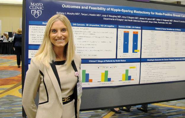
SAN FRANCISCO – Nipple-sparing mastectomy can be a safe surgical option for carefully selected patients with node-positive breast cancers, investigators reported at the 2015 ASCO Breast Cancer Symposium.
In a series of 226 patients with a total of 240 breast tumors, there was no significant difference between patients with node-positive or node-negative disease in the rate of conversion from a planned nipple-sparing procedure to a skin-sparing procedure, reported Dr. Brittany L. Murphy, a general surgery resident, and her colleagues at the Mayo Clinic in Rochester, Minn.
“Among women with node-positive breast cancer, nipple-sparing surgery may be appropriate for patients who do not have T4 or inflammatory carcinoma, patients that do not have multifocal disease, and who clinically and on imaging modalities do not appear to have nipple involvement,” Dr. Murphy said in an interview at the symposium.
In general, surgeons consider nipple-sparing mastectomies to be most appropriate for patients with early-stage disease; nodal involvement is often considered a contraindication, she said.
To see whether nipple-sparing surgery could be safely performed in patients with node-positive disease, the researchers took a retrospective look at data on 226 patients (14 with bilateral cancers) scheduled for nipple-sparing mastectomy at their center from 2009 through 2014.
In all, 182 of the cancers were lymph-node negative, and 58 were positive. Of the 58 node-positive cases, 27 (47%) were T2/T3 tumors, compared with 31 of 182 (17%) node-negative cases (P less than .0001). There were no significant differences between the groups in either estrogen receptor or HER2 receptor positivity, however.
Of the node-positive cases, 19 (33%) had cN1 (clinical) nodal involvement with pathology confirmed by fine-needle aspiration at the time of diagnosis, and 10 of these patients underwent neoadjuvant therapy followed by surgery. At the time of surgery, 6 of the 10 had pathologically confirmed positivity, and 4 were found to have ypN0 status. The remaining 9 patients in this group went on to primary surgery without neoadjuvant therapy.
Of the 39 patients who were clinically node negative, 4 had neoadjuvant therapy followed by surgery, and 35 went on to primary surgery.
The nipple-sparing procedure was successfully performed in 13 of the 14 node-positive patients who received neoadjuvant therapy and in 39 of 44 node-positive patients who went on to primary surgery. Six of the node-positive patients required conversion to a skin-sparing technique, either at the time of initial surgery based on frozen section pathology (five patients) or at a second procedure (one patient).
There were a total of seven locoregional recurrences among all patients treated with nipple-sparing mastectomy, including five in node-positive patients and two in node-negative patients. This difference was not significant.
Among the node-positive patients, three of the recurrences were in subcutaneous flaps away from the nipple at 13, 30, and 46 months of follow-up. In two cases, involved ipsilateral supraclavicular and mediastinal lymph nodes were detected at 24 and 32 months.
In the node-negative patients, one recurrence was in the nipple-areolar complex at 82 months, and one was in axillary nodes after negative sentinel lymph node biopsy at 20 months.
The 3-year locoregional disease-free estimates were 87% for lymph node positive patients compared with 99% for node negative patients (P = .007).
There were no differences between the groups regarding 3-year breast cancer–specific survival estimates, at 97% for lymph-node positive patients, and 99% for node-negative patients.
“Short-term oncologic outcomes were satisfactory. These data suggest that nipple-sparing mastectomy may be appropriate for carefully selected lymph node-positive breast cancer patients,” Dr, Murphy and colleagues wrote in a poster presented at the symposium.
SAN FRANCISCO – Nipple-sparing mastectomy can be a safe surgical option for carefully selected patients with node-positive breast cancers, investigators reported at the 2015 ASCO Breast Cancer Symposium.
In a series of 226 patients with a total of 240 breast tumors, there was no significant difference between patients with node-positive or node-negative disease in the rate of conversion from a planned nipple-sparing procedure to a skin-sparing procedure, reported Dr. Brittany L. Murphy, a general surgery resident, and her colleagues at the Mayo Clinic in Rochester, Minn.
“Among women with node-positive breast cancer, nipple-sparing surgery may be appropriate for patients who do not have T4 or inflammatory carcinoma, patients that do not have multifocal disease, and who clinically and on imaging modalities do not appear to have nipple involvement,” Dr. Murphy said in an interview at the symposium.
In general, surgeons consider nipple-sparing mastectomies to be most appropriate for patients with early-stage disease; nodal involvement is often considered a contraindication, she said.
To see whether nipple-sparing surgery could be safely performed in patients with node-positive disease, the researchers took a retrospective look at data on 226 patients (14 with bilateral cancers) scheduled for nipple-sparing mastectomy at their center from 2009 through 2014.
In all, 182 of the cancers were lymph-node negative, and 58 were positive. Of the 58 node-positive cases, 27 (47%) were T2/T3 tumors, compared with 31 of 182 (17%) node-negative cases (P less than .0001). There were no significant differences between the groups in either estrogen receptor or HER2 receptor positivity, however.
Of the node-positive cases, 19 (33%) had cN1 (clinical) nodal involvement with pathology confirmed by fine-needle aspiration at the time of diagnosis, and 10 of these patients underwent neoadjuvant therapy followed by surgery. At the time of surgery, 6 of the 10 had pathologically confirmed positivity, and 4 were found to have ypN0 status. The remaining 9 patients in this group went on to primary surgery without neoadjuvant therapy.
Of the 39 patients who were clinically node negative, 4 had neoadjuvant therapy followed by surgery, and 35 went on to primary surgery.
The nipple-sparing procedure was successfully performed in 13 of the 14 node-positive patients who received neoadjuvant therapy and in 39 of 44 node-positive patients who went on to primary surgery. Six of the node-positive patients required conversion to a skin-sparing technique, either at the time of initial surgery based on frozen section pathology (five patients) or at a second procedure (one patient).
There were a total of seven locoregional recurrences among all patients treated with nipple-sparing mastectomy, including five in node-positive patients and two in node-negative patients. This difference was not significant.
Among the node-positive patients, three of the recurrences were in subcutaneous flaps away from the nipple at 13, 30, and 46 months of follow-up. In two cases, involved ipsilateral supraclavicular and mediastinal lymph nodes were detected at 24 and 32 months.
In the node-negative patients, one recurrence was in the nipple-areolar complex at 82 months, and one was in axillary nodes after negative sentinel lymph node biopsy at 20 months.
The 3-year locoregional disease-free estimates were 87% for lymph node positive patients compared with 99% for node negative patients (P = .007).
There were no differences between the groups regarding 3-year breast cancer–specific survival estimates, at 97% for lymph-node positive patients, and 99% for node-negative patients.
“Short-term oncologic outcomes were satisfactory. These data suggest that nipple-sparing mastectomy may be appropriate for carefully selected lymph node-positive breast cancer patients,” Dr, Murphy and colleagues wrote in a poster presented at the symposium.
SAN FRANCISCO – Nipple-sparing mastectomy can be a safe surgical option for carefully selected patients with node-positive breast cancers, investigators reported at the 2015 ASCO Breast Cancer Symposium.
In a series of 226 patients with a total of 240 breast tumors, there was no significant difference between patients with node-positive or node-negative disease in the rate of conversion from a planned nipple-sparing procedure to a skin-sparing procedure, reported Dr. Brittany L. Murphy, a general surgery resident, and her colleagues at the Mayo Clinic in Rochester, Minn.
“Among women with node-positive breast cancer, nipple-sparing surgery may be appropriate for patients who do not have T4 or inflammatory carcinoma, patients that do not have multifocal disease, and who clinically and on imaging modalities do not appear to have nipple involvement,” Dr. Murphy said in an interview at the symposium.
In general, surgeons consider nipple-sparing mastectomies to be most appropriate for patients with early-stage disease; nodal involvement is often considered a contraindication, she said.
To see whether nipple-sparing surgery could be safely performed in patients with node-positive disease, the researchers took a retrospective look at data on 226 patients (14 with bilateral cancers) scheduled for nipple-sparing mastectomy at their center from 2009 through 2014.
In all, 182 of the cancers were lymph-node negative, and 58 were positive. Of the 58 node-positive cases, 27 (47%) were T2/T3 tumors, compared with 31 of 182 (17%) node-negative cases (P less than .0001). There were no significant differences between the groups in either estrogen receptor or HER2 receptor positivity, however.
Of the node-positive cases, 19 (33%) had cN1 (clinical) nodal involvement with pathology confirmed by fine-needle aspiration at the time of diagnosis, and 10 of these patients underwent neoadjuvant therapy followed by surgery. At the time of surgery, 6 of the 10 had pathologically confirmed positivity, and 4 were found to have ypN0 status. The remaining 9 patients in this group went on to primary surgery without neoadjuvant therapy.
Of the 39 patients who were clinically node negative, 4 had neoadjuvant therapy followed by surgery, and 35 went on to primary surgery.
The nipple-sparing procedure was successfully performed in 13 of the 14 node-positive patients who received neoadjuvant therapy and in 39 of 44 node-positive patients who went on to primary surgery. Six of the node-positive patients required conversion to a skin-sparing technique, either at the time of initial surgery based on frozen section pathology (five patients) or at a second procedure (one patient).
There were a total of seven locoregional recurrences among all patients treated with nipple-sparing mastectomy, including five in node-positive patients and two in node-negative patients. This difference was not significant.
Among the node-positive patients, three of the recurrences were in subcutaneous flaps away from the nipple at 13, 30, and 46 months of follow-up. In two cases, involved ipsilateral supraclavicular and mediastinal lymph nodes were detected at 24 and 32 months.
In the node-negative patients, one recurrence was in the nipple-areolar complex at 82 months, and one was in axillary nodes after negative sentinel lymph node biopsy at 20 months.
The 3-year locoregional disease-free estimates were 87% for lymph node positive patients compared with 99% for node negative patients (P = .007).
There were no differences between the groups regarding 3-year breast cancer–specific survival estimates, at 97% for lymph-node positive patients, and 99% for node-negative patients.
“Short-term oncologic outcomes were satisfactory. These data suggest that nipple-sparing mastectomy may be appropriate for carefully selected lymph node-positive breast cancer patients,” Dr, Murphy and colleagues wrote in a poster presented at the symposium.
AT THE 2015 ASCO BREAST CANCER SYMPOSIUM
Key clinical point: Some women with lymph-node positive early breast cancer may safely undergo nipple-sparing mastectomy.
Major finding: Three-year breast cancer-specific survival estimates were 97% for node positive patients and 99% for node-negative patients.
Data source: Retrospective review of data on 226 women with 240 early breast cancers.
Disclosures: The study was institutionally supported. Dr. Murphy reported no conflicts of interest.
Positive lumpectomy margin risk rises with breast density
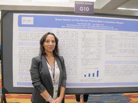
SAN FRANCISCO – Breast density is an independent risk factor for positive lumpectomy margins, pointing to a need for better methods of intraoperative margin assessment, researchers contend.
Data from a randomized clinical trial evaluating intraoperative tumor margin detection techniques indicate that for every increase in breast density category, the risk of positive margins on the main lumpectomy specimen increases by 46%, reported Dr. Tanir Allweis of Hebrew University Medical Center in Jerusalem, Israel.

“The use of newer technology and advanced techniques for intraoperative margin assessment, more extensive preop evaluation of these patients with MRI, or more liberal reshaving during the time of initial lumpectomy might be able to decrease the rate of reoperations in women with dense breasts,” Dr. Allweis said in an interview at the 2015 ASCO Breast Cancer Symposium.
She and colleague Dr. Freya Schnabel of New York University Langone Medical Center in New York City reviewed data on women enrolled in a clinical trial in which patients were randomized to lumpectomy with standard margin assessment or the use of an intraoperative radiofrequency spectroscopy device (MarginProbe). Of the 664 women enrolled in the trial, information on breast density was available for 450, and these women were included in the current study.The authors looked at data on breast density, patient and tumor characteristics, and the margin status of the primary lumpectomy specimen prior to randomization (that is, before the use of the device or the surgeon’s customary margin assessment technique).
They defined positive margins as ink on tumor. Breast density was rated on a scale of 1 (mostly fatty) to 4 (extremely dense) according to the American College of Radiology BI-RADS breast density descriptors.
Higher breast density was associated with younger age at diagnosis, lower body mass index, smaller breasts, and smaller specimen volume. Women with dense breasts were more likely to have had preoperative MRI (odds ratio [OR] 2. P less than .0001).
Each increase in breast density category was associated with an OR of 1.46 for positive margins in the main lumpectomy specimen. Thus, while women with mostly fatty breasts had a 14% risk for positive margins, women with extremely dense breasts had a 40% risk for positive margins.
The association between breast density and margin positivity remained significant after the researchers controlled for age, BMI, breast volume, and specimen volume (adjusted OR 1.39-1.52, P less than .036).
The investigators plan to explore whether the tumors in denser breasts may be larger than initially suspected because of the documented difficulties in imaging extremely dense tissues.
“These results suggest that the use of adjunctive methods for intraoperative margin assessment may be particularly helpful in this patient population. Further research will be important to clarify the benefit of various methods to decrease the rate for reexcision procedures in patients with increased breast density,” the investigators wrote in a poster presented at the symposium.
SAN FRANCISCO – Breast density is an independent risk factor for positive lumpectomy margins, pointing to a need for better methods of intraoperative margin assessment, researchers contend.
Data from a randomized clinical trial evaluating intraoperative tumor margin detection techniques indicate that for every increase in breast density category, the risk of positive margins on the main lumpectomy specimen increases by 46%, reported Dr. Tanir Allweis of Hebrew University Medical Center in Jerusalem, Israel.
“The use of newer technology and advanced techniques for intraoperative margin assessment, more extensive preop evaluation of these patients with MRI, or more liberal reshaving during the time of initial lumpectomy might be able to decrease the rate of reoperations in women with dense breasts,” Dr. Allweis said in an interview at the 2015 ASCO Breast Cancer Symposium.
She and colleague Dr. Freya Schnabel of New York University Langone Medical Center in New York City reviewed data on women enrolled in a clinical trial in which patients were randomized to lumpectomy with standard margin assessment or the use of an intraoperative radiofrequency spectroscopy device (MarginProbe). Of the 664 women enrolled in the trial, information on breast density was available for 450, and these women were included in the current study.The authors looked at data on breast density, patient and tumor characteristics, and the margin status of the primary lumpectomy specimen prior to randomization (that is, before the use of the device or the surgeon’s customary margin assessment technique).
They defined positive margins as ink on tumor. Breast density was rated on a scale of 1 (mostly fatty) to 4 (extremely dense) according to the American College of Radiology BI-RADS breast density descriptors.
Higher breast density was associated with younger age at diagnosis, lower body mass index, smaller breasts, and smaller specimen volume. Women with dense breasts were more likely to have had preoperative MRI (odds ratio [OR] 2. P less than .0001).
Each increase in breast density category was associated with an OR of 1.46 for positive margins in the main lumpectomy specimen. Thus, while women with mostly fatty breasts had a 14% risk for positive margins, women with extremely dense breasts had a 40% risk for positive margins.
The association between breast density and margin positivity remained significant after the researchers controlled for age, BMI, breast volume, and specimen volume (adjusted OR 1.39-1.52, P less than .036).
The investigators plan to explore whether the tumors in denser breasts may be larger than initially suspected because of the documented difficulties in imaging extremely dense tissues.
“These results suggest that the use of adjunctive methods for intraoperative margin assessment may be particularly helpful in this patient population. Further research will be important to clarify the benefit of various methods to decrease the rate for reexcision procedures in patients with increased breast density,” the investigators wrote in a poster presented at the symposium.
SAN FRANCISCO – Breast density is an independent risk factor for positive lumpectomy margins, pointing to a need for better methods of intraoperative margin assessment, researchers contend.
Data from a randomized clinical trial evaluating intraoperative tumor margin detection techniques indicate that for every increase in breast density category, the risk of positive margins on the main lumpectomy specimen increases by 46%, reported Dr. Tanir Allweis of Hebrew University Medical Center in Jerusalem, Israel.
“The use of newer technology and advanced techniques for intraoperative margin assessment, more extensive preop evaluation of these patients with MRI, or more liberal reshaving during the time of initial lumpectomy might be able to decrease the rate of reoperations in women with dense breasts,” Dr. Allweis said in an interview at the 2015 ASCO Breast Cancer Symposium.
She and colleague Dr. Freya Schnabel of New York University Langone Medical Center in New York City reviewed data on women enrolled in a clinical trial in which patients were randomized to lumpectomy with standard margin assessment or the use of an intraoperative radiofrequency spectroscopy device (MarginProbe). Of the 664 women enrolled in the trial, information on breast density was available for 450, and these women were included in the current study.The authors looked at data on breast density, patient and tumor characteristics, and the margin status of the primary lumpectomy specimen prior to randomization (that is, before the use of the device or the surgeon’s customary margin assessment technique).
They defined positive margins as ink on tumor. Breast density was rated on a scale of 1 (mostly fatty) to 4 (extremely dense) according to the American College of Radiology BI-RADS breast density descriptors.
Higher breast density was associated with younger age at diagnosis, lower body mass index, smaller breasts, and smaller specimen volume. Women with dense breasts were more likely to have had preoperative MRI (odds ratio [OR] 2. P less than .0001).
Each increase in breast density category was associated with an OR of 1.46 for positive margins in the main lumpectomy specimen. Thus, while women with mostly fatty breasts had a 14% risk for positive margins, women with extremely dense breasts had a 40% risk for positive margins.
The association between breast density and margin positivity remained significant after the researchers controlled for age, BMI, breast volume, and specimen volume (adjusted OR 1.39-1.52, P less than .036).
The investigators plan to explore whether the tumors in denser breasts may be larger than initially suspected because of the documented difficulties in imaging extremely dense tissues.
“These results suggest that the use of adjunctive methods for intraoperative margin assessment may be particularly helpful in this patient population. Further research will be important to clarify the benefit of various methods to decrease the rate for reexcision procedures in patients with increased breast density,” the investigators wrote in a poster presented at the symposium.
AT THE 2015 ASCO BREAST CANCER SYMPOSIUM
Key clinical point: The risk for positive lumpectomy margins rises as breast density increases.
Major finding: .Women with extremely dense breasts had a 40% risk for ink on tumor in the main lumpectomy specimen.
Data source: Review of data on 450 of 664 women enrolled in a randomized clinical trial.
Disclosures: Dune Medical Devices supported the trial. Dr. Allweis and Dr. Schnabel reported no conflicts of interest.
Dose-dense breast chemotherapy is also cost dense
SAN FRANCISCO – Despite the shorter course of care, dose-dense adjuvant chemotherapy for breast cancer may be considerably more costly than standard-dose chemotherapy, authors of a small study caution.
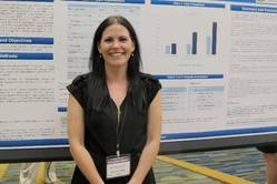
Among patients treated with four cycles of doxorubicin and cyclophosphamide (the AC regimen), the total cost for those treated with one cycle every 2 weeks (dose-dense schedule) was 77% higher than for patients treated once every 3 weeks, reported Helen O’Donovan, a medical student from Bon Secours Hospital and University College Cork, Ireland, and her associates.
The difference in cost was largely accounted for by the necessity for neutropenia prophylaxis with granulocytic colony stimulating factor (GCSF) among patients who received the dose-dense schedule, Ms. O’Donovan said in an interview at the 2015 ASCO Breast Cancer Symposium.
The mean cost of four doses of GCSF at her center was € 4,176 ($4,703). In the United States, a single syringe (one dose) of the long-acting GCSF pegfilgrastim (Neulasta) costs approximately $5,000 at retail pharmacies.
“We wanted to see whether GCSF in the long run might be cheaper if it could reduce admissions. It turns out that it wasn’t,” said Ms. O’Donovan.
To determine this, the investigators conducted a retrospective study comparing costs for patients treated with dose-dense chemotherapy with those treated once every 3 weeks for the same number of cycles. Because the hospital’s standard of care for several years has been the dose-dense regimen, the researchers were able to identify only 13 patients who had received the 3-week regimen. They matched these patients by age and year of diagnosis to 13 controls treated with dose-dense therapy.
All patients had early-stage breast cancer with no evidence of metastases and all were treated with AC-based chemotherapy with or without a taxane.
In all, six patients in the dose-dense group and five in the 3-week group required hospitalization for complications during chemotherapy. The mean duration of hospitalization was similar, at 4.7 and 4.6 days, respectively.
All patients in the dose-dense group received GCSF, compared with seven patients in the 3-week group. As noted, the mean costs for GCSF among patients with dose-dense chemotherapy were € 4,176 ($4,703), compared with € 1,900 ($2,139) for patients treated in the longer protocol (P = .0005).
Although the mean costs of hospitalization were not significantly different between the groups, adding in the cost of GCSF boosted the total costs to €6,163 ($6,937) vs. €3,486 ($3,923), and this difference was significant (P = .048).
Despite the GCSF prophylaxis and 100% compliance, febrile neutropenia was the most common reason for hospitalization in each group, with two patients in the dose-dense requiring hospitalization for 3 and 9 days, and three patients in the 3-week group needing admission, one for 4 days, and two for 8 days.
The authors acknowledged that the sample size was small, and that there may have been selection bias in the assignment of patients to the 3-week group.
SAN FRANCISCO – Despite the shorter course of care, dose-dense adjuvant chemotherapy for breast cancer may be considerably more costly than standard-dose chemotherapy, authors of a small study caution.
Among patients treated with four cycles of doxorubicin and cyclophosphamide (the AC regimen), the total cost for those treated with one cycle every 2 weeks (dose-dense schedule) was 77% higher than for patients treated once every 3 weeks, reported Helen O’Donovan, a medical student from Bon Secours Hospital and University College Cork, Ireland, and her associates.
The difference in cost was largely accounted for by the necessity for neutropenia prophylaxis with granulocytic colony stimulating factor (GCSF) among patients who received the dose-dense schedule, Ms. O’Donovan said in an interview at the 2015 ASCO Breast Cancer Symposium.
The mean cost of four doses of GCSF at her center was € 4,176 ($4,703). In the United States, a single syringe (one dose) of the long-acting GCSF pegfilgrastim (Neulasta) costs approximately $5,000 at retail pharmacies.
“We wanted to see whether GCSF in the long run might be cheaper if it could reduce admissions. It turns out that it wasn’t,” said Ms. O’Donovan.
To determine this, the investigators conducted a retrospective study comparing costs for patients treated with dose-dense chemotherapy with those treated once every 3 weeks for the same number of cycles. Because the hospital’s standard of care for several years has been the dose-dense regimen, the researchers were able to identify only 13 patients who had received the 3-week regimen. They matched these patients by age and year of diagnosis to 13 controls treated with dose-dense therapy.
All patients had early-stage breast cancer with no evidence of metastases and all were treated with AC-based chemotherapy with or without a taxane.
In all, six patients in the dose-dense group and five in the 3-week group required hospitalization for complications during chemotherapy. The mean duration of hospitalization was similar, at 4.7 and 4.6 days, respectively.
All patients in the dose-dense group received GCSF, compared with seven patients in the 3-week group. As noted, the mean costs for GCSF among patients with dose-dense chemotherapy were € 4,176 ($4,703), compared with € 1,900 ($2,139) for patients treated in the longer protocol (P = .0005).
Although the mean costs of hospitalization were not significantly different between the groups, adding in the cost of GCSF boosted the total costs to €6,163 ($6,937) vs. €3,486 ($3,923), and this difference was significant (P = .048).
Despite the GCSF prophylaxis and 100% compliance, febrile neutropenia was the most common reason for hospitalization in each group, with two patients in the dose-dense requiring hospitalization for 3 and 9 days, and three patients in the 3-week group needing admission, one for 4 days, and two for 8 days.
The authors acknowledged that the sample size was small, and that there may have been selection bias in the assignment of patients to the 3-week group.
SAN FRANCISCO – Despite the shorter course of care, dose-dense adjuvant chemotherapy for breast cancer may be considerably more costly than standard-dose chemotherapy, authors of a small study caution.
Among patients treated with four cycles of doxorubicin and cyclophosphamide (the AC regimen), the total cost for those treated with one cycle every 2 weeks (dose-dense schedule) was 77% higher than for patients treated once every 3 weeks, reported Helen O’Donovan, a medical student from Bon Secours Hospital and University College Cork, Ireland, and her associates.
The difference in cost was largely accounted for by the necessity for neutropenia prophylaxis with granulocytic colony stimulating factor (GCSF) among patients who received the dose-dense schedule, Ms. O’Donovan said in an interview at the 2015 ASCO Breast Cancer Symposium.
The mean cost of four doses of GCSF at her center was € 4,176 ($4,703). In the United States, a single syringe (one dose) of the long-acting GCSF pegfilgrastim (Neulasta) costs approximately $5,000 at retail pharmacies.
“We wanted to see whether GCSF in the long run might be cheaper if it could reduce admissions. It turns out that it wasn’t,” said Ms. O’Donovan.
To determine this, the investigators conducted a retrospective study comparing costs for patients treated with dose-dense chemotherapy with those treated once every 3 weeks for the same number of cycles. Because the hospital’s standard of care for several years has been the dose-dense regimen, the researchers were able to identify only 13 patients who had received the 3-week regimen. They matched these patients by age and year of diagnosis to 13 controls treated with dose-dense therapy.
All patients had early-stage breast cancer with no evidence of metastases and all were treated with AC-based chemotherapy with or without a taxane.
In all, six patients in the dose-dense group and five in the 3-week group required hospitalization for complications during chemotherapy. The mean duration of hospitalization was similar, at 4.7 and 4.6 days, respectively.
All patients in the dose-dense group received GCSF, compared with seven patients in the 3-week group. As noted, the mean costs for GCSF among patients with dose-dense chemotherapy were € 4,176 ($4,703), compared with € 1,900 ($2,139) for patients treated in the longer protocol (P = .0005).
Although the mean costs of hospitalization were not significantly different between the groups, adding in the cost of GCSF boosted the total costs to €6,163 ($6,937) vs. €3,486 ($3,923), and this difference was significant (P = .048).
Despite the GCSF prophylaxis and 100% compliance, febrile neutropenia was the most common reason for hospitalization in each group, with two patients in the dose-dense requiring hospitalization for 3 and 9 days, and three patients in the 3-week group needing admission, one for 4 days, and two for 8 days.
The authors acknowledged that the sample size was small, and that there may have been selection bias in the assignment of patients to the 3-week group.
AT THE 2015 ASCO BREAST CANCER SYMPOSIUM
Key clinical point: Total costs for hospitalization and GCSF are higher with dose-dense chemotherapy compared with the same regimen delivered every 3 weeks.
Major finding: The total costs of hospitalization plus GCSF were $6,937 for the dose-dense schedule vs. $3,923 for chemotherapy every 3 weeks.
Data source: Retrospective single-center study of 26 patients scheduled for adjuvant chemotherapy for early-stage breast cancer.
Disclosures: Funding for the study was not disclosed. Ms. O’Donovan reported no conflicts of interest.
Two heads plus four hands equal safe, shorter bilateral mastectomies
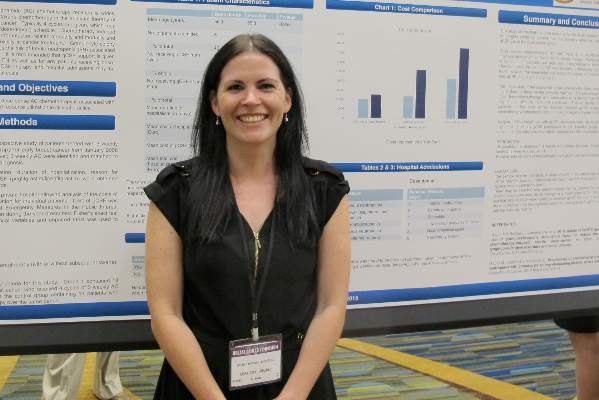
SAN FRANCISCO – Two surgeons working in tandem, one on each side of the patient, can significantly reduce the total operative time for bilateral mastectomies without compromising safety or quality of results, according to a study presented at the 2015 ASCO Breast Cancer Symposium.
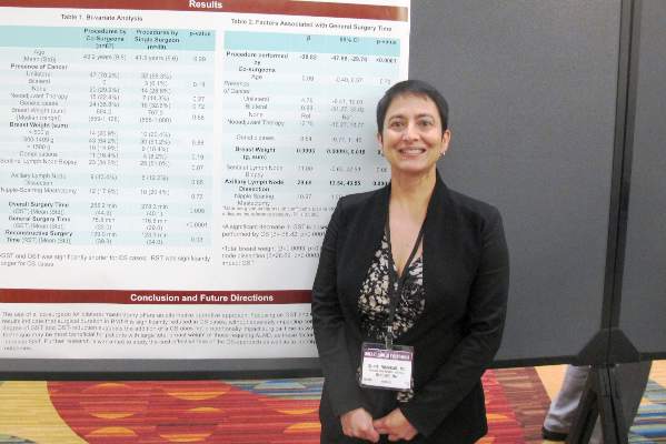
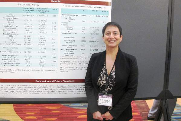
A review of records on consecutive cases of bilateral mastectomy with tissue expander reconstruction (BMTR) showed that mean overall surgery time (start of incision to closure) was about 23 minutes shorter when two surgeons were working at once, and mean general surgery time (start of incision to end of mastectomy procedure) was 41 minutes shorter with two surgeons vs. a solo operator, reported Dr. Suniti Nimbkar of the department of surgery at Brigham and Women’s Hospital, Boston.
“We did find that there was a significant reduction in time when two surgeons work together, and that reduction is particularly emphasized when you have a patient who is larger, perhaps a heavier patient, and when you have to do more extensive surgery,” she said in an interview at the symposium.
Dual surgeons did take slightly longer to perform the reconstructive surgery portion of the procedure, however.
Effective cosurgery is like a healthy marriage or a successful restaurant kitchen, with partners learning how to anticipate each other’s needs, knowing when to lend a hand, and intuitively grasping when to get out of the way, Dr. Nimbkar suggested.
“As two surgeons work together more and more, they just inevitably grow together in how they operate. I know for example that my direct partner and I operate very similarly, whereas sometimes if I do a cosurgery with a second surgeon that I don’t always operate with we have to work together to understand each other’s approach,” she said.
The investigators scanned the charts of 116 consecutive women who underwent BMTR done by eight breast surgeons at their center, looking for potential differences in operative time and 30-day postoperative complications. In all, 67 of the procedures were cosurgeries, and 49 were done by a single surgeon.
They found that in bivariate analysis, mean general surgery time was 75.8 minutes for the surgical duos, compared with 116.8 minutes for the solo surgeons (P less than .0001). Overall surgery time was also shorter, at 255.2 minutes vs. 278.3 minutes, respectively (P = .005).
Although there were numerically more complications among patients of cosurgeons, there was no statistically significant difference in postoperative complications rates between the twin and singleton surgeons.
A linear regression model showed that factors significantly associated with general surgery time were cosurgeries (P less than .0001), total breast weight (P =.03) and axillary dissection (P = .0003).
The authors noted that although two surgeons cut operative times significantly, the amount of time savings was not proportional to what they expected.
“We also want to know if the plastic surgeons are happy with the results if there are two different surgeons. Do they feel that there is too much of a difference in the way the mastectomy comes out, or is it something they’re fine with? So one of the next steps we’re going to take is to survey the plastic surgeons as to whether they’re comfortable with what we’re doing or if they have ideas about how we can be more uniform,” Dr. Nimbkar said.
SAN FRANCISCO – Two surgeons working in tandem, one on each side of the patient, can significantly reduce the total operative time for bilateral mastectomies without compromising safety or quality of results, according to a study presented at the 2015 ASCO Breast Cancer Symposium.
A review of records on consecutive cases of bilateral mastectomy with tissue expander reconstruction (BMTR) showed that mean overall surgery time (start of incision to closure) was about 23 minutes shorter when two surgeons were working at once, and mean general surgery time (start of incision to end of mastectomy procedure) was 41 minutes shorter with two surgeons vs. a solo operator, reported Dr. Suniti Nimbkar of the department of surgery at Brigham and Women’s Hospital, Boston.
“We did find that there was a significant reduction in time when two surgeons work together, and that reduction is particularly emphasized when you have a patient who is larger, perhaps a heavier patient, and when you have to do more extensive surgery,” she said in an interview at the symposium.
Dual surgeons did take slightly longer to perform the reconstructive surgery portion of the procedure, however.
Effective cosurgery is like a healthy marriage or a successful restaurant kitchen, with partners learning how to anticipate each other’s needs, knowing when to lend a hand, and intuitively grasping when to get out of the way, Dr. Nimbkar suggested.
“As two surgeons work together more and more, they just inevitably grow together in how they operate. I know for example that my direct partner and I operate very similarly, whereas sometimes if I do a cosurgery with a second surgeon that I don’t always operate with we have to work together to understand each other’s approach,” she said.
The investigators scanned the charts of 116 consecutive women who underwent BMTR done by eight breast surgeons at their center, looking for potential differences in operative time and 30-day postoperative complications. In all, 67 of the procedures were cosurgeries, and 49 were done by a single surgeon.
They found that in bivariate analysis, mean general surgery time was 75.8 minutes for the surgical duos, compared with 116.8 minutes for the solo surgeons (P less than .0001). Overall surgery time was also shorter, at 255.2 minutes vs. 278.3 minutes, respectively (P = .005).
Although there were numerically more complications among patients of cosurgeons, there was no statistically significant difference in postoperative complications rates between the twin and singleton surgeons.
A linear regression model showed that factors significantly associated with general surgery time were cosurgeries (P less than .0001), total breast weight (P =.03) and axillary dissection (P = .0003).
The authors noted that although two surgeons cut operative times significantly, the amount of time savings was not proportional to what they expected.
“We also want to know if the plastic surgeons are happy with the results if there are two different surgeons. Do they feel that there is too much of a difference in the way the mastectomy comes out, or is it something they’re fine with? So one of the next steps we’re going to take is to survey the plastic surgeons as to whether they’re comfortable with what we’re doing or if they have ideas about how we can be more uniform,” Dr. Nimbkar said.
SAN FRANCISCO – Two surgeons working in tandem, one on each side of the patient, can significantly reduce the total operative time for bilateral mastectomies without compromising safety or quality of results, according to a study presented at the 2015 ASCO Breast Cancer Symposium.
A review of records on consecutive cases of bilateral mastectomy with tissue expander reconstruction (BMTR) showed that mean overall surgery time (start of incision to closure) was about 23 minutes shorter when two surgeons were working at once, and mean general surgery time (start of incision to end of mastectomy procedure) was 41 minutes shorter with two surgeons vs. a solo operator, reported Dr. Suniti Nimbkar of the department of surgery at Brigham and Women’s Hospital, Boston.
“We did find that there was a significant reduction in time when two surgeons work together, and that reduction is particularly emphasized when you have a patient who is larger, perhaps a heavier patient, and when you have to do more extensive surgery,” she said in an interview at the symposium.
Dual surgeons did take slightly longer to perform the reconstructive surgery portion of the procedure, however.
Effective cosurgery is like a healthy marriage or a successful restaurant kitchen, with partners learning how to anticipate each other’s needs, knowing when to lend a hand, and intuitively grasping when to get out of the way, Dr. Nimbkar suggested.
“As two surgeons work together more and more, they just inevitably grow together in how they operate. I know for example that my direct partner and I operate very similarly, whereas sometimes if I do a cosurgery with a second surgeon that I don’t always operate with we have to work together to understand each other’s approach,” she said.
The investigators scanned the charts of 116 consecutive women who underwent BMTR done by eight breast surgeons at their center, looking for potential differences in operative time and 30-day postoperative complications. In all, 67 of the procedures were cosurgeries, and 49 were done by a single surgeon.
They found that in bivariate analysis, mean general surgery time was 75.8 minutes for the surgical duos, compared with 116.8 minutes for the solo surgeons (P less than .0001). Overall surgery time was also shorter, at 255.2 minutes vs. 278.3 minutes, respectively (P = .005).
Although there were numerically more complications among patients of cosurgeons, there was no statistically significant difference in postoperative complications rates between the twin and singleton surgeons.
A linear regression model showed that factors significantly associated with general surgery time were cosurgeries (P less than .0001), total breast weight (P =.03) and axillary dissection (P = .0003).
The authors noted that although two surgeons cut operative times significantly, the amount of time savings was not proportional to what they expected.
“We also want to know if the plastic surgeons are happy with the results if there are two different surgeons. Do they feel that there is too much of a difference in the way the mastectomy comes out, or is it something they’re fine with? So one of the next steps we’re going to take is to survey the plastic surgeons as to whether they’re comfortable with what we’re doing or if they have ideas about how we can be more uniform,” Dr. Nimbkar said.
AT THE 2015 ASCO BREAST CANCER SYMPOSIUM
Key clinical point:Bilateral mastectomy performed by two surgeons shortens operative time without increasing complications.
Major finding: Mean general surgery time was 75.8 minutes for two surgeons vs. 116.8 minutes for solo surgeons (P less than .0001).
Data source: Retrospective review of records on 116 consecutive patients undergoing bilateral mastectomies.
Disclosures: The study was internally funded. The authors reported no conflicts of interest.
Computer-aided detection fails to improve mammographic accuracy
Augmenting digital screening mammography with computer-aided detection failed to improve diagnostic accuracy in every performance measure and every subgroup of women studied in a series of 625,625 exams performed across the United States during a 7-year period, according to a report published online Sept. 28 in JAMA Internal Medicine.
In fact, the sensitivity of mammography was actually decreased by computer-aided detection (CAD) in a subgroup of radiologists who practiced at some sites that used CAD and others that did not.
“CAD is a technology that does not seem to warrant added compensation beyond coverage of the mammographic examination,” wrote Dr. Constance D. Lehman of the department of radiology, Massachusetts General Hospital and the Avon Comprehensive Breast Evaluation Center, both in Boston. “The results of our comprehensive study lend no support for continued reimbursement for CAD as a method to increase mammography performance or improve patient outcomes.”
Measuring the real-world impact of CAD on mammographic accuracy has been difficult and has yielded inconsistent and contradictory findings; most studies to date have been relatively small, have focused on older women only, or haven’t taken into account the early part of radiologists’ learning curves. To circumvent these problems, the investigators pooled data from five mammographic registries to include more than 625,000 full-field digital mammograms, included a demographically diverse population of women aged 40-89 years, and excluded the first year of CAD use for every radiologist in the study.

They assessed outcomes after routine screening mammography with CAD (495,818) or without CAD (129,807), which were interpreted by 271 radiologists. Breast cancer was diagnosed in 3,159 women within 1 year of these screening mammograms.
The overall sensitivity of mammography was 85.3% with CAD and 87.3% without it; sensitivity for invasive cancer was 82.1% with CAD and 85.0% without it, all of which are nonsignificant differences. Also similar were mammography’s specificity, at 91.6% with CAD and 91.4% without CAD.
The overall cancer detection rate was exactly the same regardless of the use of CAD, at 4.1 cancers per 1,000 women screened. And the invasive cancer detection rate was nearly the same, at 2.9 cancers with CAD versus 3.0 cancers without CAD (JAMA Intern Med. 2015 Sep 28. doi:10.1001/jamainternmed.2015.5231).
Also, diagnostic accuracy was the same with or without CAD regardless of patient age, patient ethnicity, breast density, patient menopausal status, family history, or interval since the last mammogram. In the subgroup of 107 radiologists who sometimes used CAD and sometimes did not, the use of CAD actually decreased the sensitivity of mammography (83.3% with CAD and 89.6% without CAD).
“Given that the evidence of the current application of CAD in community practice does not show an improvement in diagnostic accuracy, we question the policy of continuing to charge for a technology that provides no established benefits to women,” Dr. Lehman and her associates wrote.
But the researchers noted that CAD may offer advantages beyond diagnostic accuracy, such as improved work flow or shorter times spent assessing faint calcifications. CAD also may be useful in guiding treatment decisions, perhaps reducing unnecessary biopsies of lesions that have specific benign features or ensuring biopsy of lesions that have specific malignant features, they added.
The study was supported by the National Cancer Institute, the Breast Cancer Surveillance Consortium, several state public health departments, and cancer registries throughout the United States. Dr. Lehman reported receiving grant support from General Electric Healthcare and serving as a member of the company’s Comparative Effectiveness Research Advisory Board.
This study is another large-sample, real-world evaluation suggesting that CAD yields no clinically significant benefits in typical mammography practice.
Congress should rescind Medicare coverage of CAD use. And in the future, broad societal investment in new medical technologies should be withheld until large-sample assessments prove their real-world effectiveness and justify their costs.
Dr. Joshua J. Fenton is in the department of family and community medicine, the Center for Healthcare Policy and Research, and the Cancer Center at the University of California, Davis Health System, Sacramento. He reported having no relevant financial disclosures. These comments are adapted from a commentary accompanying Dr. Lehman’s report (JAMA Intern Med. 2015 Sep 28. doi:10.1001/jamainternmed.2015.5319).
This study is another large-sample, real-world evaluation suggesting that CAD yields no clinically significant benefits in typical mammography practice.
Congress should rescind Medicare coverage of CAD use. And in the future, broad societal investment in new medical technologies should be withheld until large-sample assessments prove their real-world effectiveness and justify their costs.
Dr. Joshua J. Fenton is in the department of family and community medicine, the Center for Healthcare Policy and Research, and the Cancer Center at the University of California, Davis Health System, Sacramento. He reported having no relevant financial disclosures. These comments are adapted from a commentary accompanying Dr. Lehman’s report (JAMA Intern Med. 2015 Sep 28. doi:10.1001/jamainternmed.2015.5319).
This study is another large-sample, real-world evaluation suggesting that CAD yields no clinically significant benefits in typical mammography practice.
Congress should rescind Medicare coverage of CAD use. And in the future, broad societal investment in new medical technologies should be withheld until large-sample assessments prove their real-world effectiveness and justify their costs.
Dr. Joshua J. Fenton is in the department of family and community medicine, the Center for Healthcare Policy and Research, and the Cancer Center at the University of California, Davis Health System, Sacramento. He reported having no relevant financial disclosures. These comments are adapted from a commentary accompanying Dr. Lehman’s report (JAMA Intern Med. 2015 Sep 28. doi:10.1001/jamainternmed.2015.5319).
Augmenting digital screening mammography with computer-aided detection failed to improve diagnostic accuracy in every performance measure and every subgroup of women studied in a series of 625,625 exams performed across the United States during a 7-year period, according to a report published online Sept. 28 in JAMA Internal Medicine.
In fact, the sensitivity of mammography was actually decreased by computer-aided detection (CAD) in a subgroup of radiologists who practiced at some sites that used CAD and others that did not.
“CAD is a technology that does not seem to warrant added compensation beyond coverage of the mammographic examination,” wrote Dr. Constance D. Lehman of the department of radiology, Massachusetts General Hospital and the Avon Comprehensive Breast Evaluation Center, both in Boston. “The results of our comprehensive study lend no support for continued reimbursement for CAD as a method to increase mammography performance or improve patient outcomes.”
Measuring the real-world impact of CAD on mammographic accuracy has been difficult and has yielded inconsistent and contradictory findings; most studies to date have been relatively small, have focused on older women only, or haven’t taken into account the early part of radiologists’ learning curves. To circumvent these problems, the investigators pooled data from five mammographic registries to include more than 625,000 full-field digital mammograms, included a demographically diverse population of women aged 40-89 years, and excluded the first year of CAD use for every radiologist in the study.
They assessed outcomes after routine screening mammography with CAD (495,818) or without CAD (129,807), which were interpreted by 271 radiologists. Breast cancer was diagnosed in 3,159 women within 1 year of these screening mammograms.
The overall sensitivity of mammography was 85.3% with CAD and 87.3% without it; sensitivity for invasive cancer was 82.1% with CAD and 85.0% without it, all of which are nonsignificant differences. Also similar were mammography’s specificity, at 91.6% with CAD and 91.4% without CAD.
The overall cancer detection rate was exactly the same regardless of the use of CAD, at 4.1 cancers per 1,000 women screened. And the invasive cancer detection rate was nearly the same, at 2.9 cancers with CAD versus 3.0 cancers without CAD (JAMA Intern Med. 2015 Sep 28. doi:10.1001/jamainternmed.2015.5231).
Also, diagnostic accuracy was the same with or without CAD regardless of patient age, patient ethnicity, breast density, patient menopausal status, family history, or interval since the last mammogram. In the subgroup of 107 radiologists who sometimes used CAD and sometimes did not, the use of CAD actually decreased the sensitivity of mammography (83.3% with CAD and 89.6% without CAD).
“Given that the evidence of the current application of CAD in community practice does not show an improvement in diagnostic accuracy, we question the policy of continuing to charge for a technology that provides no established benefits to women,” Dr. Lehman and her associates wrote.
But the researchers noted that CAD may offer advantages beyond diagnostic accuracy, such as improved work flow or shorter times spent assessing faint calcifications. CAD also may be useful in guiding treatment decisions, perhaps reducing unnecessary biopsies of lesions that have specific benign features or ensuring biopsy of lesions that have specific malignant features, they added.
The study was supported by the National Cancer Institute, the Breast Cancer Surveillance Consortium, several state public health departments, and cancer registries throughout the United States. Dr. Lehman reported receiving grant support from General Electric Healthcare and serving as a member of the company’s Comparative Effectiveness Research Advisory Board.
Augmenting digital screening mammography with computer-aided detection failed to improve diagnostic accuracy in every performance measure and every subgroup of women studied in a series of 625,625 exams performed across the United States during a 7-year period, according to a report published online Sept. 28 in JAMA Internal Medicine.
In fact, the sensitivity of mammography was actually decreased by computer-aided detection (CAD) in a subgroup of radiologists who practiced at some sites that used CAD and others that did not.
“CAD is a technology that does not seem to warrant added compensation beyond coverage of the mammographic examination,” wrote Dr. Constance D. Lehman of the department of radiology, Massachusetts General Hospital and the Avon Comprehensive Breast Evaluation Center, both in Boston. “The results of our comprehensive study lend no support for continued reimbursement for CAD as a method to increase mammography performance or improve patient outcomes.”
Measuring the real-world impact of CAD on mammographic accuracy has been difficult and has yielded inconsistent and contradictory findings; most studies to date have been relatively small, have focused on older women only, or haven’t taken into account the early part of radiologists’ learning curves. To circumvent these problems, the investigators pooled data from five mammographic registries to include more than 625,000 full-field digital mammograms, included a demographically diverse population of women aged 40-89 years, and excluded the first year of CAD use for every radiologist in the study.
They assessed outcomes after routine screening mammography with CAD (495,818) or without CAD (129,807), which were interpreted by 271 radiologists. Breast cancer was diagnosed in 3,159 women within 1 year of these screening mammograms.
The overall sensitivity of mammography was 85.3% with CAD and 87.3% without it; sensitivity for invasive cancer was 82.1% with CAD and 85.0% without it, all of which are nonsignificant differences. Also similar were mammography’s specificity, at 91.6% with CAD and 91.4% without CAD.
The overall cancer detection rate was exactly the same regardless of the use of CAD, at 4.1 cancers per 1,000 women screened. And the invasive cancer detection rate was nearly the same, at 2.9 cancers with CAD versus 3.0 cancers without CAD (JAMA Intern Med. 2015 Sep 28. doi:10.1001/jamainternmed.2015.5231).
Also, diagnostic accuracy was the same with or without CAD regardless of patient age, patient ethnicity, breast density, patient menopausal status, family history, or interval since the last mammogram. In the subgroup of 107 radiologists who sometimes used CAD and sometimes did not, the use of CAD actually decreased the sensitivity of mammography (83.3% with CAD and 89.6% without CAD).
“Given that the evidence of the current application of CAD in community practice does not show an improvement in diagnostic accuracy, we question the policy of continuing to charge for a technology that provides no established benefits to women,” Dr. Lehman and her associates wrote.
But the researchers noted that CAD may offer advantages beyond diagnostic accuracy, such as improved work flow or shorter times spent assessing faint calcifications. CAD also may be useful in guiding treatment decisions, perhaps reducing unnecessary biopsies of lesions that have specific benign features or ensuring biopsy of lesions that have specific malignant features, they added.
The study was supported by the National Cancer Institute, the Breast Cancer Surveillance Consortium, several state public health departments, and cancer registries throughout the United States. Dr. Lehman reported receiving grant support from General Electric Healthcare and serving as a member of the company’s Comparative Effectiveness Research Advisory Board.
FROM JAMA INTERNAL MEDICINE
Key clinical point: Augmenting digital screening mammography with computer-aided detection failed to improve diagnostic accuracy in every performance measure and every subgroup of women studied.
Major finding: Overall sensitivity of mammography was 85.3% with CAD and 87.3% without it; sensitivity for invasive cancer was 82.1% with CAD and 85.0% without it.
Data source: A large observational study comparing breast cancer detection between 495,818 mammograms that used CAD and 129,807 that did not.
Disclosures: The study was supported by the National Cancer Institute, the Breast Cancer Surveillance Consortium, several state public health departments, and cancer registries throughout the United States. Dr. Lehman reported receiving grant support from General Electric Healthcare and serving as a member of the company’s Comparative Effectiveness Research Advisory Board.

ASCO: Potentially targetable biomarkers identified in geriatric breast cancer tumors
SAN FRANCISCO – Using multiplatform profiling, potentially targetable biomarker aberrations were identified in a large sample of tumors taken from older breast cancer patients, reported researchers at the 2015 Breast Cancer Symposium.
In this cross sectional study, 1,189 tumors were collected from breast cancer patients between the ages of 70-97 years, and the samples were assayed for potential targetable biomarkers, and then the findings were compared with samples obtained from younger patients.
“Most of the differences we found were in triple negative patients, and they are related to PIK3CA mutations,” said study author Dr. Paula Pohlmann, from the Lombardi Comprehensive Cancer Center, MedStar Georgetown University Hospital, Washington.

“Also, I am very puzzled that the BRCA1 mutations were not found in older triple negative patients, as compared with the younger patients,” she noted. “Even taking into account the limitations of the study, it was still interesting.”
ERBB2 mutations were identified in non-amplifed breast cancers and less frequently in amplified ones. “And another interesting point was that both PD-1 and PD-L1 positivity by immunohistochemistry were found in all groups, and not only triple negative,” said Dr. Pohlmann. “The checkpoints are similar for both older and younger populations.”
Dr. Pohlmann explained that while she expected to see the PD-1 and PD-L1 positivity, it is worth mentioning since all the treatments focusing on PD-1 and PD-L1 in breast cancer are with triple negative disease. “But we found this expression in Her2 positive and hormone receptor positive disease.”
However, she emphasized that this doesn’t mean that they are necessarily a target. “If they will respond to that therapy, we don’t know, that’s a different story, but the markers are there,” Dr. Pohlmann said.
In this study a total of 1,189 tumors were collected from breast cancer patients (male= 21/female = 1,168) who were aged 70 years and older. The tumors were analyzed (breast biopsy n = 512; metastatic site n = 677), and of 1,088 tumors with available immunohistochemistry (IHC) of ER, PR and IHC and/or in situ hybridization (ISH) of HER2, 613 (56%) were HR+/HER2-, 72 (7%) were HR+/HER2+, 346 (32%) were triple negative, and 57 (5%) were HR-/HER2+.
Overall, 39 of 47 genes that were sequenced carried mutations with frequencies ranging from 0.2% to 37%. The highest mutation rates were seen in PIK3CA (37%), TP53 (37%), BRCA2 (12%), PTEN (5.8%), AKT1 (4.2%), cMET (3.9%), ERBB2 (3.5%), BRCA1 (3.3%), and ATM (3.2%).
Among 13 patients with ERBB2 mutation, 3 had it amplified. PD-L1 expression on tumor cells was detected in 13% of tumors and PD-1 expression on tumor-infiltrating lymphocytes was seen in 46%, but the triple negative subtype had the highest expression: 20% and 60%, respectively.
The researchers also noted that TOPO1, TLE3, AR, TOP2A, and SPARC were overexpressed in 61%, 58%, 55%, 51%, and 37% of tumors, respectively. These observations suggest that there is potential sensitivity to irinotecan, taxanes, anthracyclines, and nab-paclitaxel.
In addition, TS, RRM1, and ERCC1 were under-expressed in 69%, 69%, and 53% of tumors, respectively, which suggests that there may be potential sensitivity to fluoropyrimidines, gemcitabine, and platinums
“This study provides key elements for the design of clinical trials focusing on geriatric patient population, particularly in the subgroup of triple negative breast cancer,” said Dr. Pohlmann. “In addition, ERBB2 abnormalities in older patients may also warrant further investigation.”
Dr. Charles E. Geyer, Jr., of Virginia Commonwealth University Massey Cancer Center, Richmond, in his discussion of the paper, pointed out that while there were some differences in the groups, it wasn’t clear that they are clinically significant differences. “The most interesting were PD-1 and PD-L1, which were in higher numbers than are being reported.”
But the data from this study suggest that focus needs to be on inclusion of geriatric patients in targeted therapy trials, he said.
He also noted that in general, patients frequently have some unrealistic expectations when it comes to genetic testing. “The advocacy often gets a little ahead of the science,” Dr. Geyer said.
SAN FRANCISCO – Using multiplatform profiling, potentially targetable biomarker aberrations were identified in a large sample of tumors taken from older breast cancer patients, reported researchers at the 2015 Breast Cancer Symposium.
In this cross sectional study, 1,189 tumors were collected from breast cancer patients between the ages of 70-97 years, and the samples were assayed for potential targetable biomarkers, and then the findings were compared with samples obtained from younger patients.
“Most of the differences we found were in triple negative patients, and they are related to PIK3CA mutations,” said study author Dr. Paula Pohlmann, from the Lombardi Comprehensive Cancer Center, MedStar Georgetown University Hospital, Washington.
“Also, I am very puzzled that the BRCA1 mutations were not found in older triple negative patients, as compared with the younger patients,” she noted. “Even taking into account the limitations of the study, it was still interesting.”
ERBB2 mutations were identified in non-amplifed breast cancers and less frequently in amplified ones. “And another interesting point was that both PD-1 and PD-L1 positivity by immunohistochemistry were found in all groups, and not only triple negative,” said Dr. Pohlmann. “The checkpoints are similar for both older and younger populations.”
Dr. Pohlmann explained that while she expected to see the PD-1 and PD-L1 positivity, it is worth mentioning since all the treatments focusing on PD-1 and PD-L1 in breast cancer are with triple negative disease. “But we found this expression in Her2 positive and hormone receptor positive disease.”
However, she emphasized that this doesn’t mean that they are necessarily a target. “If they will respond to that therapy, we don’t know, that’s a different story, but the markers are there,” Dr. Pohlmann said.
In this study a total of 1,189 tumors were collected from breast cancer patients (male= 21/female = 1,168) who were aged 70 years and older. The tumors were analyzed (breast biopsy n = 512; metastatic site n = 677), and of 1,088 tumors with available immunohistochemistry (IHC) of ER, PR and IHC and/or in situ hybridization (ISH) of HER2, 613 (56%) were HR+/HER2-, 72 (7%) were HR+/HER2+, 346 (32%) were triple negative, and 57 (5%) were HR-/HER2+.
Overall, 39 of 47 genes that were sequenced carried mutations with frequencies ranging from 0.2% to 37%. The highest mutation rates were seen in PIK3CA (37%), TP53 (37%), BRCA2 (12%), PTEN (5.8%), AKT1 (4.2%), cMET (3.9%), ERBB2 (3.5%), BRCA1 (3.3%), and ATM (3.2%).
Among 13 patients with ERBB2 mutation, 3 had it amplified. PD-L1 expression on tumor cells was detected in 13% of tumors and PD-1 expression on tumor-infiltrating lymphocytes was seen in 46%, but the triple negative subtype had the highest expression: 20% and 60%, respectively.
The researchers also noted that TOPO1, TLE3, AR, TOP2A, and SPARC were overexpressed in 61%, 58%, 55%, 51%, and 37% of tumors, respectively. These observations suggest that there is potential sensitivity to irinotecan, taxanes, anthracyclines, and nab-paclitaxel.
In addition, TS, RRM1, and ERCC1 were under-expressed in 69%, 69%, and 53% of tumors, respectively, which suggests that there may be potential sensitivity to fluoropyrimidines, gemcitabine, and platinums
“This study provides key elements for the design of clinical trials focusing on geriatric patient population, particularly in the subgroup of triple negative breast cancer,” said Dr. Pohlmann. “In addition, ERBB2 abnormalities in older patients may also warrant further investigation.”
Dr. Charles E. Geyer, Jr., of Virginia Commonwealth University Massey Cancer Center, Richmond, in his discussion of the paper, pointed out that while there were some differences in the groups, it wasn’t clear that they are clinically significant differences. “The most interesting were PD-1 and PD-L1, which were in higher numbers than are being reported.”
But the data from this study suggest that focus needs to be on inclusion of geriatric patients in targeted therapy trials, he said.
He also noted that in general, patients frequently have some unrealistic expectations when it comes to genetic testing. “The advocacy often gets a little ahead of the science,” Dr. Geyer said.
SAN FRANCISCO – Using multiplatform profiling, potentially targetable biomarker aberrations were identified in a large sample of tumors taken from older breast cancer patients, reported researchers at the 2015 Breast Cancer Symposium.
In this cross sectional study, 1,189 tumors were collected from breast cancer patients between the ages of 70-97 years, and the samples were assayed for potential targetable biomarkers, and then the findings were compared with samples obtained from younger patients.
“Most of the differences we found were in triple negative patients, and they are related to PIK3CA mutations,” said study author Dr. Paula Pohlmann, from the Lombardi Comprehensive Cancer Center, MedStar Georgetown University Hospital, Washington.
“Also, I am very puzzled that the BRCA1 mutations were not found in older triple negative patients, as compared with the younger patients,” she noted. “Even taking into account the limitations of the study, it was still interesting.”
ERBB2 mutations were identified in non-amplifed breast cancers and less frequently in amplified ones. “And another interesting point was that both PD-1 and PD-L1 positivity by immunohistochemistry were found in all groups, and not only triple negative,” said Dr. Pohlmann. “The checkpoints are similar for both older and younger populations.”
Dr. Pohlmann explained that while she expected to see the PD-1 and PD-L1 positivity, it is worth mentioning since all the treatments focusing on PD-1 and PD-L1 in breast cancer are with triple negative disease. “But we found this expression in Her2 positive and hormone receptor positive disease.”
However, she emphasized that this doesn’t mean that they are necessarily a target. “If they will respond to that therapy, we don’t know, that’s a different story, but the markers are there,” Dr. Pohlmann said.
In this study a total of 1,189 tumors were collected from breast cancer patients (male= 21/female = 1,168) who were aged 70 years and older. The tumors were analyzed (breast biopsy n = 512; metastatic site n = 677), and of 1,088 tumors with available immunohistochemistry (IHC) of ER, PR and IHC and/or in situ hybridization (ISH) of HER2, 613 (56%) were HR+/HER2-, 72 (7%) were HR+/HER2+, 346 (32%) were triple negative, and 57 (5%) were HR-/HER2+.
Overall, 39 of 47 genes that were sequenced carried mutations with frequencies ranging from 0.2% to 37%. The highest mutation rates were seen in PIK3CA (37%), TP53 (37%), BRCA2 (12%), PTEN (5.8%), AKT1 (4.2%), cMET (3.9%), ERBB2 (3.5%), BRCA1 (3.3%), and ATM (3.2%).
Among 13 patients with ERBB2 mutation, 3 had it amplified. PD-L1 expression on tumor cells was detected in 13% of tumors and PD-1 expression on tumor-infiltrating lymphocytes was seen in 46%, but the triple negative subtype had the highest expression: 20% and 60%, respectively.
The researchers also noted that TOPO1, TLE3, AR, TOP2A, and SPARC were overexpressed in 61%, 58%, 55%, 51%, and 37% of tumors, respectively. These observations suggest that there is potential sensitivity to irinotecan, taxanes, anthracyclines, and nab-paclitaxel.
In addition, TS, RRM1, and ERCC1 were under-expressed in 69%, 69%, and 53% of tumors, respectively, which suggests that there may be potential sensitivity to fluoropyrimidines, gemcitabine, and platinums
“This study provides key elements for the design of clinical trials focusing on geriatric patient population, particularly in the subgroup of triple negative breast cancer,” said Dr. Pohlmann. “In addition, ERBB2 abnormalities in older patients may also warrant further investigation.”
Dr. Charles E. Geyer, Jr., of Virginia Commonwealth University Massey Cancer Center, Richmond, in his discussion of the paper, pointed out that while there were some differences in the groups, it wasn’t clear that they are clinically significant differences. “The most interesting were PD-1 and PD-L1, which were in higher numbers than are being reported.”
But the data from this study suggest that focus needs to be on inclusion of geriatric patients in targeted therapy trials, he said.
He also noted that in general, patients frequently have some unrealistic expectations when it comes to genetic testing. “The advocacy often gets a little ahead of the science,” Dr. Geyer said.
FROM THE ASCO BREAST CANCER SYMPOSIUM 2015
Key clinical point: Multiplatform profiling identified potentially targetable biomarker aberrations in a large sample of tumor taken from older breast cancer patients.
Major finding: Most of the differences found between younger and older patients were in triple negative patients, and these were primarily related to PIK3CA mutations.
Data source: A cross sectional study comprised of 1189 tumors collected from breast cancer patients aged 70-97 years, and assayed for potential targetable biomarkers.
Disclosures: Dr. Pohlmann has relationships with Immunonet BioSciences, OncoPlex Diagnostics; Personalized Cancer Therapy and patents/royalties and other intellectual property for Immunological Compositions as Cancer Therapeutics. Dr. Joanne Xiu and Dr. Sandeep K. Reddy are employees of Caris Life Sciences, who conducted the genetic analysis.

ASCO: Neoadjuvant chemo does not increase breast cancer surgery complications
SAN FRANCISCO – Breast cancer surgery within 30 days of neoadjuvant chemotherapy appears to be safe, with no significant increase in risk of postoperative complications compared with surgery performed in women who did not receive chemotherapy.
An analysis of data on more than 3,500 patients who underwent surgery for invasive breast cancer after receiving neoadjuvant chemotherapy showed that in an analysis adjusted to better balance patient characteristics, there were no significant differences in postoperative complication rates for patients who received chemotherapy and those who did not, reported Dr. Erin Cordeiro from the Ottawa Hospital in Ontario, Canada, and associates, at a breast cancer symposium sponsored jointly by the American Society of Clinical Oncology, American Society for Radiation Oncology, and Society of Surgical Oncology.

“I really think this should put to rest the concern that surgeons might have about neoadjuvant chemotherapy particularly because this [study] compressed people within 30 days of the surgery date,” commented Dr. Charles E. Geyer, Jr., from the Virginia Commonwealth University School of Medicine in Richmond. Dr. Geyer was the invited discussant.
The use of neoadjuvant chemotherapy in the treatment of patients with breast cancer continues to increase, growing from 14% in 2006, to 20% in 2011, Dr. Cordeiro said.
But because myelosuppression, particularly neutropenia, is a common side of chemotherapy, surgeons are often concerned that operating too soon after a patient completes a course of chemotherapy could result in increased post-operative complications, she said.
To determine whether neoadjuvant chemotherapy would increase the proportion of overall 30-day postoperative complications among patients undergoing surgery for invasive breast cancer, Dr. Cordeiro drew on the American College of Surgeons National Surgical Quality Improvement Program (ACS-NSQIP) database, which contains validated, risk-adjusted surgical outcomes data from more than 600 hospitals in 49 states, Canada, and Europe.

She identified data on 67,685 patients who underwent surgery for invasive breast cancer from 2005 through 2012. Of this group, 3,624 (5.5%) had received neoadjuvant chemotherapy. Investigators excludedpPatients with high-risk comorbidities and those who underwent other surgery concurrent with breast surgery (e.g., oophorectomy).
In unadjusted analysis, the rate of a composite of overall 30-day post-operative complications, the primary outcome, was 4.9% for patients who had undergone neoadjuvant chemotherapy, compared with 3.7% for patients who did not and this difference was significant (P= .0003). The higher rate among patient who had received chemotherapy recently was largely due to infections, Dr. Cordeiro said.
However, in a multivariate analysis adjusted for propensity score – a measure of the probability of receiving neoeadjuvant chemotherapy by age, diabetes, chronic obstructive pulmonary disease, hypertension, bilateral surgery, and/or year of surgery – the differences between patients who underwent neoadjuvant chemo and those who did not vanished (odds ratio 1.16; 95% confidence interval, 0.98-1.36).
Dr. Cordeiro acknowledged that the study was limited by the use of retrospective data, a lack of knowledge of exactly when chemotherapy was stopped in relation to the surgery data, lack of data on which regimens were used, and no information about prophylatic antibiotics or thromboprophylaxis.
The investigators plan to perform additional subgroup analyses to determine whether there might be differences in complication rates in patients who did or did not receive neoadjuvant chemo by type of breast surgery (breast-conserving vs. mastectomy), type of axillary surgery (sentinel node biopsy vs. axillary lymph node dissection) or among patients who undergo immediate reconstruction.
The study was presented as a poster and in an oral abstract session.
SAN FRANCISCO – Breast cancer surgery within 30 days of neoadjuvant chemotherapy appears to be safe, with no significant increase in risk of postoperative complications compared with surgery performed in women who did not receive chemotherapy.
An analysis of data on more than 3,500 patients who underwent surgery for invasive breast cancer after receiving neoadjuvant chemotherapy showed that in an analysis adjusted to better balance patient characteristics, there were no significant differences in postoperative complication rates for patients who received chemotherapy and those who did not, reported Dr. Erin Cordeiro from the Ottawa Hospital in Ontario, Canada, and associates, at a breast cancer symposium sponsored jointly by the American Society of Clinical Oncology, American Society for Radiation Oncology, and Society of Surgical Oncology.
“I really think this should put to rest the concern that surgeons might have about neoadjuvant chemotherapy particularly because this [study] compressed people within 30 days of the surgery date,” commented Dr. Charles E. Geyer, Jr., from the Virginia Commonwealth University School of Medicine in Richmond. Dr. Geyer was the invited discussant.
The use of neoadjuvant chemotherapy in the treatment of patients with breast cancer continues to increase, growing from 14% in 2006, to 20% in 2011, Dr. Cordeiro said.
But because myelosuppression, particularly neutropenia, is a common side of chemotherapy, surgeons are often concerned that operating too soon after a patient completes a course of chemotherapy could result in increased post-operative complications, she said.
To determine whether neoadjuvant chemotherapy would increase the proportion of overall 30-day postoperative complications among patients undergoing surgery for invasive breast cancer, Dr. Cordeiro drew on the American College of Surgeons National Surgical Quality Improvement Program (ACS-NSQIP) database, which contains validated, risk-adjusted surgical outcomes data from more than 600 hospitals in 49 states, Canada, and Europe.
She identified data on 67,685 patients who underwent surgery for invasive breast cancer from 2005 through 2012. Of this group, 3,624 (5.5%) had received neoadjuvant chemotherapy. Investigators excludedpPatients with high-risk comorbidities and those who underwent other surgery concurrent with breast surgery (e.g., oophorectomy).
In unadjusted analysis, the rate of a composite of overall 30-day post-operative complications, the primary outcome, was 4.9% for patients who had undergone neoadjuvant chemotherapy, compared with 3.7% for patients who did not and this difference was significant (P= .0003). The higher rate among patient who had received chemotherapy recently was largely due to infections, Dr. Cordeiro said.
However, in a multivariate analysis adjusted for propensity score – a measure of the probability of receiving neoeadjuvant chemotherapy by age, diabetes, chronic obstructive pulmonary disease, hypertension, bilateral surgery, and/or year of surgery – the differences between patients who underwent neoadjuvant chemo and those who did not vanished (odds ratio 1.16; 95% confidence interval, 0.98-1.36).
Dr. Cordeiro acknowledged that the study was limited by the use of retrospective data, a lack of knowledge of exactly when chemotherapy was stopped in relation to the surgery data, lack of data on which regimens were used, and no information about prophylatic antibiotics or thromboprophylaxis.
The investigators plan to perform additional subgroup analyses to determine whether there might be differences in complication rates in patients who did or did not receive neoadjuvant chemo by type of breast surgery (breast-conserving vs. mastectomy), type of axillary surgery (sentinel node biopsy vs. axillary lymph node dissection) or among patients who undergo immediate reconstruction.
The study was presented as a poster and in an oral abstract session.
SAN FRANCISCO – Breast cancer surgery within 30 days of neoadjuvant chemotherapy appears to be safe, with no significant increase in risk of postoperative complications compared with surgery performed in women who did not receive chemotherapy.
An analysis of data on more than 3,500 patients who underwent surgery for invasive breast cancer after receiving neoadjuvant chemotherapy showed that in an analysis adjusted to better balance patient characteristics, there were no significant differences in postoperative complication rates for patients who received chemotherapy and those who did not, reported Dr. Erin Cordeiro from the Ottawa Hospital in Ontario, Canada, and associates, at a breast cancer symposium sponsored jointly by the American Society of Clinical Oncology, American Society for Radiation Oncology, and Society of Surgical Oncology.
“I really think this should put to rest the concern that surgeons might have about neoadjuvant chemotherapy particularly because this [study] compressed people within 30 days of the surgery date,” commented Dr. Charles E. Geyer, Jr., from the Virginia Commonwealth University School of Medicine in Richmond. Dr. Geyer was the invited discussant.
The use of neoadjuvant chemotherapy in the treatment of patients with breast cancer continues to increase, growing from 14% in 2006, to 20% in 2011, Dr. Cordeiro said.
But because myelosuppression, particularly neutropenia, is a common side of chemotherapy, surgeons are often concerned that operating too soon after a patient completes a course of chemotherapy could result in increased post-operative complications, she said.
To determine whether neoadjuvant chemotherapy would increase the proportion of overall 30-day postoperative complications among patients undergoing surgery for invasive breast cancer, Dr. Cordeiro drew on the American College of Surgeons National Surgical Quality Improvement Program (ACS-NSQIP) database, which contains validated, risk-adjusted surgical outcomes data from more than 600 hospitals in 49 states, Canada, and Europe.
She identified data on 67,685 patients who underwent surgery for invasive breast cancer from 2005 through 2012. Of this group, 3,624 (5.5%) had received neoadjuvant chemotherapy. Investigators excludedpPatients with high-risk comorbidities and those who underwent other surgery concurrent with breast surgery (e.g., oophorectomy).
In unadjusted analysis, the rate of a composite of overall 30-day post-operative complications, the primary outcome, was 4.9% for patients who had undergone neoadjuvant chemotherapy, compared with 3.7% for patients who did not and this difference was significant (P= .0003). The higher rate among patient who had received chemotherapy recently was largely due to infections, Dr. Cordeiro said.
However, in a multivariate analysis adjusted for propensity score – a measure of the probability of receiving neoeadjuvant chemotherapy by age, diabetes, chronic obstructive pulmonary disease, hypertension, bilateral surgery, and/or year of surgery – the differences between patients who underwent neoadjuvant chemo and those who did not vanished (odds ratio 1.16; 95% confidence interval, 0.98-1.36).
Dr. Cordeiro acknowledged that the study was limited by the use of retrospective data, a lack of knowledge of exactly when chemotherapy was stopped in relation to the surgery data, lack of data on which regimens were used, and no information about prophylatic antibiotics or thromboprophylaxis.
The investigators plan to perform additional subgroup analyses to determine whether there might be differences in complication rates in patients who did or did not receive neoadjuvant chemo by type of breast surgery (breast-conserving vs. mastectomy), type of axillary surgery (sentinel node biopsy vs. axillary lymph node dissection) or among patients who undergo immediate reconstruction.
The study was presented as a poster and in an oral abstract session.
AT THE ASCO BREAST CANCER SYMPOSIUM
Key clinical point: Neoadjuvant chemotherapy does not appear to increase the risk for 30-day post-operative complications following surgery for invasive breast cancer.
Major finding: The odds ratio for postoperative complications among patients who underwent chemotherapy was 1.16 but was not statistically significant.
Data source: Retrospective review from a prospective surgical outcomes database on 67,685 patients, 3,624 of whom had neoadjuvant chemotherapy.
Disclosures: Funding for the study was not disclosed. Dr. Cordeiro reported having no conflicts of interest. Dr. Geyer reported a consulting or advisory role with Stennion, research funding from Incyte, and travel expenses from Abbvie, AstraZeneca, and Genetech/Roche.

ASCO: Many women with triple-negative breast cancer aren’t screened for BRCA
SAN FRANCISCO – Many younger women diagnosed with triple-negative breast cancers do not get tested for BRCA, despite guideline recommendations, investigators report.
Among 173 women with triple-negative tumors -- lacking the HER2, estrogen and progesterone receptors –17% of those who should have been tested for BRCA according to National Comprehensive Cancer Network (NCCN) guidelines, were not tested.
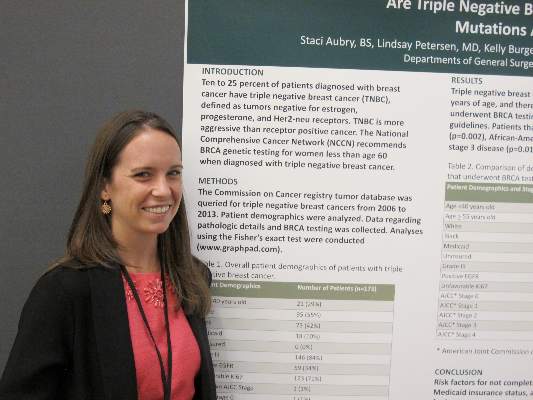
Women less likely to be tested were those 55 years or older, African Americans, those who list Medicaid as their primary form of insurance, and those with stage 3 disease, reported Staci Aubry, a 4th-year medical student at Rush University Medical Center in Chicago, and her associates.
In an interview, Ms. Aubry said that some of the women who were eligible for genetic testing under the guidelines simply declined it.
“Often patients, if they didn’t have a daughter or if they were older and were in the 50 to 60 [year-old] range, even though they’re still included in the NCCN guidelines to be screened, still refused to be tested,” she said.
NCCN guidelines on genetic and familial high-risk assessment for breast and ovarian cancer susceptibility recommend BRCA genetic testing for all women below age 60 years who are diagnosed with triple-negative breast cancer.
“Some histopathologic features have reported to occur more frequently in breast cancers characterized by a BRCA1 or BRCA2 mutation. For example, several studies have shown that BRCA1 breast cancer is more likely to be characterized as ER-/PR-negative and HER2-negative (i.e., “triple negative”), the guidelines note.
To see whether clinicians were adhering to this recommendation, the investigators searched the Commission on Cancer registry tumor database for cases of triple-negative breast cancers diagnosed from 2006 through 2013.
They identified 173 patients, 105 of whom were younger than 60 and thus recommended for screening. Of this group, 87 (83%) were tested for BRCA, and 15 were found to be BRCA positive, but the remaining 18 patients (17%) were not tested.
When the authors looked at demographic and clinical factors that might have accounted for the differences between women who were tested for the gene and those who were not, four factors stood out. Women who did not get tested were more likely to be 55 or older (P = .002), African American (P = .001), Medicaid insured (P = .021) or to have American Joint Commission on Cancer stage 3 disease (P = .014).
“The main message for clinicians is to be aware that these disparities exist, and keep in mind that these four groups – people who are under Medicaid insurance, between 55 and 60 years of age, African American, or who have higher disease stage – should be targeted for screening,” Ms. Aubry said.
SAN FRANCISCO – Many younger women diagnosed with triple-negative breast cancers do not get tested for BRCA, despite guideline recommendations, investigators report.
Among 173 women with triple-negative tumors -- lacking the HER2, estrogen and progesterone receptors –17% of those who should have been tested for BRCA according to National Comprehensive Cancer Network (NCCN) guidelines, were not tested.
Women less likely to be tested were those 55 years or older, African Americans, those who list Medicaid as their primary form of insurance, and those with stage 3 disease, reported Staci Aubry, a 4th-year medical student at Rush University Medical Center in Chicago, and her associates.
In an interview, Ms. Aubry said that some of the women who were eligible for genetic testing under the guidelines simply declined it.
“Often patients, if they didn’t have a daughter or if they were older and were in the 50 to 60 [year-old] range, even though they’re still included in the NCCN guidelines to be screened, still refused to be tested,” she said.
NCCN guidelines on genetic and familial high-risk assessment for breast and ovarian cancer susceptibility recommend BRCA genetic testing for all women below age 60 years who are diagnosed with triple-negative breast cancer.
“Some histopathologic features have reported to occur more frequently in breast cancers characterized by a BRCA1 or BRCA2 mutation. For example, several studies have shown that BRCA1 breast cancer is more likely to be characterized as ER-/PR-negative and HER2-negative (i.e., “triple negative”), the guidelines note.
To see whether clinicians were adhering to this recommendation, the investigators searched the Commission on Cancer registry tumor database for cases of triple-negative breast cancers diagnosed from 2006 through 2013.
They identified 173 patients, 105 of whom were younger than 60 and thus recommended for screening. Of this group, 87 (83%) were tested for BRCA, and 15 were found to be BRCA positive, but the remaining 18 patients (17%) were not tested.
When the authors looked at demographic and clinical factors that might have accounted for the differences between women who were tested for the gene and those who were not, four factors stood out. Women who did not get tested were more likely to be 55 or older (P = .002), African American (P = .001), Medicaid insured (P = .021) or to have American Joint Commission on Cancer stage 3 disease (P = .014).
“The main message for clinicians is to be aware that these disparities exist, and keep in mind that these four groups – people who are under Medicaid insurance, between 55 and 60 years of age, African American, or who have higher disease stage – should be targeted for screening,” Ms. Aubry said.
SAN FRANCISCO – Many younger women diagnosed with triple-negative breast cancers do not get tested for BRCA, despite guideline recommendations, investigators report.
Among 173 women with triple-negative tumors -- lacking the HER2, estrogen and progesterone receptors –17% of those who should have been tested for BRCA according to National Comprehensive Cancer Network (NCCN) guidelines, were not tested.
Women less likely to be tested were those 55 years or older, African Americans, those who list Medicaid as their primary form of insurance, and those with stage 3 disease, reported Staci Aubry, a 4th-year medical student at Rush University Medical Center in Chicago, and her associates.
In an interview, Ms. Aubry said that some of the women who were eligible for genetic testing under the guidelines simply declined it.
“Often patients, if they didn’t have a daughter or if they were older and were in the 50 to 60 [year-old] range, even though they’re still included in the NCCN guidelines to be screened, still refused to be tested,” she said.
NCCN guidelines on genetic and familial high-risk assessment for breast and ovarian cancer susceptibility recommend BRCA genetic testing for all women below age 60 years who are diagnosed with triple-negative breast cancer.
“Some histopathologic features have reported to occur more frequently in breast cancers characterized by a BRCA1 or BRCA2 mutation. For example, several studies have shown that BRCA1 breast cancer is more likely to be characterized as ER-/PR-negative and HER2-negative (i.e., “triple negative”), the guidelines note.
To see whether clinicians were adhering to this recommendation, the investigators searched the Commission on Cancer registry tumor database for cases of triple-negative breast cancers diagnosed from 2006 through 2013.
They identified 173 patients, 105 of whom were younger than 60 and thus recommended for screening. Of this group, 87 (83%) were tested for BRCA, and 15 were found to be BRCA positive, but the remaining 18 patients (17%) were not tested.
When the authors looked at demographic and clinical factors that might have accounted for the differences between women who were tested for the gene and those who were not, four factors stood out. Women who did not get tested were more likely to be 55 or older (P = .002), African American (P = .001), Medicaid insured (P = .021) or to have American Joint Commission on Cancer stage 3 disease (P = .014).
“The main message for clinicians is to be aware that these disparities exist, and keep in mind that these four groups – people who are under Medicaid insurance, between 55 and 60 years of age, African American, or who have higher disease stage – should be targeted for screening,” Ms. Aubry said.
AT THE ASCO BREAST CANCER SYMPOSIUM
Key clinical point: Guidelines recommend screening for BRCA in women under 60 years of age with triple-negative breast cancer.
Major finding: Of 105 women eligible for BRCA screening, 18 (17%) were not tested.
Data source: Review of registry data on 173 women with triple-negative breast cancer, including 105 under age 60 years and therefore recommended for BRCA testing.
Disclosures: The investigators did not disclosed a funding source. Ms. Aubry reported having no conflicts of interest.
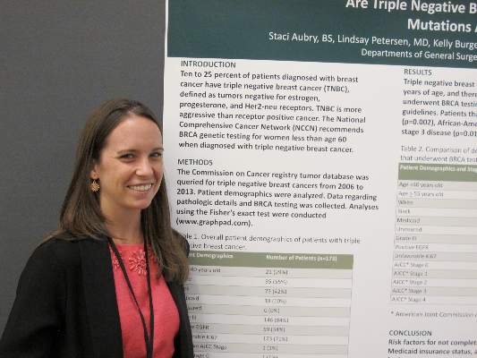
ASCO: Model predicts risk for breast cancer from atypical hyperplasia
SAN FRANCISCO – A woman’s age at biopsy and the number of atypical hyperplasia foci appear to be good predictors of risk for subsequent breast cancer, investigators say.
A review of pathology records and medical history on more than 13,000 women with benign breast disease showed that a predictive model including age and atypia effectively identified those women with atypical hyperplasia at highest risk for developing breast cancer, reported Dr. Amy C. Degnim from the Mayo Clinic in Rochester, Minnesota.

“In our country, approximately 10% of all benign breast biopsies are showing atypical hyperplasia, and with about a million women who undergo biopsy every year, this about 100,000 women every year who are diagnosed with atypical hyperplasia. These women are known to have an increased risk of breast cancer,” Dr. Degnim said at a breast symposium here jointly sponsored by the American Society of Clinical Oncology, American Society For Radiation Oncology, and Society of Surgical Oncology.
Current risk prediction models such as the Breast Cancer Risk Assessment Tool and IBIS Breast Cancer Risk Evaluation Tool tend to underestimate or overestimate risk of breast cancer in women with atypical hyperplasia, prompting the investigators to explore developing a reliable prediction tool, she said
The investigators identified a cohort of 13,538 women diagnosed with benign breast disease at the Mayo Clinic from 1967 through 2001, and they found data on 699 with atypical hyperplasia confirmed by pathology review blinded to outcomes. They collected data on clinical and histologic features and identified breast cancer event through review of medical records and questionnaires.
In addition to the Mayo patients used to develop the model, the authors tested it in a validation sample of 461 women with atypical hyperplasia treated at Vanderibilt University in Nashville, Tennessee.
They rounded up data on potential contributors to a risk-prediction model using Lasso-identified variables that they then plugged into a Cox regression model.
They found that of all possible co-variates, including body-mass index at biopsy, age at menarche, indication for biopsy, number of live births, breastfeeding, family features, and histologic features such as involution or calcifications, only the age at biopsy and number of foci of atypical hyperplasia remained as robust predictors for breast cancer risk.
They then tested the model on data from the 699 women in the development set, who had had a total of 142 breast cancer events over a median follow-up of 8.1 years, and in the external validation set of 461 women who had a total of 114 breast cancer events over a median follow-up of 11.4 years.

They found that the concordance between the prediction model and the actual outcomes at 5, 10, and 30 years was 0.607. 0.633, and 0.607, respectively, for the development set.
The model performed a little less well for the validation set, with 5-, 10-, and 30-year concordance of 0.557, 0.584, and 0.557.
Based on their findings, they developed a risk-prediction table showing relative risk for women by age and number of foci (1, 2, or 3 or more). For example, the table shows a 5-year absolute risk of 9.69% for a woman 70-74 years in age at the time of biopsy with 3 or more foci of atypia, compared with just 4.5% for a woman of the same age with only 1 focus. There are risk prediction tables for 5-, 10-, and 30-year absolute risk.
In her commentary on the study, Dr. A. Marilyn Leitch from the University of Texas Southwestern Medical Center in Dallas, noted that the risk-prediction model relies on specific detail in the pathology report for the description of the number of foci of atypical hyperplasia.
“This model can provide estimates that are more informative to the patient than ‘4.5 times risk of breast cancer.’ However, while a high score might motivate a patient for intervention, most patients in the less than 20% risk category, and so we may not have as much persuasion from this model,” she said.
SAN FRANCISCO – A woman’s age at biopsy and the number of atypical hyperplasia foci appear to be good predictors of risk for subsequent breast cancer, investigators say.
A review of pathology records and medical history on more than 13,000 women with benign breast disease showed that a predictive model including age and atypia effectively identified those women with atypical hyperplasia at highest risk for developing breast cancer, reported Dr. Amy C. Degnim from the Mayo Clinic in Rochester, Minnesota.
“In our country, approximately 10% of all benign breast biopsies are showing atypical hyperplasia, and with about a million women who undergo biopsy every year, this about 100,000 women every year who are diagnosed with atypical hyperplasia. These women are known to have an increased risk of breast cancer,” Dr. Degnim said at a breast symposium here jointly sponsored by the American Society of Clinical Oncology, American Society For Radiation Oncology, and Society of Surgical Oncology.
Current risk prediction models such as the Breast Cancer Risk Assessment Tool and IBIS Breast Cancer Risk Evaluation Tool tend to underestimate or overestimate risk of breast cancer in women with atypical hyperplasia, prompting the investigators to explore developing a reliable prediction tool, she said
The investigators identified a cohort of 13,538 women diagnosed with benign breast disease at the Mayo Clinic from 1967 through 2001, and they found data on 699 with atypical hyperplasia confirmed by pathology review blinded to outcomes. They collected data on clinical and histologic features and identified breast cancer event through review of medical records and questionnaires.
In addition to the Mayo patients used to develop the model, the authors tested it in a validation sample of 461 women with atypical hyperplasia treated at Vanderibilt University in Nashville, Tennessee.
They rounded up data on potential contributors to a risk-prediction model using Lasso-identified variables that they then plugged into a Cox regression model.
They found that of all possible co-variates, including body-mass index at biopsy, age at menarche, indication for biopsy, number of live births, breastfeeding, family features, and histologic features such as involution or calcifications, only the age at biopsy and number of foci of atypical hyperplasia remained as robust predictors for breast cancer risk.
They then tested the model on data from the 699 women in the development set, who had had a total of 142 breast cancer events over a median follow-up of 8.1 years, and in the external validation set of 461 women who had a total of 114 breast cancer events over a median follow-up of 11.4 years.
They found that the concordance between the prediction model and the actual outcomes at 5, 10, and 30 years was 0.607. 0.633, and 0.607, respectively, for the development set.
The model performed a little less well for the validation set, with 5-, 10-, and 30-year concordance of 0.557, 0.584, and 0.557.
Based on their findings, they developed a risk-prediction table showing relative risk for women by age and number of foci (1, 2, or 3 or more). For example, the table shows a 5-year absolute risk of 9.69% for a woman 70-74 years in age at the time of biopsy with 3 or more foci of atypia, compared with just 4.5% for a woman of the same age with only 1 focus. There are risk prediction tables for 5-, 10-, and 30-year absolute risk.
In her commentary on the study, Dr. A. Marilyn Leitch from the University of Texas Southwestern Medical Center in Dallas, noted that the risk-prediction model relies on specific detail in the pathology report for the description of the number of foci of atypical hyperplasia.
“This model can provide estimates that are more informative to the patient than ‘4.5 times risk of breast cancer.’ However, while a high score might motivate a patient for intervention, most patients in the less than 20% risk category, and so we may not have as much persuasion from this model,” she said.
SAN FRANCISCO – A woman’s age at biopsy and the number of atypical hyperplasia foci appear to be good predictors of risk for subsequent breast cancer, investigators say.
A review of pathology records and medical history on more than 13,000 women with benign breast disease showed that a predictive model including age and atypia effectively identified those women with atypical hyperplasia at highest risk for developing breast cancer, reported Dr. Amy C. Degnim from the Mayo Clinic in Rochester, Minnesota.
“In our country, approximately 10% of all benign breast biopsies are showing atypical hyperplasia, and with about a million women who undergo biopsy every year, this about 100,000 women every year who are diagnosed with atypical hyperplasia. These women are known to have an increased risk of breast cancer,” Dr. Degnim said at a breast symposium here jointly sponsored by the American Society of Clinical Oncology, American Society For Radiation Oncology, and Society of Surgical Oncology.
Current risk prediction models such as the Breast Cancer Risk Assessment Tool and IBIS Breast Cancer Risk Evaluation Tool tend to underestimate or overestimate risk of breast cancer in women with atypical hyperplasia, prompting the investigators to explore developing a reliable prediction tool, she said
The investigators identified a cohort of 13,538 women diagnosed with benign breast disease at the Mayo Clinic from 1967 through 2001, and they found data on 699 with atypical hyperplasia confirmed by pathology review blinded to outcomes. They collected data on clinical and histologic features and identified breast cancer event through review of medical records and questionnaires.
In addition to the Mayo patients used to develop the model, the authors tested it in a validation sample of 461 women with atypical hyperplasia treated at Vanderibilt University in Nashville, Tennessee.
They rounded up data on potential contributors to a risk-prediction model using Lasso-identified variables that they then plugged into a Cox regression model.
They found that of all possible co-variates, including body-mass index at biopsy, age at menarche, indication for biopsy, number of live births, breastfeeding, family features, and histologic features such as involution or calcifications, only the age at biopsy and number of foci of atypical hyperplasia remained as robust predictors for breast cancer risk.
They then tested the model on data from the 699 women in the development set, who had had a total of 142 breast cancer events over a median follow-up of 8.1 years, and in the external validation set of 461 women who had a total of 114 breast cancer events over a median follow-up of 11.4 years.
They found that the concordance between the prediction model and the actual outcomes at 5, 10, and 30 years was 0.607. 0.633, and 0.607, respectively, for the development set.
The model performed a little less well for the validation set, with 5-, 10-, and 30-year concordance of 0.557, 0.584, and 0.557.
Based on their findings, they developed a risk-prediction table showing relative risk for women by age and number of foci (1, 2, or 3 or more). For example, the table shows a 5-year absolute risk of 9.69% for a woman 70-74 years in age at the time of biopsy with 3 or more foci of atypia, compared with just 4.5% for a woman of the same age with only 1 focus. There are risk prediction tables for 5-, 10-, and 30-year absolute risk.
In her commentary on the study, Dr. A. Marilyn Leitch from the University of Texas Southwestern Medical Center in Dallas, noted that the risk-prediction model relies on specific detail in the pathology report for the description of the number of foci of atypical hyperplasia.
“This model can provide estimates that are more informative to the patient than ‘4.5 times risk of breast cancer.’ However, while a high score might motivate a patient for intervention, most patients in the less than 20% risk category, and so we may not have as much persuasion from this model,” she said.
AT THE ASCO BREAST CANCER SYMPOSIUM
Key clinical point: Age at biopsy and number of atypical hyperplasia foci may predict risk for subsequent breast cancer.
Major finding: A 70-74 year-old woman with three foci of atypia has double the risk for breast cancer as a same-age peer with just one focus.
Data source: Retrospective review of data on 699 women in a model-development set and 461 in a validation set.
Disclosures: The study was supported by grants from the Mayo Clinic and Komen Foundation. Dr. Degnim and Dr. Leitch reported no relevant conflicts of interest.

ASCO: 80-gene profile pegs breast tumors resistant to trastuzumab
SAN FRANCISCO – Molecular subtyping of breast tumors with an 80-gene panel appears to be a better predictor of response or resistance to neoadjuvant therapy than do standard techniques and may identify patients who could benefit from dual HER-2 blockade, according to data presented by investigators.
In a prospective trial, molecular subtyping with the BluePrint 80-gene profile (Agendia) classified 23% of tumors into a different subgroup than did immunohistochemical staining and fluorescent in-situ hybridization (IHC/FISH) and identified a sub-group of so-called triple-positive tumors (positive for the HER-2, estrogen and progresterone receptors) with luminal features.
“This triple-positive, 80-gene luminal subtype, is relatively resistant to neoadjuvant chemotherapy with trastuzumab [Herceptin] alone. Pertuzumab [Perjeta] overcame resistance to chemotherapy with trastuzumab in a substantial proportion of the triple-positive 80-gene luminal subtype,” said Dr. Peter Beitsch from the Dallas (Texas) Surgical group, in an oral session at a breast cancer meeting sponsored jointly by the American Society of Clinical Oncology, American Society of Radiation Oncology, and Society of Surgical Oncology. The data were also presented in a poster session.
“This trial was about 4 years long, and over the course of the trial, we saw the triple-positive luminal group get more and more pathologically complete responses, and we finally figured it out. What had happened was that about midway through the trial, pertuzumab became the standard,” Dr. Beitsch said in an interview.
The investigators used the gene profile to subtype tumors from 889 women from the ages of 18 years through 90 years with histologically proven breast cancer who had not yet undergone excision biopsy, axillary dissection, or breast cancer therapy.
Patients were treated with neoadjuvant therapy at the discretion of their oncologists, provided that the regimen was peer-reviewed or accepted by the National Comprehensive Cancer Network.

As noted before, 23% of tumors were classified into a different subgroup than those assigned by ICH/FISH.
The gene profile re-classified 82 of 167 patients (49%) who were classified by IHC/FISH as triple-positive as BluePrint luminal instead. In this subgroup of patients, the pathologic complete response (pCR) rate was 17%. In contrast, of the 72 patients (43%) classified as triple-positive by IHC/FISH and as BluePrint HER2 by the gene profile, the pCR rate was 59%, a statistically significantly better response rate than patients with the BluePrint luminal designation (P = .0001).
The remaining 13 patients (8%) were reclassified as BluePrint basal.
“We looked at the patients who were triple-positive, luminal subtype, and they had an absymal [chance of achieving pathologic complete response] with just trastuzumab and chemotherapy, but, you add in pertuzumab, and it goes up 10-fold,” Dr. Beitsch said.
“Using this particular molecular platform may indeed allow us to better categorize patients over what ICH alone is able to do,” said Dr. William J. Gradishar from the Lurie Comprehensive Cancer Center at Northwestern University in Chicago, Illinois, the invited discussant.
He noted that therapy guided by intrinsic subtype could be even more potent and might result in superior outcomes.
“Of course, in the end, the only way we’re going to be able to definitively address this is by rigorously looking at this in a prospective manner,” he said.
SAN FRANCISCO – Molecular subtyping of breast tumors with an 80-gene panel appears to be a better predictor of response or resistance to neoadjuvant therapy than do standard techniques and may identify patients who could benefit from dual HER-2 blockade, according to data presented by investigators.
In a prospective trial, molecular subtyping with the BluePrint 80-gene profile (Agendia) classified 23% of tumors into a different subgroup than did immunohistochemical staining and fluorescent in-situ hybridization (IHC/FISH) and identified a sub-group of so-called triple-positive tumors (positive for the HER-2, estrogen and progresterone receptors) with luminal features.
“This triple-positive, 80-gene luminal subtype, is relatively resistant to neoadjuvant chemotherapy with trastuzumab [Herceptin] alone. Pertuzumab [Perjeta] overcame resistance to chemotherapy with trastuzumab in a substantial proportion of the triple-positive 80-gene luminal subtype,” said Dr. Peter Beitsch from the Dallas (Texas) Surgical group, in an oral session at a breast cancer meeting sponsored jointly by the American Society of Clinical Oncology, American Society of Radiation Oncology, and Society of Surgical Oncology. The data were also presented in a poster session.
“This trial was about 4 years long, and over the course of the trial, we saw the triple-positive luminal group get more and more pathologically complete responses, and we finally figured it out. What had happened was that about midway through the trial, pertuzumab became the standard,” Dr. Beitsch said in an interview.
The investigators used the gene profile to subtype tumors from 889 women from the ages of 18 years through 90 years with histologically proven breast cancer who had not yet undergone excision biopsy, axillary dissection, or breast cancer therapy.
Patients were treated with neoadjuvant therapy at the discretion of their oncologists, provided that the regimen was peer-reviewed or accepted by the National Comprehensive Cancer Network.
As noted before, 23% of tumors were classified into a different subgroup than those assigned by ICH/FISH.
The gene profile re-classified 82 of 167 patients (49%) who were classified by IHC/FISH as triple-positive as BluePrint luminal instead. In this subgroup of patients, the pathologic complete response (pCR) rate was 17%. In contrast, of the 72 patients (43%) classified as triple-positive by IHC/FISH and as BluePrint HER2 by the gene profile, the pCR rate was 59%, a statistically significantly better response rate than patients with the BluePrint luminal designation (P = .0001).
The remaining 13 patients (8%) were reclassified as BluePrint basal.
“We looked at the patients who were triple-positive, luminal subtype, and they had an absymal [chance of achieving pathologic complete response] with just trastuzumab and chemotherapy, but, you add in pertuzumab, and it goes up 10-fold,” Dr. Beitsch said.
“Using this particular molecular platform may indeed allow us to better categorize patients over what ICH alone is able to do,” said Dr. William J. Gradishar from the Lurie Comprehensive Cancer Center at Northwestern University in Chicago, Illinois, the invited discussant.
He noted that therapy guided by intrinsic subtype could be even more potent and might result in superior outcomes.
“Of course, in the end, the only way we’re going to be able to definitively address this is by rigorously looking at this in a prospective manner,” he said.
SAN FRANCISCO – Molecular subtyping of breast tumors with an 80-gene panel appears to be a better predictor of response or resistance to neoadjuvant therapy than do standard techniques and may identify patients who could benefit from dual HER-2 blockade, according to data presented by investigators.
In a prospective trial, molecular subtyping with the BluePrint 80-gene profile (Agendia) classified 23% of tumors into a different subgroup than did immunohistochemical staining and fluorescent in-situ hybridization (IHC/FISH) and identified a sub-group of so-called triple-positive tumors (positive for the HER-2, estrogen and progresterone receptors) with luminal features.
“This triple-positive, 80-gene luminal subtype, is relatively resistant to neoadjuvant chemotherapy with trastuzumab [Herceptin] alone. Pertuzumab [Perjeta] overcame resistance to chemotherapy with trastuzumab in a substantial proportion of the triple-positive 80-gene luminal subtype,” said Dr. Peter Beitsch from the Dallas (Texas) Surgical group, in an oral session at a breast cancer meeting sponsored jointly by the American Society of Clinical Oncology, American Society of Radiation Oncology, and Society of Surgical Oncology. The data were also presented in a poster session.
“This trial was about 4 years long, and over the course of the trial, we saw the triple-positive luminal group get more and more pathologically complete responses, and we finally figured it out. What had happened was that about midway through the trial, pertuzumab became the standard,” Dr. Beitsch said in an interview.
The investigators used the gene profile to subtype tumors from 889 women from the ages of 18 years through 90 years with histologically proven breast cancer who had not yet undergone excision biopsy, axillary dissection, or breast cancer therapy.
Patients were treated with neoadjuvant therapy at the discretion of their oncologists, provided that the regimen was peer-reviewed or accepted by the National Comprehensive Cancer Network.
As noted before, 23% of tumors were classified into a different subgroup than those assigned by ICH/FISH.
The gene profile re-classified 82 of 167 patients (49%) who were classified by IHC/FISH as triple-positive as BluePrint luminal instead. In this subgroup of patients, the pathologic complete response (pCR) rate was 17%. In contrast, of the 72 patients (43%) classified as triple-positive by IHC/FISH and as BluePrint HER2 by the gene profile, the pCR rate was 59%, a statistically significantly better response rate than patients with the BluePrint luminal designation (P = .0001).
The remaining 13 patients (8%) were reclassified as BluePrint basal.
“We looked at the patients who were triple-positive, luminal subtype, and they had an absymal [chance of achieving pathologic complete response] with just trastuzumab and chemotherapy, but, you add in pertuzumab, and it goes up 10-fold,” Dr. Beitsch said.
“Using this particular molecular platform may indeed allow us to better categorize patients over what ICH alone is able to do,” said Dr. William J. Gradishar from the Lurie Comprehensive Cancer Center at Northwestern University in Chicago, Illinois, the invited discussant.
He noted that therapy guided by intrinsic subtype could be even more potent and might result in superior outcomes.
“Of course, in the end, the only way we’re going to be able to definitively address this is by rigorously looking at this in a prospective manner,” he said.
AT THE ASCO BREAST CANCER SYMPOSIUM
Key clinical point: An 80-gene profile identified a subtype of breast cancer that may require dual HER2 blockade for effective treatment.
Major finding: A subgroup of patients with triple-positive breast cancer and luminal subtype had a pathologic complete response rate of 17%, compared with 59% of patients with tumors classified as HER2-driven.
Data source: Prospective study of 889 women enrolled in a gene profiling trial.
Disclosures: The study was sponsored by Agendia. Dr. Beitsch disclosed receiving research support and serving as a speaker for the company. Dr. Gradishar reported no relevant conflicts of interest.