User login
Endocuff safely cut nearly 1 minute off colonoscopy time
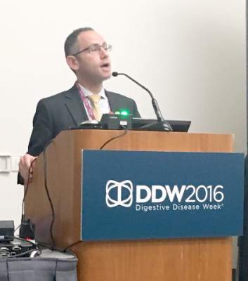
SAN DIEGO – A disposable Endocuff cut colonoscopic withdrawal times by nearly a minute and slightly improved polyp detection, compared with standard colonoscopy, according to a randomized, prospective trial of 562 patients.
The Endocuff caused no known adverse effects except for superficial mucosal trauma, Dr. Paul Feuerstadt said at the annual Digestive Disease Week. The study, which is the first of its kind in the United States, suggests that the Endocuff can improve the efficiency of colonoscopies without undermining detection rates, he added.
The plastic, flexible Endocuff slides onto the tip of a standard colonoscope, and has phalanges that press on the colonic mucosa “to improve polyp and adenoma detection rates, at least in theory,” said Dr. Feuerstadt, who is at the Gastroenterology Center of Connecticut in Hamden, Conn.
Use of the device improved the polyp detection rate by 63% and adenoma detection by 86% in a previous study in Germany.

For the current study, Dr. Feuerstadt and his associates screened 1,067 consecutive patients at two endoscopy centers in Connecticut, and excluded those with colitis, inflammatory bowel disease, diarrhea, chronic splenomegaly, and a history of surgical resection or colonic stricture. The 562 remaining patients were randomized to either Endocuff-assisted or standard colonoscopies performed by eight endoscopists with historically high adenoma detection rates of nearly 44%.
Use of the Endocuff seemed to slightly improve polyp detection, though none of the primary comparisons reached statistical significance, despite sufficient study power, Dr. Feuerstadt said. The rate of polyp detection was 63% for Endocuff-assisted colonoscopy and 60% for standard colonoscopy (P = .41), while rates of adenoma detection were 42% and 45%, respectively. There was a nonsignificant trend toward higher detection of sessile serrated adenomas (11% versus 9%; P = .37).
Notably, average withdrawal times were 9.9 minutes with the Endocuff (standard deviation, 5.5 minutes), versus 11.1 minutes without it (standard deviation, 5.9 minutes; P = .02). There were no perforations or other major adverse events, no instances of the Endocuff coming off the scope, and no difference in bleeding rates between the two groups.
However, 8% of Endocuff patients had mild mucosal trauma, compared with none of the control group, Dr. Feuerstadt reported.
The two groups resembled one another demographically, clinically, and in terms of their family history of colonic polyps. However, the Endocuff group had a higher frequency of first-degree relatives younger than age 50 years with colon cancer, Dr. Feuerstadt noted.
The endoscopists had an average historical ADR of 43.6%, “very similar to the 44.7% we saw in the study,” he added. “The device yields similar adenoma detection rates overall, with shorter withdrawal times, thereby increasing colonoscopic efficiency.”
Dr. Feuerstadt did not report funding sources. He disclosed consulting fees from Medivators, which makes endoscope reprocessing and related products.
SAN DIEGO – A disposable Endocuff cut colonoscopic withdrawal times by nearly a minute and slightly improved polyp detection, compared with standard colonoscopy, according to a randomized, prospective trial of 562 patients.
The Endocuff caused no known adverse effects except for superficial mucosal trauma, Dr. Paul Feuerstadt said at the annual Digestive Disease Week. The study, which is the first of its kind in the United States, suggests that the Endocuff can improve the efficiency of colonoscopies without undermining detection rates, he added.
The plastic, flexible Endocuff slides onto the tip of a standard colonoscope, and has phalanges that press on the colonic mucosa “to improve polyp and adenoma detection rates, at least in theory,” said Dr. Feuerstadt, who is at the Gastroenterology Center of Connecticut in Hamden, Conn.
Use of the device improved the polyp detection rate by 63% and adenoma detection by 86% in a previous study in Germany.
For the current study, Dr. Feuerstadt and his associates screened 1,067 consecutive patients at two endoscopy centers in Connecticut, and excluded those with colitis, inflammatory bowel disease, diarrhea, chronic splenomegaly, and a history of surgical resection or colonic stricture. The 562 remaining patients were randomized to either Endocuff-assisted or standard colonoscopies performed by eight endoscopists with historically high adenoma detection rates of nearly 44%.
Use of the Endocuff seemed to slightly improve polyp detection, though none of the primary comparisons reached statistical significance, despite sufficient study power, Dr. Feuerstadt said. The rate of polyp detection was 63% for Endocuff-assisted colonoscopy and 60% for standard colonoscopy (P = .41), while rates of adenoma detection were 42% and 45%, respectively. There was a nonsignificant trend toward higher detection of sessile serrated adenomas (11% versus 9%; P = .37).
Notably, average withdrawal times were 9.9 minutes with the Endocuff (standard deviation, 5.5 minutes), versus 11.1 minutes without it (standard deviation, 5.9 minutes; P = .02). There were no perforations or other major adverse events, no instances of the Endocuff coming off the scope, and no difference in bleeding rates between the two groups.
However, 8% of Endocuff patients had mild mucosal trauma, compared with none of the control group, Dr. Feuerstadt reported.
The two groups resembled one another demographically, clinically, and in terms of their family history of colonic polyps. However, the Endocuff group had a higher frequency of first-degree relatives younger than age 50 years with colon cancer, Dr. Feuerstadt noted.
The endoscopists had an average historical ADR of 43.6%, “very similar to the 44.7% we saw in the study,” he added. “The device yields similar adenoma detection rates overall, with shorter withdrawal times, thereby increasing colonoscopic efficiency.”
Dr. Feuerstadt did not report funding sources. He disclosed consulting fees from Medivators, which makes endoscope reprocessing and related products.
AT DDW® 2016
Key clinical point: Attaching a disposable Endocuff to the tip of a colonoscope enabled endoscopists to cut about 1 minute off withdrawal times and slightly increase their polyp detection rates.
Major finding: The rate of polyp detection was 63% for Endocuff-assisted colonoscopy and 60% for standard colonoscopy (P = .41). Average withdrawal times were 9.9 minutes with the Endocuff and 11.1 minutes without it (P = .02).
Data source: A prospective, randomized, controlled trial of 562 patients from two endoscopy centers.
Disclosures: Dr. Feuerstadt disclosed consulting fees from Medivators, which makes endoscope reprocessing and related products.
Additional D1 biopsy increased diagnostic yield for celiac disease
Among a large cohort of patients referred for endoscopy for suspected celiac disease as well as all upper gastrointestinal symptoms, a single additional D1 biopsy specimen from any site significantly increased the diagnostic yield for celiac disease, according to researchers.
Of 1,378 patients who had D2 and D1 biopsy specimens taken, 268 were newly diagnosed with celiac disease, and 26 had villous atrophy confined to D1, defined as ultrashort celiac disease (USCD). Compared with a standard D2 biopsy, an additional D1 biopsy increased the diagnostic yield by 9.7% (P less than .0001). Among the 26 diagnosed with USCD, 7 had normal D2 biopsy specimens, and 4 others had negative tests for endomysial antibodies (EMAs), totaling 11 patients for whom celiac disease would have been missed in the absence of a D1 biopsy.
“The addition of a D1 biopsy specimen to diagnose celiac disease may reduce the known delay in diagnosis that many patients with celiac disease experience. This may allow earlier institution of a gluten-free diet, potentially prevent nutritional deficiencies, and reduce the symptomatic burden of celiac disease,” wrote Dr. Peter Mooney of Royal Hallamshire Hospital, Sheffield, England, and his colleagues. (Gastroenterology 2016 April 7. doi: 10.1053/j-gastro.2016.01.029).
The prospective study recruited 1,378 consecutive patients referred to a single teaching hospital for endoscopy from 2008 to 2014. In total, 268 were newly diagnosed with celiac disease, and 26 were diagnosed with USCD.
To investigate the optimal site for targeted D1 sampling, 171 patients underwent quadrantic D1 biopsy, 61 of whom were diagnosed with celiac disease. Biopsy specimens from any topographical area resulted in high sensitivity, a fact that increases the feasibility of a D1 biopsy policy, since no specific target area is required, according to the researchers. Nonceliac abnormalities such as peptic duodenitis or gastric heterotopia have been suggested to impede interpretation of D1 biopsies, but these were rare in the study and did not interfere with the analysis.
USCD may be an early form of conventional celiac disease, an idea supported by the findings. Compared with patients diagnosed with conventional celiac disease, patients diagnosed with USCD were younger and had a much lower rate of diarrhea, which by decision-tree analysis was the single factor discriminating between the two groups. Compared with healthy controls, individuals with conventional celiac disease, but not USCD, were more likely to present with anemia, diarrhea, a family history of celiac disease, lethargy, and osteoporosis. Patients with USCD and conventional disease had similar rates of IgA tissue transglutaminase antibodies (tTG), but USCD patients had lower titers (P less than .001). The USCD group also had fewer ferritin and folate deficiencies.
The researchers suggested that clinical phenotypic differences may be due to minimal loss of absorptive capacity associated with a short segment of villous atrophy. Given the younger average age at diagnosis of USCD and lower tTG titers, USCD may represent an early stage of celiac disease, resulting in fewer nutritional deficiencies observed because of a shorter lead time to diagnosis.
Although USCD patients had a milder clinical phenotype, which has raised concerns that a strict gluten-free diet may be unnecessary, follow-up data demonstrated that a gluten-free diet produced improvement in symptoms and a significant decrease in the tTG titer. These results may indicate that the immune cascade was switched off, according to the researchers, and that early diagnosis may present a unique opportunity to prevent further micronutrient deficiency.
Dr. Mooney and his coauthors reported having no relevant financial disclosures.
FROM GASTROENTEROLOGY
Key clinical point: When added to a standard D2 biopsy, a single D1 biopsy from any site significantly increased the diagnostic yield for celiac disease.
Major finding: In total, 26 of 268 patients diagnosed with celiac disease had villous atrophy confined to D1 (ultrashort celiac disease); an additional D1 biopsy increased the diagnostic yield by 9.7% (P less than .0001), compared with a standard D2 biopsy.
Data source: A prospective study of 1,378 consecutive patients referred to a single teaching hospital for endoscopy from 2008 to 2014, 268 of whom were newly diagnosed with celiac disease and 26 with USCD.
Disclosures: Dr. Mooney and his coauthors reported having no relevant financial disclosures.
Surveillance finds pancreatic ductal carcinoma in situ at resectable stage
Surveillance of CDNK2A mutation carriers detected most pancreatic ductal carcinoma in situ (PDAC) at a resectable stage, while the surveillance benefit was lower for those with familial prostate cancer.
Among 178 CDKN2A mutation carriers, PDAC was detected in 13 (7.3%), 9 of whom underwent surgery. Compared with previously reported rates of 15%-20% for symptomatic PDAC, this 70% resection rate represents a substantial increase. The 5-year survival rate of 24% for screen-detected PDAC was higher than 4%-7% reported for symptomatic sporadic PDAC. Among individuals with familial prostate cancer (FPC), 13 of 214 individuals (6.1%) underwent surgery, but with a higher proportion of precursor lesions detected, just four high-risk lesions (1.9% of screened FPC patients) were removed.
Whether surveillance improved prognosis for FPC families was difficult to determine, according to the investigators. The yield of PDAC was low at 0.9%, as was the yield of relevant precursor lesions (grade 3 PanIN and high-grade IPMN) at 1.9%.
“However, if surgical removal of multifocal grade 2 PanIN and multifocal BD-IPMNs is regarded as beneficial, the diagnostic yield increases to 3.7% (eight of 214 patients), and surveillance of FPC might also be considered effective,” wrote Dr. Hans Vasen, professor in the department of gastroenterology and hepatology at the Leiden University Medical Center, the Netherlands, and colleagues. “The value of surveillance of FPC is still not clear, and the main effect seems to be prevention of PDAC by removal of” precursor lesions, they added (J Clin Oncol. 2016 Apr 25. doi: 10.1200/JCO.2015.64.0730).
The retrospective evaluation of an ongoing prospective follow-up study included 411 high-risk individuals: 178 with CDKN2A mutations, 214 with familial pancreatic cancer, and 19 with BRCA1/2 or PALB2 mutations. The study was conducted at three expert centers in Marburg, Germany; Leiden, the Netherlands; and Madrid.
In the BRCA1/2 and PALB2 mutation cohort, one individual (3.8%) with a BRCA2 mutation developed PDAC and underwent surgery; 17 months after the surgery this patient died of liver metastasis. Two others underwent surgery for cystic lesions and are in good health at 10 and 21 months after surgery.
In the cohort of CDKN2A mutation carriers, the mean age at the start of surveillance was 56 years (range, 37-75) and the mean follow-up time was 53 months (range, 0-169): in total, 866 MRIs and 106 endoscopic ultrasounds were conducted. In the FPC group, the mean age was 48 years (range, 27-81), and the mean follow up was 2.8 years (range, 0-10.8): 618 MRIs and 402 endoscopic ultrasounds were conducted. Among BRCA1/2 and PALB2 mutation carriers, the mean age was 52.6 years (range, 25-70), and the mean follow up was 32.7 months (range, 1-119).
Given the difficulty of detecting precursor lesions and distinguishing incipient neoplasia from lower grade or nonneoplastic cystic lesions, the authors of the accompanying study achieved impressive results in improving cancer outcomes among high-risk individuals.
Several strategies for earlier cancer detection can be gleaned from the study. Improved outcomes may depend on expert centers running the surveillance. The detection rate of 2%-7%, depending on the cohort studied and the surveillance protocol, may have room for improvement with better risk stratification and refined protocols for cost effectiveness. The age at the start of surveillance may be one place to start: the mean age of pancreatic ductal carcinoma in situ detection was 53-68 years, depending on the center, and it may be possible to shift the starting age upward to improve yield.
The type of mutation conferring susceptibility may aid in risk stratification. For example, CDKN2A mutation carriers had a higher cancer rate (16%) than BRCA/PALB2 mutation carriers (5%). Other factors that could mitigate risk upward include diabetes, family history, and smoking history. A composite risk assessment could aid in identifying the highest-risk patients. Lastly, future studies are needed to determine which surveillance protocols are best. To make valid comparisons, several surveillance protocols must be tested.
These results impact not only high-risk individuals, but the general population as well. The data support that early detection improves outcomes and highlights the need for developing better biomarkers and tests for early detection of PDAC.
Dr. Teresa A. Brentnall is professor in the department of medicine, division of gastroenterology, University of Washington, Seattle. These remarks were part of an accompanying editorial (J Clin Oncol. 2016 Apr 25. doi: 10.1200/JCO.2015.64.0730).
Surveillance of CDNK2A mutation carriers detected most pancreatic ductal carcinoma in situ (PDAC) at a resectable stage, while the surveillance benefit was lower for those with familial prostate cancer.
Among 178 CDKN2A mutation carriers, PDAC was detected in 13 (7.3%), 9 of whom underwent surgery. Compared with previously reported rates of 15%-20% for symptomatic PDAC, this 70% resection rate represents a substantial increase. The 5-year survival rate of 24% for screen-detected PDAC was higher than 4%-7% reported for symptomatic sporadic PDAC. Among individuals with familial prostate cancer (FPC), 13 of 214 individuals (6.1%) underwent surgery, but with a higher proportion of precursor lesions detected, just four high-risk lesions (1.9% of screened FPC patients) were removed.
Whether surveillance improved prognosis for FPC families was difficult to determine, according to the investigators. The yield of PDAC was low at 0.9%, as was the yield of relevant precursor lesions (grade 3 PanIN and high-grade IPMN) at 1.9%.
“However, if surgical removal of multifocal grade 2 PanIN and multifocal BD-IPMNs is regarded as beneficial, the diagnostic yield increases to 3.7% (eight of 214 patients), and surveillance of FPC might also be considered effective,” wrote Dr. Hans Vasen, professor in the department of gastroenterology and hepatology at the Leiden University Medical Center, the Netherlands, and colleagues. “The value of surveillance of FPC is still not clear, and the main effect seems to be prevention of PDAC by removal of” precursor lesions, they added (J Clin Oncol. 2016 Apr 25. doi: 10.1200/JCO.2015.64.0730).
The retrospective evaluation of an ongoing prospective follow-up study included 411 high-risk individuals: 178 with CDKN2A mutations, 214 with familial pancreatic cancer, and 19 with BRCA1/2 or PALB2 mutations. The study was conducted at three expert centers in Marburg, Germany; Leiden, the Netherlands; and Madrid.
In the BRCA1/2 and PALB2 mutation cohort, one individual (3.8%) with a BRCA2 mutation developed PDAC and underwent surgery; 17 months after the surgery this patient died of liver metastasis. Two others underwent surgery for cystic lesions and are in good health at 10 and 21 months after surgery.
In the cohort of CDKN2A mutation carriers, the mean age at the start of surveillance was 56 years (range, 37-75) and the mean follow-up time was 53 months (range, 0-169): in total, 866 MRIs and 106 endoscopic ultrasounds were conducted. In the FPC group, the mean age was 48 years (range, 27-81), and the mean follow up was 2.8 years (range, 0-10.8): 618 MRIs and 402 endoscopic ultrasounds were conducted. Among BRCA1/2 and PALB2 mutation carriers, the mean age was 52.6 years (range, 25-70), and the mean follow up was 32.7 months (range, 1-119).
FROM THE JOURNAL OF CLINICAL ONCOLOGY
Key clinical point: Surveillance of high-risk individuals was relatively successful in detecting pancreatic ductal carcinoma in situ (PDAC) at a resectable stage.
Major finding: The detection rate in CDKN2A mutation carriers was 7.3% and the resection rate for screen-detected PDAC was 75%, compared with previous reports of 15%-20% for symptomatic PDAC; the PDAC detection rate in individuals with familial prostate cancer was much lower at 0.9%.
Data source: Evaluation of an ongoing prospective follow-up study at three European centers included 411 individuals: 178 with CDKN2A mutations, 214 with familial pancreatic cancer, and 19 with BRCA1/2 or PALB2 mutations.
Disclosures: Dr. Vasen and most coauthors reported having no disclosures. Five coauthors reported financial ties to industry sources.
VIDEO: Rectal indomethacin does not prevent pancreatitis post ERCP
Patients who receive rectal indomethacin after undergoing endoscopic retrograde cholangiopancreatography (ERCP) are not any less likely to develop pancreatitis than individuals who don’t, according to the findings of a recent study published in Gastroenterology (2016 Jan 9. doi: 10.1053/j.gastro.2015.12.018).
“These results are in contrast to recent studies highlighting the benefit of rectal NSAIDS to prevent PEP [post-ECRP pancreatitis] in high-risk patients [and] counter the guidelines espoused by the European Society for Gastrointestinal Endoscopy, which recently recommended giving rectal indomethacin to prevent PEP in all patients undergoing ERCP,” said the study authors, led by Dr. John M. Levenick of Penn State University in Hershey, Pa.
SOURCE: AMERICAN GASTROENTEROLOGICAL ASSOCIATION
Dr. Levenick and his coinvestigators screened 604 consecutive patients undergoing ERCP, with and without endoscopic ultrasound, at the Dartmouth-Hitchcock Medical Center between March 2013 and December 2014, eventually enrolling and randomizing 449 subjects into two cohorts: one in which subjects were given indomethacin after undergoing ERCP (n = 223), and one in which subjects were simply given a placebo (n = 226). Randomization happened after subjects’ major papilla had been reached, and cannulation attempts were started.
Individuals were excluded if they had active acute pancreatitis or had undergone ERCP to treat or diagnose acute pancreatitis, if they had any contraindications or allergies to NSAIDs, or were younger than 18 years of age, among other factors. The mean age of the indomethacin cohort was 64.9 years, with 118 (52.9%) females; in the placebo cohort, mean age was 64.3 years and 118 (52.2%) were female.
Pancreatitis occurred in 27 subjects overall, 16 (7.2%) of whom were in the indomethacin cohort and the other 11 (4.9%) were on placebo followed ERCP (P = .33). No subjects receiving indomethacin had severe or moderately severe PEP, but one subject had severe PEP and one had moderately severe PEP in the placebo cohort (P = 1.0). There was no necrotizing pancreatitis in either cohort, nor were there any significant differences in gastrointestinal bleeding (P = .75), death (P = .25), or 30-day hospital readmission (P = .1) between the two cohorts.
“Prophylactic rectal indomethacin did not reduce the incidence or severity of PEP in consecutive patients undergoing ERCP,” Dr. Levenick and his coauthors concluded, adding that “guidelines that recommend the administration of rectal indomethacin in all patients undergoing ERCP should be reconsidered.”
This study was funded by the National Pancreas Foundation and a grant from the National Institutes of Health. Dr. Levenick and his coauthors did not report any financial disclosures.
Acute pancreatitis is the most common and feared complication of endoscopic retrograde cholangiopancreatography (ERCP). The incidence of post-ERCP pancreatitis is around 10% with a mortality of 0.7% (Gastrointest Endosc. 2015;81:143-9). Recent advances in noninvasive pancreaticobiliary imaging, risk stratification before ERCP, prophylactic pancreatic stent placement, and administration of nonsteroidal anti-inflammatory drugs (NSAIDs) have improved the overall risk benefit ratio of ERCP.
NSAIDs are potent inhibitors of phospholipase A2, cyclooxygenase, and of the activation of platelets and endothelium, all of which play a central role in the pathogenesis of post-ERCP pancreatitis. NSAIDs constitute an attractive option in clinical practice, because they are inexpensive and widely available with a favorable risk profile. A recent multicenter randomized controlled trial (RCT) of 602 patients at high-risk for post-ERCP pancreatitis showed that rectal indomethacin is associated with a 7.7% absolute and a 46% relative risk reduction of post-ERCP pancreatitis (N Engl J Med. 2012;366:1414-22). These findings have been broadly adapted in endoscopic practice in the United States.
|
| Dr. Georgios Papachristou |
The presented RCT by Dr. Levenick and his colleagues evaluated the efficacy of rectal indomethacin in preventing post-ERCP pancreatitis among consecutive patients undergoing ERCP in a single U.S. center. This study was a well designed and conducted RCT following the CONSORT guidelines and utilizing an independent data and safety monitoring board.
The authors reported that rectal indomethacin did not result in reduction of post-ERCP pancreatitis (7.2%) when compared with placebo (4.9%). Of importance, 70% of patients included were at average risk for post-ERCP pancreatitis. Furthermore, despite a calculated sample size of 1,398 patients, the study was terminated early after enrolling only 449 patients based on the interim analysis showing futility to reach a statistically different outcome.
This well executed RCT reports no benefit in administering rectal indomethacin in all patients undergoing ERCP. Evidence strongly supports that rectal indomethacin remains an important advancement in preventing post-ERCP pancreatitis. However, its benefit is likely limited to a selected group of patients, those at high-risk for post-ERCP pancreatitis. Further studies are under way to clarify whether rectal indomethacin alone vs. indomethacin plus prophylactic pancreatic stenting is more effective in preventing post-ERCP pancreatitis in high-risk patients.
Dr. Georgios Papachristou is associate professor of medicine at the University of Pittsburgh. He is a consultant for Shire and has received funding from the National Institutes of Health and the VA Health System.
Acute pancreatitis is the most common and feared complication of endoscopic retrograde cholangiopancreatography (ERCP). The incidence of post-ERCP pancreatitis is around 10% with a mortality of 0.7% (Gastrointest Endosc. 2015;81:143-9). Recent advances in noninvasive pancreaticobiliary imaging, risk stratification before ERCP, prophylactic pancreatic stent placement, and administration of nonsteroidal anti-inflammatory drugs (NSAIDs) have improved the overall risk benefit ratio of ERCP.
NSAIDs are potent inhibitors of phospholipase A2, cyclooxygenase, and of the activation of platelets and endothelium, all of which play a central role in the pathogenesis of post-ERCP pancreatitis. NSAIDs constitute an attractive option in clinical practice, because they are inexpensive and widely available with a favorable risk profile. A recent multicenter randomized controlled trial (RCT) of 602 patients at high-risk for post-ERCP pancreatitis showed that rectal indomethacin is associated with a 7.7% absolute and a 46% relative risk reduction of post-ERCP pancreatitis (N Engl J Med. 2012;366:1414-22). These findings have been broadly adapted in endoscopic practice in the United States.
|
|
| Dr. Georgios Papachristou |
The presented RCT by Dr. Levenick and his colleagues evaluated the efficacy of rectal indomethacin in preventing post-ERCP pancreatitis among consecutive patients undergoing ERCP in a single U.S. center. This study was a well designed and conducted RCT following the CONSORT guidelines and utilizing an independent data and safety monitoring board.
The authors reported that rectal indomethacin did not result in reduction of post-ERCP pancreatitis (7.2%) when compared with placebo (4.9%). Of importance, 70% of patients included were at average risk for post-ERCP pancreatitis. Furthermore, despite a calculated sample size of 1,398 patients, the study was terminated early after enrolling only 449 patients based on the interim analysis showing futility to reach a statistically different outcome.
This well executed RCT reports no benefit in administering rectal indomethacin in all patients undergoing ERCP. Evidence strongly supports that rectal indomethacin remains an important advancement in preventing post-ERCP pancreatitis. However, its benefit is likely limited to a selected group of patients, those at high-risk for post-ERCP pancreatitis. Further studies are under way to clarify whether rectal indomethacin alone vs. indomethacin plus prophylactic pancreatic stenting is more effective in preventing post-ERCP pancreatitis in high-risk patients.
Dr. Georgios Papachristou is associate professor of medicine at the University of Pittsburgh. He is a consultant for Shire and has received funding from the National Institutes of Health and the VA Health System.
Patients who receive rectal indomethacin after undergoing endoscopic retrograde cholangiopancreatography (ERCP) are not any less likely to develop pancreatitis than individuals who don’t, according to the findings of a recent study published in Gastroenterology (2016 Jan 9. doi: 10.1053/j.gastro.2015.12.018).
“These results are in contrast to recent studies highlighting the benefit of rectal NSAIDS to prevent PEP [post-ECRP pancreatitis] in high-risk patients [and] counter the guidelines espoused by the European Society for Gastrointestinal Endoscopy, which recently recommended giving rectal indomethacin to prevent PEP in all patients undergoing ERCP,” said the study authors, led by Dr. John M. Levenick of Penn State University in Hershey, Pa.
SOURCE: AMERICAN GASTROENTEROLOGICAL ASSOCIATION
Dr. Levenick and his coinvestigators screened 604 consecutive patients undergoing ERCP, with and without endoscopic ultrasound, at the Dartmouth-Hitchcock Medical Center between March 2013 and December 2014, eventually enrolling and randomizing 449 subjects into two cohorts: one in which subjects were given indomethacin after undergoing ERCP (n = 223), and one in which subjects were simply given a placebo (n = 226). Randomization happened after subjects’ major papilla had been reached, and cannulation attempts were started.
Individuals were excluded if they had active acute pancreatitis or had undergone ERCP to treat or diagnose acute pancreatitis, if they had any contraindications or allergies to NSAIDs, or were younger than 18 years of age, among other factors. The mean age of the indomethacin cohort was 64.9 years, with 118 (52.9%) females; in the placebo cohort, mean age was 64.3 years and 118 (52.2%) were female.
Pancreatitis occurred in 27 subjects overall, 16 (7.2%) of whom were in the indomethacin cohort and the other 11 (4.9%) were on placebo followed ERCP (P = .33). No subjects receiving indomethacin had severe or moderately severe PEP, but one subject had severe PEP and one had moderately severe PEP in the placebo cohort (P = 1.0). There was no necrotizing pancreatitis in either cohort, nor were there any significant differences in gastrointestinal bleeding (P = .75), death (P = .25), or 30-day hospital readmission (P = .1) between the two cohorts.
“Prophylactic rectal indomethacin did not reduce the incidence or severity of PEP in consecutive patients undergoing ERCP,” Dr. Levenick and his coauthors concluded, adding that “guidelines that recommend the administration of rectal indomethacin in all patients undergoing ERCP should be reconsidered.”
This study was funded by the National Pancreas Foundation and a grant from the National Institutes of Health. Dr. Levenick and his coauthors did not report any financial disclosures.
FROM GASTROENTEROLOGY
Key clinical point: Rectal indomethacin does not prevent pancreatitis in patients who undergo endoscopic retrograde cholangiopancreatography (ERCP).
Major finding: 7.2% of subjects on indomethacin and 4.9% on placebo developed post-ERCP pancreatitis, indicating no significant difference between the two cohorts (P = .33).
Data source: Prospective, double-blind, placebo-controlled study of 449 ERCP patients between March 2013 and December 2014.
Disclosures: Study funded by National Pancreas Foundation and National Institutes of Health. Dr. Levenick and his coauthors did not report any relevant financial disclosures.

VIDEO: Newer MRI hardware, software significantly better at detecting pancreatic cysts
As magnetic resonance imaging technology continues to advance year after year, so does MRI’s ability to accurately detect pancreatic cysts, according to a new study published in the April issue of Clinical Gastroenterology and Hepatology (doi: 10.1016/j.cgh.2015.08.038).
“To our knowledge, this is the first study to analyze the relationship between the technical improvements in imaging techniques (specifically, MRI) and the presence of incidentally found PCLs [pancreatic cystic lesions],” said the study authors, led by Dr. Michael B. Wallace of the Mayo Clinic in Jacksonville, Fla.

Dr. Wallace and his coinvestigators launched this retrospective descriptive study selecting the first 50 consecutive abdominal MRI patients at the Jacksonville Mayo Clinic during January and February of each year from 2005 through 2014, for a total of 500 cases who met inclusion criteria included in the study. Patients were excluded if they had preexisting symptomatic or asymptomatic pancreatitis, either acute or chronic, pancreatic masses, pancreatic cysts, pancreatic surgery, pancreatic symptoms, or any pancreas-related indications found by MRI.
The clinic underwent periodic MRI updates over the course of the 10-year study, along with requisite software updates to “take advantage of the new hardware technology,” the study explains. Major hardware improvements, provided by Siemens Medical Solutions USA, were Symphony/Sonata, Espree/Avanto, and Aera/Skyra, while software updates corresponding to each hardware update were VA, VB, and VD, respectively.
SOURCE: AMERICAN GASTROENTEROLOGICAL ASSOCIATION
Furthermore, each software update had other, smaller upgrades, leading to a total of 20 combinations of MRI hardware and software on which MRIs were performed over the 10 years. Every MRI taken included “an axial and a coronal T2-weighted single-shot (HASTE) pulse sequence [with] TR 1400-1500 ms, TE 82-99 ms, and slice thickness 5-7 mm (gap, 0.5-0.7 mm).” Each MRI was analyzed by a pancreatic MRI specialist to find incidental cysts.
The number of patients found with pancreatic cysts increased incrementally from 2005 to 2014, with 2010 being the year with the highest number. A total of 208 subjects (41.6%) were found to have incidental cysts, but only 44 of these cases were discovered in the original MRI. The presence of cysts was associated with older age in patients who had them; only 20% of all subjects under 50 years of age had cysts, compared to 32.4% of those between 50 and 60 years, 54.9% of those between 60 and 70 years, and 61.5% of those over the age of 70 years (P less than .01).
Additionally, 56.4% of all subjects with diabetes (P less than .01), 59.0% of subjects with nonmelanoma skin cancer (P less than .03), and 58.1% of those with hepatocarcinoma (P less than .02) were also found to have cysts. Most striking, however, is that newer hardware and software permutations were able to detect cysts in 56.3% (Skyra) of patients who had them, compared with only 30.3% (Symphony) of patients who underwent MRI on older technology.
“The variable field strength” (1.5 T vs. 3 T) was not significantly related to the presence of PCLs,” Dr. Wallace and his coauthors concluded. “We believe this may be secondary to the lack of power of the analysis, because only 6% of the examinations were 3-T studies. Therefore, we speculate that this relationship may be confirmed if the number of 3-T studies increased.”
Males and females each made up roughly 50% of the study population, with a median age of 60 years and 85% being white. Additionally, 4% of subjects had a family history of pancreatic cancer, 12% had a personal history of solid organ transplant, and 53% had a personal history of smoking.
This study was funded by the Mayo Clinic. Dr. Wallace disclosed that he has received grant funding from Olympus, Boston Scientific, and Cosmo Pharmaceuticals, and travel support from Olympus. No other authors reported any financial disclosures.
The increasing prevalence of pancreatic cystic lesions on MRI scanning may provide an important opportunity for detection of early precursors of pancreatic cancer – or may represent just another insignificant incidental finding. What is the implication of a small asymptomatic cyst?
MRI scanning of the pancreas has revolutionized our ability to detect early cystic neoplasms of the pancreas. Pancreatic cysts appear as well-defined, small, round fluid-filled structures within the pancreas. The inner structures – such as septations, nodules, and adjacent masses – offer clues as to the type of cyst and the risk of malignancy. But the real strength of pancreatic MRI scanning is the ability to detect and portray small cysts and the adjacent main pancreatic duct.
The size, number, and distribution of cysts over time can be tracked with MRI surveillance. By tracking the diameter of cysts and calculating the rate of growth of cysts, clinicians may be able to predict the development of malignancy in intraductal papillary mucinous neoplasms.
How should these patients be managed clinically? Once a cyst has been identified, are clinicians obligated to notify the patient, monitor the cyst with an established surveillance program, or biopsy the cyst? If the cyst is very small and benign appearing, can the clinician ignore the finding and perhaps not notify the patient?
Once again, we are watching dilemmas unfold as technology outstrips our understanding of diseases and their management. We are going to need some good correlations between imaging and tissue of pancreatic cystic lesions. In the meantime, it is important to reserve the use of pancreatic MRI scanning to high-risk patients or patients with CT scan abnormalities.
Dr. William R. Brugge, AGAF, is professor of medicine, Harvard Medical School, and director, Pancreas Biliary Center, Massachusetts General Hospital, both in Boston. He is a consultant with Boston Scientific.
As magnetic resonance imaging technology continues to advance year after year, so does MRI’s ability to accurately detect pancreatic cysts, according to a new study published in the April issue of Clinical Gastroenterology and Hepatology (doi: 10.1016/j.cgh.2015.08.038).
“To our knowledge, this is the first study to analyze the relationship between the technical improvements in imaging techniques (specifically, MRI) and the presence of incidentally found PCLs [pancreatic cystic lesions],” said the study authors, led by Dr. Michael B. Wallace of the Mayo Clinic in Jacksonville, Fla.
Dr. Wallace and his coinvestigators launched this retrospective descriptive study selecting the first 50 consecutive abdominal MRI patients at the Jacksonville Mayo Clinic during January and February of each year from 2005 through 2014, for a total of 500 cases who met inclusion criteria included in the study. Patients were excluded if they had preexisting symptomatic or asymptomatic pancreatitis, either acute or chronic, pancreatic masses, pancreatic cysts, pancreatic surgery, pancreatic symptoms, or any pancreas-related indications found by MRI.
The clinic underwent periodic MRI updates over the course of the 10-year study, along with requisite software updates to “take advantage of the new hardware technology,” the study explains. Major hardware improvements, provided by Siemens Medical Solutions USA, were Symphony/Sonata, Espree/Avanto, and Aera/Skyra, while software updates corresponding to each hardware update were VA, VB, and VD, respectively.
SOURCE: AMERICAN GASTROENTEROLOGICAL ASSOCIATION
Furthermore, each software update had other, smaller upgrades, leading to a total of 20 combinations of MRI hardware and software on which MRIs were performed over the 10 years. Every MRI taken included “an axial and a coronal T2-weighted single-shot (HASTE) pulse sequence [with] TR 1400-1500 ms, TE 82-99 ms, and slice thickness 5-7 mm (gap, 0.5-0.7 mm).” Each MRI was analyzed by a pancreatic MRI specialist to find incidental cysts.
The number of patients found with pancreatic cysts increased incrementally from 2005 to 2014, with 2010 being the year with the highest number. A total of 208 subjects (41.6%) were found to have incidental cysts, but only 44 of these cases were discovered in the original MRI. The presence of cysts was associated with older age in patients who had them; only 20% of all subjects under 50 years of age had cysts, compared to 32.4% of those between 50 and 60 years, 54.9% of those between 60 and 70 years, and 61.5% of those over the age of 70 years (P less than .01).
Additionally, 56.4% of all subjects with diabetes (P less than .01), 59.0% of subjects with nonmelanoma skin cancer (P less than .03), and 58.1% of those with hepatocarcinoma (P less than .02) were also found to have cysts. Most striking, however, is that newer hardware and software permutations were able to detect cysts in 56.3% (Skyra) of patients who had them, compared with only 30.3% (Symphony) of patients who underwent MRI on older technology.
“The variable field strength” (1.5 T vs. 3 T) was not significantly related to the presence of PCLs,” Dr. Wallace and his coauthors concluded. “We believe this may be secondary to the lack of power of the analysis, because only 6% of the examinations were 3-T studies. Therefore, we speculate that this relationship may be confirmed if the number of 3-T studies increased.”
Males and females each made up roughly 50% of the study population, with a median age of 60 years and 85% being white. Additionally, 4% of subjects had a family history of pancreatic cancer, 12% had a personal history of solid organ transplant, and 53% had a personal history of smoking.
This study was funded by the Mayo Clinic. Dr. Wallace disclosed that he has received grant funding from Olympus, Boston Scientific, and Cosmo Pharmaceuticals, and travel support from Olympus. No other authors reported any financial disclosures.
FROM CLINICAL GASTROENTEROLOGY AND HEPATOLOGY
Key clinical point: Newer MRI technology is more effective at detecting pancreatic cysts, particularly in patients with diabetes or advanced age.
Major finding: Newer MRI hardware and software detected pancreatic cysts in 56.3% of patients, compared with only 30.3% on older MRI hardware and software.
Data source: Retrospective, descriptive study of 500 patients undergoing MRI for nonpancreatic indications during January and February of 2005-2014.
Disclosures: Study funded by the Mayo Clinic. Dr. Michael B. Wallace disclosed relationships with Olympus, Boston Scientific, and Cosmo Pharmaceuticals.

Endoscopic techniques provide organ-sparing options for myotomy, resection, and more
Minimally invasive endoscopic procedures are transforming gastrointestinal surgery by offering organ-sparing approaches for a range of pathologies. At the 2016 AGA Tech Summit, Dr. David Rattner updated attendees on three of these techniques – endoscopic submucosal dissection (ESD), peroral endoscopic myotomy (POEM), and endoscopic full-thickness resection (EFTR).
“It is likely that endoluminal treatments, combined with better understanding of molecular pathology, will decrease the need for radical surgical procedures for some common gastrointestinal conditions,” he said in an interview before the meeting, which is sponsored by the AGA Center for GI Innovation and Technology. Dr. Rattner is chief of the division of gastrointestinal and general surgery at Massachusetts General Hospital in Boston.

Dr. Jose Martinez put transorifice surgery in historical perspective and pointed the way forward in an accompanying presentation. “The collaboration with physicians and industry has really started to come along with the natural orifice approach,” said Dr. Martinez, professor of surgery at the University of Miami Health System.
Developed in the 1980s and substantially refined in the mid-2000s, ESD pioneered the endoscopic resection of entire intramucosal tumors without the need for open surgery. In a recent meta-analysis, ESD achieved significantly higher rates of en bloc resection than endoscopic mucosal resection (EMR), with significantly lower rates of local recurrence and comparable complication rates (World J Gastroenterol. 2014;20:8282-7).
ESD involves resecting the mucosa and much of the submucosa, while conserving the muscular layer of the esophagus. In contrast, during POEM, the surgeon intentionally divides the muscular layer, while the intact mucosa provides the barrier between the mediastinum or peritoneum and the esophageal lumen. The defining feature of POEM is the creation of a submucosal tunnel to access the muscle layer for myotomy without incising the skin.
POEM has been found highly successful for treating esophageal achalasia. In fact, in a recently published series of 500 cases, the success rate was 100%, even though the cohort included elderly patients and those with sigmoid esophagus and complex treatment histories (J Am Coll Surg. 2015;221:256-64). Two months post-treatment, average Eckardt scores had decreased by an average of 5.0, and lower esophageal sphincter pressures had fallen by nearly 50%. The adverse event rate was 3%, there were no fatalities, and improvements persisted 3 years later. “Nationwide, numerous centers have incorporated this technique and gotten significant positive results,” said Dr. Martinez.
“POEM will be shown to be as effective as any other currently existing therapy for the treatment of achalasia,” emphasized Dr. Rattner. Indeed, its success has sparked investigations of submucosal tunneling for other indications, such as tumor resection and pyloromyotomy for gastroparesis (World J Gastroenterol. 2014;20:17746-55).
Another cutting-edge technique is EFTR, described as a powerful tool not only for acquiring diagnostic tissue, but also for sparing some patients from surgery. Compared with ESD, it has the potential for higher diagnostic yield of full-thickness specimens, such as in cases of nonlifting adenomas, adenomas at difficult anatomic locations, T1-carcinomas, or submucosal colorectal tumors. For years, a lack of safe techniques and devices kept EFTR from entering routine clinical practice. But intensive research on natural orifice translumenal endoscopic surgery (NOTES) has helped propel innovations such as over-the-scope clips for closing incisions, and smaller, more maneuverable, over-the-scope full-thickness resection devices. Large-scale trials of EFTR are lacking, but in a recent report of 19 consecutive colonic submucosal tumors, EFTR enabled the removal of the entire tumor with capsule intact in 18 instances (Endoscopy. 2013;45[09]:770-3). Notably, colonic wall defects could be closed endoscopically in 16 of 18 cases.
“We are still a ways away from being able to perform EFTR on a routine basis, but new technologies are under development that will make this possible,” said Dr. Rattner. Agreeing that the future of transorifice surgery is bright and growing, Dr. Martinez said, “Strictures, obstructions, bleeding, foreign bodies are now fully resolved through endoscopic approaches.”
Minimally invasive endoscopic procedures are transforming gastrointestinal surgery by offering organ-sparing approaches for a range of pathologies. At the 2016 AGA Tech Summit, Dr. David Rattner updated attendees on three of these techniques – endoscopic submucosal dissection (ESD), peroral endoscopic myotomy (POEM), and endoscopic full-thickness resection (EFTR).
“It is likely that endoluminal treatments, combined with better understanding of molecular pathology, will decrease the need for radical surgical procedures for some common gastrointestinal conditions,” he said in an interview before the meeting, which is sponsored by the AGA Center for GI Innovation and Technology. Dr. Rattner is chief of the division of gastrointestinal and general surgery at Massachusetts General Hospital in Boston.
Dr. Jose Martinez put transorifice surgery in historical perspective and pointed the way forward in an accompanying presentation. “The collaboration with physicians and industry has really started to come along with the natural orifice approach,” said Dr. Martinez, professor of surgery at the University of Miami Health System.
Developed in the 1980s and substantially refined in the mid-2000s, ESD pioneered the endoscopic resection of entire intramucosal tumors without the need for open surgery. In a recent meta-analysis, ESD achieved significantly higher rates of en bloc resection than endoscopic mucosal resection (EMR), with significantly lower rates of local recurrence and comparable complication rates (World J Gastroenterol. 2014;20:8282-7).
ESD involves resecting the mucosa and much of the submucosa, while conserving the muscular layer of the esophagus. In contrast, during POEM, the surgeon intentionally divides the muscular layer, while the intact mucosa provides the barrier between the mediastinum or peritoneum and the esophageal lumen. The defining feature of POEM is the creation of a submucosal tunnel to access the muscle layer for myotomy without incising the skin.
POEM has been found highly successful for treating esophageal achalasia. In fact, in a recently published series of 500 cases, the success rate was 100%, even though the cohort included elderly patients and those with sigmoid esophagus and complex treatment histories (J Am Coll Surg. 2015;221:256-64). Two months post-treatment, average Eckardt scores had decreased by an average of 5.0, and lower esophageal sphincter pressures had fallen by nearly 50%. The adverse event rate was 3%, there were no fatalities, and improvements persisted 3 years later. “Nationwide, numerous centers have incorporated this technique and gotten significant positive results,” said Dr. Martinez.
“POEM will be shown to be as effective as any other currently existing therapy for the treatment of achalasia,” emphasized Dr. Rattner. Indeed, its success has sparked investigations of submucosal tunneling for other indications, such as tumor resection and pyloromyotomy for gastroparesis (World J Gastroenterol. 2014;20:17746-55).
Another cutting-edge technique is EFTR, described as a powerful tool not only for acquiring diagnostic tissue, but also for sparing some patients from surgery. Compared with ESD, it has the potential for higher diagnostic yield of full-thickness specimens, such as in cases of nonlifting adenomas, adenomas at difficult anatomic locations, T1-carcinomas, or submucosal colorectal tumors. For years, a lack of safe techniques and devices kept EFTR from entering routine clinical practice. But intensive research on natural orifice translumenal endoscopic surgery (NOTES) has helped propel innovations such as over-the-scope clips for closing incisions, and smaller, more maneuverable, over-the-scope full-thickness resection devices. Large-scale trials of EFTR are lacking, but in a recent report of 19 consecutive colonic submucosal tumors, EFTR enabled the removal of the entire tumor with capsule intact in 18 instances (Endoscopy. 2013;45[09]:770-3). Notably, colonic wall defects could be closed endoscopically in 16 of 18 cases.
“We are still a ways away from being able to perform EFTR on a routine basis, but new technologies are under development that will make this possible,” said Dr. Rattner. Agreeing that the future of transorifice surgery is bright and growing, Dr. Martinez said, “Strictures, obstructions, bleeding, foreign bodies are now fully resolved through endoscopic approaches.”
AT THE 2016 AGA TECH SUMMIT

Leading scope manufacturer to pay hefty settlement for alleged kickbacks, bribery
Embattled scope maker Olympus Corp., already under federal investigation for its role in superbug outbreaks, has agreed to pay $646 million to resolve criminal and civil probes into illegal kickbacks and bribes to doctors and hospitals.
Federal prosecutors said Tuesday that the company’s settlement is the largest ever for violations of the U.S. anti-kickback law. A portion of the company’s payout, $22.8 million, will resolve foreign bribery allegations in Latin America.
U.S. investigators said the company’s “greed-fueled kickback scheme” from 2006 to 2011 used grants, consulting deals, trips to Japan, lavish gifts, and free equipment to induce influential doctors to order more Olympus devices at prominent hospitals and keep out competitors.
After a key doctor for a Midwest hospital system took a week-long trip to Japan and received a company grant, an Olympus vice president wrote an internal email in 2006 that said, “We have received all of the orders expected and have kept (a competitor) completely out of the (Midwestern hospital) system. Hooray!”
The federal complaint against Olympus mentioned similar scenarios at a prominent California institution and a New York medical center, but the exact names were redacted.
The violations produced about $600 million in sales and $230 million in gross profits for Olympus, federal officials said.
Federal prosecutors credited the help of a former compliance officer at Olympus, John Slowik, who filed a whistle-blower case against the company under seal in 2010.
Olympus agreed to a corporate-integrity agreement and the appointment of an independent monitor.
“Olympus leadership acknowledges the company’s responsibility for the past conduct, which does not represent the values of Olympus or its employees,” Nacho Abia, chief executive of the Olympus Corp. of the Americas unit in Center Valley, Pa., said in a statement. “Olympus is committed to complying with all laws and regulations and to adhering to our own rigorous code of conduct.”
Olympus has come under fire for failing to alert U.S. regulators and hospitals sooner about the risks of infection from its duodenoscopes, a gastrointestinal scope that has proven difficult to clean of dangerous bacteria because of its intricate design.
A Senate investigation released in January identified 19 scope-related outbreaks at U.S. medical centers during 2012-2015 that sickened nearly 200 patients with drug-resistant infections.
Olympus announced in January it would recall its duodenoscopes nationwide and make repairs to improve patient safety. The company controls 85% of the U.S. market for gastrointestinal scopes.
Last year, the Tokyo company disclosed it had received a subpoena from federal prosecutors in March 2015 seeking “information relating to duodenoscopes that Olympus manufactures and sells.”
This article was originally published by Kaiser Health News, a national health policy news service that is part of the nonpartisan Henry J. Kaiser Family Foundation.
Manufacturer issues new reprocessing instructions for ED-3490TK video duodenoscope
PENTAX, the manufacturer of the ED-3490TK video duodenoscope, has issued updated validated manual reprocessing instructions to replace those provided in the original device labeling in response to a Food and Drug Administration Safety Communication released last February concerning the design of endoscopic retrograde cholangiopancreatography (ERCP) duodenoscopes and the risk of multidrug-resistant bacterial infections.
The FDA has reviewed these updated reprocessing instructions and recommends that staff be trained to implement them as soon as possible. Several changes have been made to the protocol for precleaning, manual cleaning, and high-level disinfection that the FDA found to be adequate.
![]()
Olympus, the manufacturer of the TJF-Q180V duodenoscope has also issued updated manual reprocessing instructions.
In February 2015, the FDA first announced that the agency had received reports of multidrug-resistant bacterial infections in patients who had undergone ERCP with duodenoscopes that had been cleaned and disinfected properly (according to manufacturer instructions) and determined that the “complex design of ERCP endoscopes (also called duodenoscopes) may impede effective reprocessing.”
Since then, the FDA has been working with duodenoscope manufacturers to revise their manual reprocessing instructions to devise standard procedures to eliminate the risk of spreading infection between patients and better survey any contamination of the duodenoscopes.
The American Gastroenterological Association's Center for GI Innovation and Technology has been working closely with the FDA and device manufacturers to develop a path forward with zero device-associated infections.
Adverse events associated with duodenoscopes should be reported to the FDA’s MedWatch program at 800-332-1088 or www.accessdata.fda.gov/scripts/medwatch.
Increased prevalence of pancreatic cysts due to MRI improvements
The apparent increase in the prevalence of incidental pancreatic cysts in recent years may be tied to improvements in MRI scanning technology, results of a study suggest.
Researchers conducted a retrospective analysis of data from 500 patients who underwent an MRI for nonpancreatic indications at a single center between 2005 and 2014; 50 were sampled from each year in chronological order.

A total of 208 patients (41.6%) were found to have an incidental cyst, of which less than a quarter were described in the original MRI report, according to a paper published online in Clinical Gastroenterology and Hepatology.
Analysis showed a very strong association between the type of imaging hardware and software, and the presence of cysts; older hardware found pancreatic cysts in 30.3% of patients and the newest hardware found cysts in 56.3% of patients.
However, MRI field strength was not associated with the frequency of lesion discovery (Clin Gastro Hepatol. 2015 Sep 11. doi: 10.1016/j.cgh.2015.08.038).
Most cysts were relatively small, with a median size of 4 mm. Nearly half of the patients with a cyst only had one described.
Nearly two-thirds of these cysts (62%) had an uncertain diagnosis, but 35% of patients were diagnosed with an intraductal papillary mucinous neoplasm, and one patient showed radiologic evidence of subacute pancreatitis.
When compared to the rest of the cohort, individuals with pancreatic cysts were more likely to be older, have diabetes mellitus, or have a personal history of cancer, particularly nonmelanoma skin cancer and hepatocellular carcinoma.
“Our study demonstrates the relationship between the higher trend of incidental pancreatic cysts observed in the recent years and the improvements in the technical features of MRIs,” wrote Dr. Maria Moris and colleagues from the Mayo Clinic, Jacksonville.
The authors said the real prevalence of pancreatic cystic lesions is estimated to range from 0.2% to 44.7%.
Their finding of a prevalence of 41.6% was higher than that found in similar imaging studies, but the authors suggested some of this may be due to the lack of a size cutoff in their study, as opposed to a 5-mm cutoff used in one earlier study that found a prevalence of 10%.
“Moreover, we believe that we may have even underestimated the real prevalence because of the absence of magnetic resonance cholangiopancreatography sequences (only 19% of the examinations), and the lack of 3-T studies (6% of the MRIs),” they wrote.
The median size of the lesions was lower than those found in previous studies.
“This smaller size was unexpected because, as a result of the exclusion criteria applied, the technical features of the MRIs were not the most specific for [pancreatic cystic lesion] PCL visualization,” the authors wrote, suggesting that this may have been due to the radiologist’s experience in this field.
The study was funded by the Joyce E. Baker Foundation for Research at Mayo Clinic in Jacksonville. One author disclosed grants or travel support from Olympus, Boston Scientific, and Cosmo Pharmaceuticals. There were no other conflicts of interest declared.
AGA Resource
Review the AGA guideline on the management of asymptomatic pancreatic neoplastic cysts at http://www.gastro.org/guidelines/2015/5/29/pancreatic-cysts.
The apparent increase in the prevalence of incidental pancreatic cysts in recent years may be tied to improvements in MRI scanning technology, results of a study suggest.
Researchers conducted a retrospective analysis of data from 500 patients who underwent an MRI for nonpancreatic indications at a single center between 2005 and 2014; 50 were sampled from each year in chronological order.
A total of 208 patients (41.6%) were found to have an incidental cyst, of which less than a quarter were described in the original MRI report, according to a paper published online in Clinical Gastroenterology and Hepatology.
Analysis showed a very strong association between the type of imaging hardware and software, and the presence of cysts; older hardware found pancreatic cysts in 30.3% of patients and the newest hardware found cysts in 56.3% of patients.
However, MRI field strength was not associated with the frequency of lesion discovery (Clin Gastro Hepatol. 2015 Sep 11. doi: 10.1016/j.cgh.2015.08.038).
Most cysts were relatively small, with a median size of 4 mm. Nearly half of the patients with a cyst only had one described.
Nearly two-thirds of these cysts (62%) had an uncertain diagnosis, but 35% of patients were diagnosed with an intraductal papillary mucinous neoplasm, and one patient showed radiologic evidence of subacute pancreatitis.
When compared to the rest of the cohort, individuals with pancreatic cysts were more likely to be older, have diabetes mellitus, or have a personal history of cancer, particularly nonmelanoma skin cancer and hepatocellular carcinoma.
“Our study demonstrates the relationship between the higher trend of incidental pancreatic cysts observed in the recent years and the improvements in the technical features of MRIs,” wrote Dr. Maria Moris and colleagues from the Mayo Clinic, Jacksonville.
The authors said the real prevalence of pancreatic cystic lesions is estimated to range from 0.2% to 44.7%.
Their finding of a prevalence of 41.6% was higher than that found in similar imaging studies, but the authors suggested some of this may be due to the lack of a size cutoff in their study, as opposed to a 5-mm cutoff used in one earlier study that found a prevalence of 10%.
“Moreover, we believe that we may have even underestimated the real prevalence because of the absence of magnetic resonance cholangiopancreatography sequences (only 19% of the examinations), and the lack of 3-T studies (6% of the MRIs),” they wrote.
The median size of the lesions was lower than those found in previous studies.
“This smaller size was unexpected because, as a result of the exclusion criteria applied, the technical features of the MRIs were not the most specific for [pancreatic cystic lesion] PCL visualization,” the authors wrote, suggesting that this may have been due to the radiologist’s experience in this field.
The study was funded by the Joyce E. Baker Foundation for Research at Mayo Clinic in Jacksonville. One author disclosed grants or travel support from Olympus, Boston Scientific, and Cosmo Pharmaceuticals. There were no other conflicts of interest declared.
AGA Resource
Review the AGA guideline on the management of asymptomatic pancreatic neoplastic cysts at http://www.gastro.org/guidelines/2015/5/29/pancreatic-cysts.
FROM CLINICAL GASTROENTEROLOGY AND HEPATOLOGY
Key clinical point: The apparent increase of the prevalence of incidental pancreatic cysts in recent years may be tied to improvements in MRI scanning technology.
Major finding: Older MRI hardware found pancreatic cysts in 30.3% of patients and the newest hardware found cysts in 56.3% of patients.
Data source: A retrospective analysis of data from 500 patients who underwent an MRI for nonpancreatic indications.
Disclosures: The study was funded by the Joyce E. Baker Foundation for Research at Mayo Clinic in Jacksonville, Fla. One author disclosed grants or travel support from Olympus, Boston Scientific, and Cosmo Pharmaceuticals. There were no other conflicts of interest declared.
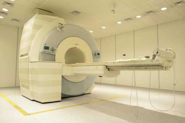
P2X7 receptor implicated in visceral pain caused by chronic pancreatitis
A subtype of purinergic receptor on spinal microglial cells mediated visceral pain hypersensitivity in rats with chronic pancreatitis, and pharmacologic or genetic inhibition of this receptor improved hyperalgesia, according to a report in the November issue of Cellular and Molecular Gastroenterology and Hepatology.
“Our study may be the first to identify that P2X7 receptors in spinal microglia are upregulated in chronic pancreatitis, and that this upregulation is associated with the development of visceral hyperalgesia,” said Dr. Pei-Yi Liu at National Yang-Ming University in Taipei, Taiwan, and her associates. A common laboratory dye known as brilliant blue G, which is an antagonist of P2X7R, “not only attenuated but also prevented CP-related chronic visceral hyperalgesia,” the researchers reported.
Chronic pancreatitis causes intense, recurrent epigastric pain that is “difficult and frustrating” to control and can lead to malnutrition, narcotic analgesic addiction, and social and financial problems, said the researchers. Previously, they had linked visceral pain in murine CP to activation of spinal microglia, the main effector immune cells in the central nervous system. The molecular pathways remained unclear, but some research had implicated extracellular adenosine triphosphate (ATP) as well as purinergic receptors in the CNS. Because a purine receptor subtype known as P2X7 had been linked to neuropathic and inflammatory pain, the researchers wondered if it also facilitated visceral pain (Cell Mol Gastroenterol Hepatol. 2015 Jul 22. doi: 10.1016/j.jcmgh.2015.07.008). To explore that question, they created a CP model by injecting 2% trinitrobenzene sulfonic acid into the pancreatic ducts of male rats. They measured behavioral responses to mechanical and electrical stimulation and quantified spinal cord P2X7R levels with the help of standard laboratory assays. They also watched for changes in pain-related behaviors after blocking spinal cord P2X7R with brilliant blue G or knocking it down with short interfering RNA (siRNA).
Spinal P2X7R expression rose significantly after CP induction, as did levels of the OX-42 microglial marker in the dorsal horn of the spinal cord, said the investigators. Brilliant blue G and genetic knock down suppressed P2X7R expression, inhibited activation of spinal microglia, and “significantly attenuated” nociceptive behaviors, they added.
The researchers also pretreated some rats with brilliant blue G before inducing CP and saw that these rats exhibited significantly lower pain responses to mechanical and electrical stimuli compared with other CP rats. In fact, the nociceptive responses of the pretreated CP rats resembled those of non-CP control rats, the investigators said. Spinal tissue from pretreated rats also lacked signs of P2X7R upregulation, they noted.
Taken together, the data “indicate a critical role of P2X7R expressed in the spinal cord in the development of chronic visceral pain in CP,” concluded the researchers. Brilliant blue G inhibits voltage-gated sodium channels, which are known to contribute to chronic visceral pain, and “may represent an effective drug for the treatment of chronic pain in chronic pancreatitis patients,” they added.
The study was funded by Taipei Veterans General Hospital, National Science Council of Taiwan, and the Taiwan Ministry of Education Aim for Top University Grant. The investigators declared no competing interests.
The traditional approach to treating pain in chronic pancreatitis is as if it were a “plumbing” problem – problems with ductal drainage. More recently, the emphasis has been on sensitization of the sensorineural system (“wiring”), in which the pain responses are greatly exaggerated. An additional consideration is whether this sensitization occurs in peripheral nerves that directly innervate the pancreas, or in the central nervous system, or both. This is clinically important because treatments directed at the periphery, e.g., pancreatectomy, may not be effective in patients in whom central sensitization is dominant.

|
Dr. Pankaj Jay Pasricha |
The findings of Dr. Lui and colleagues show that spinal (hence central) sensitization is important in chronic pancreatitis pain, and that this may be mediated by nonneuronal cells (microglia) in the spinal cord via P2X7R, a nucleotide receptor. This is not surprising, given that this signaling system has shown to be important in other forms of chronic pain. However, some questions remain – is peripheral sensitization driving these changes?
Clearly the “drug” they have used (BBG) is relatively harmless, but it is not practical because it may not be safe in humans (apart from coloring them blue). BBG also affects other channels, notably neuronal voltage-dependent sodium channels. Nevertheless, this study does offer new insight into the pathogenesis of pain in chronic pancreatitis and by itself is an important cautionary message for the growing enthusiasm for total pancreatectomy. It also identifies potential new therapeutic targets for treatment of pain and will, it is hoped, stimulate engagement from the pharmaceutical industry that is developing drugs directed toward glial activation and in particular the P2X7 receptor.
Dr. Pankaj Jay Pasricha, AGAF, is professor of medicine, Johns Hopkins University School of Medicine, director of Johns Hopkins Center for Motility Disorders and Digestive Diseases, and professor of innovation management, The Carey Business School, Johns Hopkins University, Baltimore. He has no conflicts of interest.
The traditional approach to treating pain in chronic pancreatitis is as if it were a “plumbing” problem – problems with ductal drainage. More recently, the emphasis has been on sensitization of the sensorineural system (“wiring”), in which the pain responses are greatly exaggerated. An additional consideration is whether this sensitization occurs in peripheral nerves that directly innervate the pancreas, or in the central nervous system, or both. This is clinically important because treatments directed at the periphery, e.g., pancreatectomy, may not be effective in patients in whom central sensitization is dominant.
|
|
Dr. Pankaj Jay Pasricha |
The findings of Dr. Lui and colleagues show that spinal (hence central) sensitization is important in chronic pancreatitis pain, and that this may be mediated by nonneuronal cells (microglia) in the spinal cord via P2X7R, a nucleotide receptor. This is not surprising, given that this signaling system has shown to be important in other forms of chronic pain. However, some questions remain – is peripheral sensitization driving these changes?
Clearly the “drug” they have used (BBG) is relatively harmless, but it is not practical because it may not be safe in humans (apart from coloring them blue). BBG also affects other channels, notably neuronal voltage-dependent sodium channels. Nevertheless, this study does offer new insight into the pathogenesis of pain in chronic pancreatitis and by itself is an important cautionary message for the growing enthusiasm for total pancreatectomy. It also identifies potential new therapeutic targets for treatment of pain and will, it is hoped, stimulate engagement from the pharmaceutical industry that is developing drugs directed toward glial activation and in particular the P2X7 receptor.
Dr. Pankaj Jay Pasricha, AGAF, is professor of medicine, Johns Hopkins University School of Medicine, director of Johns Hopkins Center for Motility Disorders and Digestive Diseases, and professor of innovation management, The Carey Business School, Johns Hopkins University, Baltimore. He has no conflicts of interest.
A subtype of purinergic receptor on spinal microglial cells mediated visceral pain hypersensitivity in rats with chronic pancreatitis, and pharmacologic or genetic inhibition of this receptor improved hyperalgesia, according to a report in the November issue of Cellular and Molecular Gastroenterology and Hepatology.
“Our study may be the first to identify that P2X7 receptors in spinal microglia are upregulated in chronic pancreatitis, and that this upregulation is associated with the development of visceral hyperalgesia,” said Dr. Pei-Yi Liu at National Yang-Ming University in Taipei, Taiwan, and her associates. A common laboratory dye known as brilliant blue G, which is an antagonist of P2X7R, “not only attenuated but also prevented CP-related chronic visceral hyperalgesia,” the researchers reported.
Chronic pancreatitis causes intense, recurrent epigastric pain that is “difficult and frustrating” to control and can lead to malnutrition, narcotic analgesic addiction, and social and financial problems, said the researchers. Previously, they had linked visceral pain in murine CP to activation of spinal microglia, the main effector immune cells in the central nervous system. The molecular pathways remained unclear, but some research had implicated extracellular adenosine triphosphate (ATP) as well as purinergic receptors in the CNS. Because a purine receptor subtype known as P2X7 had been linked to neuropathic and inflammatory pain, the researchers wondered if it also facilitated visceral pain (Cell Mol Gastroenterol Hepatol. 2015 Jul 22. doi: 10.1016/j.jcmgh.2015.07.008). To explore that question, they created a CP model by injecting 2% trinitrobenzene sulfonic acid into the pancreatic ducts of male rats. They measured behavioral responses to mechanical and electrical stimulation and quantified spinal cord P2X7R levels with the help of standard laboratory assays. They also watched for changes in pain-related behaviors after blocking spinal cord P2X7R with brilliant blue G or knocking it down with short interfering RNA (siRNA).
Spinal P2X7R expression rose significantly after CP induction, as did levels of the OX-42 microglial marker in the dorsal horn of the spinal cord, said the investigators. Brilliant blue G and genetic knock down suppressed P2X7R expression, inhibited activation of spinal microglia, and “significantly attenuated” nociceptive behaviors, they added.
The researchers also pretreated some rats with brilliant blue G before inducing CP and saw that these rats exhibited significantly lower pain responses to mechanical and electrical stimuli compared with other CP rats. In fact, the nociceptive responses of the pretreated CP rats resembled those of non-CP control rats, the investigators said. Spinal tissue from pretreated rats also lacked signs of P2X7R upregulation, they noted.
Taken together, the data “indicate a critical role of P2X7R expressed in the spinal cord in the development of chronic visceral pain in CP,” concluded the researchers. Brilliant blue G inhibits voltage-gated sodium channels, which are known to contribute to chronic visceral pain, and “may represent an effective drug for the treatment of chronic pain in chronic pancreatitis patients,” they added.
The study was funded by Taipei Veterans General Hospital, National Science Council of Taiwan, and the Taiwan Ministry of Education Aim for Top University Grant. The investigators declared no competing interests.
FROM CELLULAR AND MOLECULAR GASTROENTEROLOGY AND HEPATOLOGY
Key clinical point: A subtype of purinergic receptor on spinal microglial cells mediated visceral pain hypersensitivity in rats with chronic pancreatitis.
Major finding: Spinal P2X7R expression rose significantly after CP induction, and pharmacologic inhibition and genetic knock down inhibited activation of spinal microglia and “significantly attenuated” nociceptive behaviors.
Data source: Controlled, prospective, molecular and behavioral study of rats with chemically induced chronic pancreatitis.
Disclosures: The study was funded by Taipei Veterans General Hospital, National Science Council of Taiwan, and the Taiwan Ministry of Education Aim for Top University Grant. The investigators declared no competing interests.