User login
LUCENT-1: Mirikizumab sees phase 3 success in UC treatment
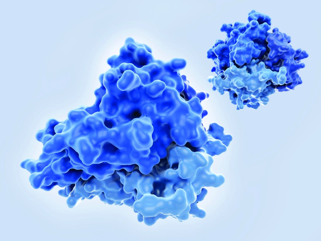
Mirikizumab is the first interleukin (IL) 23p19 to report positive phase 3 results for induction therapy in patients who have moderate to severe ulcerative colitis (UC) and have already been treated with at least one prior therapy.
Compared to placebo, almost a quarter (24.2%) of patients treated with mirikizumab versus 13% of those treated with placebo were in clinical remission at the end of the 12-week LUCENT-1 study. This was the study’s primary endpoint and it was a significant result (P = .00006), study investigator Geert D’Haens, MD, PhD, reported at the 17th congress of the European Crohn’s and Colitis Organisation.
Moreover, mirikizumab met all its secondary endpoints in the trial. At the end of the study almost two-thirds (63.5%) of patients achieved a clinical response (vs. 42.2% in the placebo arm); over one-third (36.3%) exhibited endoscopic remission (vs. 21.1%), and just over a quarter (27.1% vs. 13.9%) showed histologic-endoscopic mucosal improvement, according to a press release. The P values for all comparisons were less than .00001.
Perhaps more importantly from the patient perspective, there was greater symptomatic improvement and less bowel urgency in those treated with mirikizumab versus those randomized to placebo.
“The results confirmed [the] efficacy and safety noted in the phase 2 induction [trials], and supports mirikizumab’s potential as treatment for ulcerative colitis,” said Dr. D’Haens, professor of gastroenterology and hepatology at Amsterdam University Medical Centers.
Dominik Bettenworth, MD, who was not involved in the study, said that “the positive results from the LUCENT-1 trial are an important step towards selective IL-23 inhibition as a new mode of action for the treatment of patients with UC.
“This treatment approach has been shown efficacious and launched in other immune-mediated diseases such as psoriasis,” said Dr. Bettenworth, a specialist in internal medicine and gastroenterology in Münster, Germany.
“Noteworthy, for the first time in a phase 3 trial in IBD, [bowel] urgency has been assessed as a secondary endpoint – a clinical symptom of particular importance for patients with UC,” he added, noting that regarding safety, “the LUCENT-1 trial further confirms the previously observed overall good safety profile of IL-23 inhibitors.”
About mirikizumab and LUCENT-1
Mirikizumab is a humanized monoclonal antibody directed against the p19 subunit of IL-23 and is one of several IL23p19 antibodies currently under investigation for the treatment of IBD.
LUCENT-1 was a multicenter, randomized, double-blind, parallel group, placebo-controlled induction study in just over 1,200 patients aged 18-80 years. For inclusion, patients had to have moderately to severely active UC and had to have received at least one prior therapy, but had an inadequate or loss of response to it or been intolerant of it. Baseline data showed that 40% of patients included had been treated with corticosteroids and 24% with immunomodulators, and any current treatment remained unchanged during the study. Biologic treatment had failed in about 40% of patients and any patient taking such therapy had to discontinue it before participating in the trial.
Patients were randomized to receive 300 mg of mirikizumab or matching placebo, given intravenously at weeks 0, 4, and 8. Dr. D’Haens noted that randomization had been in a 3:1 ratio and had been stratified according to various factors such as biologic failure status, corticosteroid use, baseline disease activity, and region of the world where the patient was recruited.
Better results if biologic naive
Clinical remission was stringently defined as: Mayo stool frequency subscore of zero or one, with a reduction of one point or greater from baseline; no rectal bleeding; and a Mayo endoscopic subscore of zero to one. When looking at this primary endpoint in terms of whether patients had received prior biologic treatment, there was a higher remission rate at week 12 if patients had not previously been given any biologic therapy than if they had, but rates were still higher than placebo (30.9% vs. 15.8% and 15.2% vs. 8.5%, respectively), according to the press release. The P value was less than .001 for both comparisons although the study was not powered to look at these subgroups of patients.
Similarly, for clinical response – which was a decrease in the modified Mayo score of 2 or more points and at least a 30% decrease from initial values, as well as a decrease in rectal bleeding – there were differences between patients who had (54.6% for mirikizumab vs. 29.7% for placebo) and had not (70.1% vs. 50.3%) received a biologic previously (again, P less than .001 for both comparisons).
“Control of bowel function is one of the most debilitating, or one of the most important features for patients with ulcerative colitis,” Dr. D’Haens said when presenting the data on bowel urgency. In the trial there was an improvement in urgency – which was rated by patients – starting as early as week 2, with further improvement seen as the induction period went on.
Mirikizumab safety consistent with other trials
Dr. D’Haens noted that the safety profile of IL23 antibodies was “extremely clean.” As with other trials of mirikizumab, he said, there were similar or lower rates to placebo for many adverse events including any infection (15.1% vs. 14%) and cerebrocardiovascular events (0.6% vs. 0.1%). The overall rate of treatment emergent adverse events was 44.5% vs. 46.1% for mirikizumab and placebo. Notably, there were fewer serious adverse events (2.8% vs. 5.3%.) and discontinuations because of side effects (1.6% vs. 7.2%) in the mirikizumab group.
Are gastroenterologists now spoiled for choice?
Lots of questions followed Dr. D’Haens presentation, many picking up on the high placebo response and remission rates.
“We’ve seen it in a number of trials now,” Dr. D’Haens said. “One of the reasons is that patients are allowed to start corticosteroids as late as 2 weeks before randomization,” he observed. In LUCENT-1, half of the patients that were using steroids were receiving a dose of 20 mg of prednisone.
“Now when you start 20 mg of prednisone 2 weeks before randomization that might have an impact on your placebo readout. So I think that’s a lesson for many more trials in the future,” Dr. D’Haens said,”
A high placebo response rate was not expected and another hypothesis is that maybe additional clinical support for fatigue that was received may have played a role.
Several delegates asked for guidance on where mirikizumab and other IL-23p19 blockers might now fit in the grand scheme of patient treatment.
“It is really nice to have expanding therapeutic options but how will we choose?” said one in the online Q&A. “Which IL23 antagonist should we be using now?” and “Is ustekinumab obsolete in UC?” asked others.
“I think it’s early days to decide where the field is going,” Dr. D’Haens said after his presentation. “We don’t have head-to-head data. In GALAXI, ustekinumab was a reference arm.”
There will be further subanalyses of LUCENT-1 to come, including results from endoscopy and histology investigations, and the maintenance trial LUCENT-2 will also report soon.
“The feeling is that IL23 blockade is more specific, [more] beneficial in inflammatory bowel disease than blocking both IL12 and IL23,” Dr. D’Haens said.
Dr. D’Haens was an investigator in the LUCENT-1 trial and has acted as an adviser to the study’s sponsor, Eli Lilly, among many other big pharma companies. Dr. Bettenworth is on the advisory board or is a consultant for AbbVie, Amgen, Arena, Atheneum, BNG Service GmbH, Bristol Myers Squibb, CED-Service GmbH, Celltrion, Else Kröner-Fresenius-Foundation, Galapagos, Guidepoint, Impulze, Falk Foundation, Ferring, Janssen Cilag, Medical Tribune, MedTriX, MSD, Mylan, Onkowissen, Pharmacosmos, Pfizer, Roche, Sandoz, Takeda, Tetrameros, Thieme, Tillotts Pharma, and Vifor Pharma.
FROM ECCO 2022
Is proactive TDM the way to go?
Dear colleagues,
We shift gears from discussing GI hospitalists to focusing on the treatment of inflammatory bowel disease. The introduction of anti-TNFs brought about a paradigm shift in IBD management. With the ability to measure drug and antibody levels, we are also able to alter dose and timing to increase the efficacy of these medications. Some experts have extended this reactive drug monitoring approach to a more proactive method with the expectation that this may prevent loss of efficacy and development of adverse events. Dr. Loren G. Rabinowitz and colleagues and Dr. Hans Herfarth describe these two approaches to anti-TNF management in IBD, drawing from the current data and their own experiences. I look forward to hearing your thoughts and experiences by email ([email protected]).

Gyanprakash A. Ketwaroo, MD, MSc, is an assistant professor of medicine at Baylor College of Medicine, Houston. He is an associate editor for GI & Hepatology News.
Better outcomes than reactive TDM
By Loren G. Rabinowitz, MD; Konstantinos Papamichael, PhD, MD; and Adam S. Cheifetz, MD, AGAF
Therapeutic drug monitoring (TDM), or the practice of treatment optimization based on serum drug concentrations, is used in many settings, including solid organ transplantation, infection, and immune-mediated inflammatory diseases, including inflammatory bowel disease (IBD). In IBD, the use of TDM has been an area of keen research focus, and, in our view, should be standard practice for optimization of biologic therapy, particularly in the setting of anti–tumor necrosis factor (TNF) therapy. TDM has demonstrated utility in determining the correct timing and dosage of biologics and can provide the impetus for deescalating or discontinuing a biologic in favor of an alternative one. It also allows prescribers the ability to protect patients from severe infusion reactions if they have developed anti-drug antibodies (ADAs).

Reactive TDM refers to a strategy of assessing drug concentration and presence of ADAs in the setting of primary nonresponse (PNR) and loss of response (LOR) to a biologic agent. In this context, TDM informs possible reasons for loss or lack of response to treatment – for example, insufficient drug concentration or the development of high-titer ADAs (immunogenicity) – thus better directing the management of these unwanted outcomes.1 Insufficient anti-TNF concentrations have been associated with PNR and lack of clinical remission at 1 year in patients with IBD,2 which underscores the need for a durable strategy to ensure appropriate drug concentrations from the induction through maintenance phases of biologic administration. Reactive TDM can also be used to inform the decision to abandon a particular therapy in favor of a different biologic and to guide the selection of the next biologic agent, and has been shown to be less expensive than empiric dose escalation.2 With regard to infliximab and adalimumab, it is our practice to continue dose escalation until drug concentrations are above 10-15 mcg/mL prior to abandoning therapy.1

For a significant number of patients, reactive TDM identifies at-risk patients too late, when ADAs have already formed. Because the number of medications to treat IBD remains limited, waiting for a patient to lose response to an agent, particularly anti-TNF therapies, increases the likelihood of immunogenicity, thus rendering an agent unusable. Proactive TDM or checking drug trough concentrations preemptively and at predetermined intervals, and dosing to an appropriate concentration, can improve patient outcomes. If drug concentration is determined to be not “at target,” dosage and timing of administration can be increased with or without the addition of an immunomodulator (thiopurines or methotrexate) to optimize the biologic’s efficacy and prevent immunogenicity. This approach allows the provider to anticipate and proactively guard against PNR and future LOR.

Proactive TDM of anti-TNF therapy has been associated with better patient outcomes in both pediatric and adult populations when compared with empiric dose optimization and/or reactive TDM.2,3 In patients with immune-mediated inflammatory diseases undergoing maintenance therapy with infliximab, proactive TDM was found to be more effective than treatment without TDM in sustaining disease control without disease worsening.4 Proactive TDM has been associated with better patient outcomes, including increased rates of clinical remission in both pediatric and adult populations and decreased rates of IBD-related surgery, hospitalization, serious infusion reactions, and development of ADAs, when compared with reactive TDM or empiric optimization. Preliminary data suggest that proactive TDM can also be used to efficiently guide dose deescalation in patients in remission with drug concentrations markedly above target and to allow for optimization of infliximab monotherapy so that combination therapy can be employed more judiciously (that is, in a patient who developed rapid ADA to a different anti-TNF).1 This could potentially attenuate the risks associated with long-term immunomodulator use, which include lymphomas and higher rates of serious and opportunistic infections. A recent study using a pharmacokinetic dashboard showed that the majority of patients with IBD will need accelerated dosing by the third infusion to maintain therapeutic infliximab concentrations during induction and maintenance therapy, highlighting the urgent need for widespread adoption of early proactive TDM.5 It is likely that proactive TDM is most important early in therapy when patients are most inflamed and have more rapid drug clearance. For this reason, proactive TDM should ideally be used for all patients during the induction phase. It is our practice to continue to follow drug concentrations one or two times per year once a patient has achieved remission. A recent literature review and consensus statement highlights the utility of TDM and what is known at this time.1
TDM should be standard of care for patients with IBD. At minimum, reactive TDM has rationalized the management of PNR, is associated with better outcomes, and is more cost effective than empiric dose escalation.1,2 In this setting, however, many patients have already developed ADAs that cannot be overcome. At present, anti-TNF therapy remains the most effective agent for our sickest patients with IBD. Given the as-yet limited armamentarium of medications available, particularly for patients with fistulizing perianal Crohn’s disease (CD) and severe ulcerative colitis (UC), proactive TDM, which allows for improved optimization and long-term durability of biologics, is essential to the care of any IBD patient requiring these medications. Proactive TDM should ideally be used for all patients during the induction phase and at least once during maintenance therapy. There is also the potential for TDM-driven dose de-escalation for patients in remission and optimization of infliximab monotherapy, thus avoiding combination therapy with an immunomodulator in some cases. Future perspectives for a more precise application of TDM include the use of pharmacokinetic modeling dashboards and pharmacogenetics toward achieving truly individualized medicine.3
Dr. Rabinowitz, Dr. Papamichael, and Dr. Cheifetz are with the department of medicine and division of gastroenterology at Beth Israel Deaconess Medical Center and Harvard Medical School, both in Boston. Dr. Rabinowitz reports no conflicts of interest. Dr. Papamichael reports lecture fees from Mitsubishi Tanabe Pharma and Physicians’ Education Resource; consultancy fee from Prometheus Laboratories; and scientific advisory board fees from ProciseDx and Scipher Medicine Corporation. Dr. Cheifetz reports consulting for Janssen, AbbVie, Samsung, Arena Pharmaceuticals, Grifols, Prometheus, Bristol Myers Squibb, Artizan Biosciences, Artugan Therapeutics, and Equillium.
References
1. Cheifetz AS et al. Am J Gastroenterol. 2021 Oct 1;116(10):2014-25.
2. Kennedy NA et al. Lancet Gastroenterol Hepatol. 2019 May;4(5):341-53.
3. Papamichael K et al. Lancet Gastroenterol Hepatol. 2022;7(2):171-85.
4. Syversen SW et al. JAMA. 2021;326(23):2375-84.
5. Dubinsky MC et al. Inflamm Bowel Dis. 2022 Jan 3. doi: 10.1093/ibd/izab285.
Taking a closer look at the evidence
By Hans Herfarth, MD, PhD, AGAF
The debate of therapeutic drug monitoring (TDM) in the setting of anti–tumor necrosis factor (TNF) therapy has been ongoing for more than a decade. Reactive TDM, the measurement of drug concentrations in the context of loss of treatment response, is now generally accepted and recommended in multiple national and international inflammatory bowel disease (IBD) guidelines. Proactive TDM, defined as the systematic measurement of drug trough concentrations and anti-drug antibodies with dose adaptations to a predefined target drug concentration, seems to offer a possibility to stabilize drug levels and prevent anti-drug antibody formation due to low systemic drug levels, thus potentially preventing the well-known loss of response to anti-TNF therapy, which occurs in more than 50% of patients over time. However, proactive TDM is not endorsed by evidence-based guidelines, dividing IBD physicians into believers and nonbelievers and limiting uptake into clinical practice.

As with reactive TDM, one should assume that the framework for proactive TDM should have been reliably established based on factual data derived from prospective controlled studies and not rely on retrospective cohorts or “Expert Panel” consensus statements. And indeed, several prospective controlled studies with sizable IBD patient cohorts have been published. Of note, all TDM studies for IBD were conducted in patients on anti-TNF maintenance therapy, and currently no prospective studies in larger IBD populations are available for proactive TDM during induction therapy. Two prospective studies, the PRECISION and the NOR-DRUM trial, report that proactive TDM is better than no TDM at all.
However, in the comparison of proactive TDM and reactive TDM (including at least one drug adaptation in maintenance or drug escalation based on clinical symptoms or biomarkers), three studies have demonstrated no significant differences in drug persistence or overall maintenance of clinical remission. Only a fourth (the pediatric PAILOT study) reported a lower frequency of mild flares and less steroid exposure in the proactive TDM arm over 1 year, but it did not show differences in drug persistence or overall clinical remission compared with the reactive TDM arm. Interestingly, the differences in flare frequency were apparent only in patients on monotherapy but not in the subgroup on combination therapy with an immunomodulator, stressing the well-known beneficial effect of a combination therapy with thiopurines in CD first shown in the SONIC trial.
One problem, at least in the TDM trials with infliximab (IFX), may have been a delay in optimizing IFX levels until the next drug infusion because of the turnaround time of the drug assays. However, even the most recently published ultra-proactive TDM study with ad hoc dose adjustments based on
The value of proactive TDM in induction therapy remains an ongoing concern. There is no doubt that the severity of intestinal inflammation with subsequent loss of drug in the intestine can result in low drug serum concentrations correlating to lower clinical responses and higher rates of immunogenicity with the formation of anti-drug antibodies. A recent study including patients with chronic immune-mediated inflammatory diseases such as IBD, rheumatoid arthritis, and psoriasis did not find a value in proactive TDM of IFX in the induction phase, but more severe IBD may have been underrepresented in this study. Administration of significantly higher induction dosing of adalimumab (160 mg weeks 0, 1, 2, and 3) with significantly higher trough levels compared with a standard induction regimen in the SERENE study has not been shown to increase the short- or long-term remission rates in UC or CD patients. Therefore, higher trough levels in a patient population do not automatically result in better outcomes, but proactive TDM may still have found a few patients who may have benefited from an even higher induction regimen. The UC and CD SERENE maintenance studies also evaluated proactive TDM versus clinical adjustment based on clinical and biomarkers. After 1 year, no differences in the efficacy endpoints of clinical, endoscopic, and deep remission were found. These somewhat surprising results, which have been reported only in meeting abstracts, suggest that simply increasing trough levels to a higher target (one of the primary aims of proactive TDM) is not an effective universal approach for achieving higher remission rates in induction or better outcomes in maintenance. Instead, the SERENE data show that similar results can be achieved by regular clinical follow-up and monitoring of loss of response based on symptoms and/or biochemical markers followed by drug adaptation (which may then also be based on reactive TDM).
One unquestionable effect of proactive TDM is that the process of checking and controlling drug levels suggests for the treating physician better control over the anti-TNF therapy and for the patient reassurance that the treatment is in the intended target range. Proactive TDM also may be cost effective in the group of patients whose anti-TNF treatment regimen can be deescalated because of high drug levels. Despite the increased number of studies showing no clinical advantage of proactive TDM of every patient on anti-TNF therapy, there may be benefits for subgroups. Proactive TDM with point-of-care testing of drug levels may be helpful during induction therapy in patients with a high inflammatory burden, which results in uncontrolled drug loss via the intestine. Proactive TDM during maintenance therapy (for example, every 6-12 months) may be beneficial in subgroups of patients at risk for developing low anti-TNF levels or anti-drug antibodies, such as patients with a genetic predisposition to anti-TNF anti-drug antibody formation (such as the HLA-DQ1*05 allele), patients on a second anti-TNF therapy after loss of response to the first one, and patients on anti-TNF therapy in combination with thiopurines or methotrexate who deescalate to anti-TNF monotherapy.
In summary, there is no doubt that proactive TDM is better than no TDM (meaning no drug adjustments at all). However, nearly all controlled prospective studies show no significant benefit of proactive TDM versus reactive TDM or drug escalation based on clinical symptoms or biomarkers. Future studies should target clearly defined patient groups at risk of losing response to anti-TNF to clarify if proactive TDM is a valuable tool to achieving better therapeutic results in clinical practice.
Dr. Herfarth is professor of medicine and codirector of the UNC Multidisciplinary IBD Center at University of North Carolina at Chapel Hill. He reports serving as a consultant to Alivio Therapeutics, AMAG, Bristol Myers Squibb, Boehringer Ingleheim, ExeGi Pharma, Finch, Gilead, Janssen, Lycera, Merck, Otsuka, Pfizer, PureTech, and Seres and receiving research support from Allakos, Artizan Biosciences, and Pfizer.
Relevant resources
- Syversen SW et al. JAMA. 2021;326:2375-84.
- Strik AS et al. Scand J Gastroenterol. 2021 Feb;56(2):145-54.
- Bossuyt P et al. J Crohns Colitis. 2022 Feb 23;16(2):199-206.
- D’Haens G et al. Gastroenterology. 2018;154:1343-51.e1.
Dear colleagues,
We shift gears from discussing GI hospitalists to focusing on the treatment of inflammatory bowel disease. The introduction of anti-TNFs brought about a paradigm shift in IBD management. With the ability to measure drug and antibody levels, we are also able to alter dose and timing to increase the efficacy of these medications. Some experts have extended this reactive drug monitoring approach to a more proactive method with the expectation that this may prevent loss of efficacy and development of adverse events. Dr. Loren G. Rabinowitz and colleagues and Dr. Hans Herfarth describe these two approaches to anti-TNF management in IBD, drawing from the current data and their own experiences. I look forward to hearing your thoughts and experiences by email ([email protected]).
Gyanprakash A. Ketwaroo, MD, MSc, is an assistant professor of medicine at Baylor College of Medicine, Houston. He is an associate editor for GI & Hepatology News.
Better outcomes than reactive TDM
By Loren G. Rabinowitz, MD; Konstantinos Papamichael, PhD, MD; and Adam S. Cheifetz, MD, AGAF
Therapeutic drug monitoring (TDM), or the practice of treatment optimization based on serum drug concentrations, is used in many settings, including solid organ transplantation, infection, and immune-mediated inflammatory diseases, including inflammatory bowel disease (IBD). In IBD, the use of TDM has been an area of keen research focus, and, in our view, should be standard practice for optimization of biologic therapy, particularly in the setting of anti–tumor necrosis factor (TNF) therapy. TDM has demonstrated utility in determining the correct timing and dosage of biologics and can provide the impetus for deescalating or discontinuing a biologic in favor of an alternative one. It also allows prescribers the ability to protect patients from severe infusion reactions if they have developed anti-drug antibodies (ADAs).
Reactive TDM refers to a strategy of assessing drug concentration and presence of ADAs in the setting of primary nonresponse (PNR) and loss of response (LOR) to a biologic agent. In this context, TDM informs possible reasons for loss or lack of response to treatment – for example, insufficient drug concentration or the development of high-titer ADAs (immunogenicity) – thus better directing the management of these unwanted outcomes.1 Insufficient anti-TNF concentrations have been associated with PNR and lack of clinical remission at 1 year in patients with IBD,2 which underscores the need for a durable strategy to ensure appropriate drug concentrations from the induction through maintenance phases of biologic administration. Reactive TDM can also be used to inform the decision to abandon a particular therapy in favor of a different biologic and to guide the selection of the next biologic agent, and has been shown to be less expensive than empiric dose escalation.2 With regard to infliximab and adalimumab, it is our practice to continue dose escalation until drug concentrations are above 10-15 mcg/mL prior to abandoning therapy.1
For a significant number of patients, reactive TDM identifies at-risk patients too late, when ADAs have already formed. Because the number of medications to treat IBD remains limited, waiting for a patient to lose response to an agent, particularly anti-TNF therapies, increases the likelihood of immunogenicity, thus rendering an agent unusable. Proactive TDM or checking drug trough concentrations preemptively and at predetermined intervals, and dosing to an appropriate concentration, can improve patient outcomes. If drug concentration is determined to be not “at target,” dosage and timing of administration can be increased with or without the addition of an immunomodulator (thiopurines or methotrexate) to optimize the biologic’s efficacy and prevent immunogenicity. This approach allows the provider to anticipate and proactively guard against PNR and future LOR.
Proactive TDM of anti-TNF therapy has been associated with better patient outcomes in both pediatric and adult populations when compared with empiric dose optimization and/or reactive TDM.2,3 In patients with immune-mediated inflammatory diseases undergoing maintenance therapy with infliximab, proactive TDM was found to be more effective than treatment without TDM in sustaining disease control without disease worsening.4 Proactive TDM has been associated with better patient outcomes, including increased rates of clinical remission in both pediatric and adult populations and decreased rates of IBD-related surgery, hospitalization, serious infusion reactions, and development of ADAs, when compared with reactive TDM or empiric optimization. Preliminary data suggest that proactive TDM can also be used to efficiently guide dose deescalation in patients in remission with drug concentrations markedly above target and to allow for optimization of infliximab monotherapy so that combination therapy can be employed more judiciously (that is, in a patient who developed rapid ADA to a different anti-TNF).1 This could potentially attenuate the risks associated with long-term immunomodulator use, which include lymphomas and higher rates of serious and opportunistic infections. A recent study using a pharmacokinetic dashboard showed that the majority of patients with IBD will need accelerated dosing by the third infusion to maintain therapeutic infliximab concentrations during induction and maintenance therapy, highlighting the urgent need for widespread adoption of early proactive TDM.5 It is likely that proactive TDM is most important early in therapy when patients are most inflamed and have more rapid drug clearance. For this reason, proactive TDM should ideally be used for all patients during the induction phase. It is our practice to continue to follow drug concentrations one or two times per year once a patient has achieved remission. A recent literature review and consensus statement highlights the utility of TDM and what is known at this time.1
TDM should be standard of care for patients with IBD. At minimum, reactive TDM has rationalized the management of PNR, is associated with better outcomes, and is more cost effective than empiric dose escalation.1,2 In this setting, however, many patients have already developed ADAs that cannot be overcome. At present, anti-TNF therapy remains the most effective agent for our sickest patients with IBD. Given the as-yet limited armamentarium of medications available, particularly for patients with fistulizing perianal Crohn’s disease (CD) and severe ulcerative colitis (UC), proactive TDM, which allows for improved optimization and long-term durability of biologics, is essential to the care of any IBD patient requiring these medications. Proactive TDM should ideally be used for all patients during the induction phase and at least once during maintenance therapy. There is also the potential for TDM-driven dose de-escalation for patients in remission and optimization of infliximab monotherapy, thus avoiding combination therapy with an immunomodulator in some cases. Future perspectives for a more precise application of TDM include the use of pharmacokinetic modeling dashboards and pharmacogenetics toward achieving truly individualized medicine.3
Dr. Rabinowitz, Dr. Papamichael, and Dr. Cheifetz are with the department of medicine and division of gastroenterology at Beth Israel Deaconess Medical Center and Harvard Medical School, both in Boston. Dr. Rabinowitz reports no conflicts of interest. Dr. Papamichael reports lecture fees from Mitsubishi Tanabe Pharma and Physicians’ Education Resource; consultancy fee from Prometheus Laboratories; and scientific advisory board fees from ProciseDx and Scipher Medicine Corporation. Dr. Cheifetz reports consulting for Janssen, AbbVie, Samsung, Arena Pharmaceuticals, Grifols, Prometheus, Bristol Myers Squibb, Artizan Biosciences, Artugan Therapeutics, and Equillium.
References
1. Cheifetz AS et al. Am J Gastroenterol. 2021 Oct 1;116(10):2014-25.
2. Kennedy NA et al. Lancet Gastroenterol Hepatol. 2019 May;4(5):341-53.
3. Papamichael K et al. Lancet Gastroenterol Hepatol. 2022;7(2):171-85.
4. Syversen SW et al. JAMA. 2021;326(23):2375-84.
5. Dubinsky MC et al. Inflamm Bowel Dis. 2022 Jan 3. doi: 10.1093/ibd/izab285.
Taking a closer look at the evidence
By Hans Herfarth, MD, PhD, AGAF
The debate of therapeutic drug monitoring (TDM) in the setting of anti–tumor necrosis factor (TNF) therapy has been ongoing for more than a decade. Reactive TDM, the measurement of drug concentrations in the context of loss of treatment response, is now generally accepted and recommended in multiple national and international inflammatory bowel disease (IBD) guidelines. Proactive TDM, defined as the systematic measurement of drug trough concentrations and anti-drug antibodies with dose adaptations to a predefined target drug concentration, seems to offer a possibility to stabilize drug levels and prevent anti-drug antibody formation due to low systemic drug levels, thus potentially preventing the well-known loss of response to anti-TNF therapy, which occurs in more than 50% of patients over time. However, proactive TDM is not endorsed by evidence-based guidelines, dividing IBD physicians into believers and nonbelievers and limiting uptake into clinical practice.
As with reactive TDM, one should assume that the framework for proactive TDM should have been reliably established based on factual data derived from prospective controlled studies and not rely on retrospective cohorts or “Expert Panel” consensus statements. And indeed, several prospective controlled studies with sizable IBD patient cohorts have been published. Of note, all TDM studies for IBD were conducted in patients on anti-TNF maintenance therapy, and currently no prospective studies in larger IBD populations are available for proactive TDM during induction therapy. Two prospective studies, the PRECISION and the NOR-DRUM trial, report that proactive TDM is better than no TDM at all.
However, in the comparison of proactive TDM and reactive TDM (including at least one drug adaptation in maintenance or drug escalation based on clinical symptoms or biomarkers), three studies have demonstrated no significant differences in drug persistence or overall maintenance of clinical remission. Only a fourth (the pediatric PAILOT study) reported a lower frequency of mild flares and less steroid exposure in the proactive TDM arm over 1 year, but it did not show differences in drug persistence or overall clinical remission compared with the reactive TDM arm. Interestingly, the differences in flare frequency were apparent only in patients on monotherapy but not in the subgroup on combination therapy with an immunomodulator, stressing the well-known beneficial effect of a combination therapy with thiopurines in CD first shown in the SONIC trial.
One problem, at least in the TDM trials with infliximab (IFX), may have been a delay in optimizing IFX levels until the next drug infusion because of the turnaround time of the drug assays. However, even the most recently published ultra-proactive TDM study with ad hoc dose adjustments based on
The value of proactive TDM in induction therapy remains an ongoing concern. There is no doubt that the severity of intestinal inflammation with subsequent loss of drug in the intestine can result in low drug serum concentrations correlating to lower clinical responses and higher rates of immunogenicity with the formation of anti-drug antibodies. A recent study including patients with chronic immune-mediated inflammatory diseases such as IBD, rheumatoid arthritis, and psoriasis did not find a value in proactive TDM of IFX in the induction phase, but more severe IBD may have been underrepresented in this study. Administration of significantly higher induction dosing of adalimumab (160 mg weeks 0, 1, 2, and 3) with significantly higher trough levels compared with a standard induction regimen in the SERENE study has not been shown to increase the short- or long-term remission rates in UC or CD patients. Therefore, higher trough levels in a patient population do not automatically result in better outcomes, but proactive TDM may still have found a few patients who may have benefited from an even higher induction regimen. The UC and CD SERENE maintenance studies also evaluated proactive TDM versus clinical adjustment based on clinical and biomarkers. After 1 year, no differences in the efficacy endpoints of clinical, endoscopic, and deep remission were found. These somewhat surprising results, which have been reported only in meeting abstracts, suggest that simply increasing trough levels to a higher target (one of the primary aims of proactive TDM) is not an effective universal approach for achieving higher remission rates in induction or better outcomes in maintenance. Instead, the SERENE data show that similar results can be achieved by regular clinical follow-up and monitoring of loss of response based on symptoms and/or biochemical markers followed by drug adaptation (which may then also be based on reactive TDM).
One unquestionable effect of proactive TDM is that the process of checking and controlling drug levels suggests for the treating physician better control over the anti-TNF therapy and for the patient reassurance that the treatment is in the intended target range. Proactive TDM also may be cost effective in the group of patients whose anti-TNF treatment regimen can be deescalated because of high drug levels. Despite the increased number of studies showing no clinical advantage of proactive TDM of every patient on anti-TNF therapy, there may be benefits for subgroups. Proactive TDM with point-of-care testing of drug levels may be helpful during induction therapy in patients with a high inflammatory burden, which results in uncontrolled drug loss via the intestine. Proactive TDM during maintenance therapy (for example, every 6-12 months) may be beneficial in subgroups of patients at risk for developing low anti-TNF levels or anti-drug antibodies, such as patients with a genetic predisposition to anti-TNF anti-drug antibody formation (such as the HLA-DQ1*05 allele), patients on a second anti-TNF therapy after loss of response to the first one, and patients on anti-TNF therapy in combination with thiopurines or methotrexate who deescalate to anti-TNF monotherapy.
In summary, there is no doubt that proactive TDM is better than no TDM (meaning no drug adjustments at all). However, nearly all controlled prospective studies show no significant benefit of proactive TDM versus reactive TDM or drug escalation based on clinical symptoms or biomarkers. Future studies should target clearly defined patient groups at risk of losing response to anti-TNF to clarify if proactive TDM is a valuable tool to achieving better therapeutic results in clinical practice.
Dr. Herfarth is professor of medicine and codirector of the UNC Multidisciplinary IBD Center at University of North Carolina at Chapel Hill. He reports serving as a consultant to Alivio Therapeutics, AMAG, Bristol Myers Squibb, Boehringer Ingleheim, ExeGi Pharma, Finch, Gilead, Janssen, Lycera, Merck, Otsuka, Pfizer, PureTech, and Seres and receiving research support from Allakos, Artizan Biosciences, and Pfizer.
Relevant resources
- Syversen SW et al. JAMA. 2021;326:2375-84.
- Strik AS et al. Scand J Gastroenterol. 2021 Feb;56(2):145-54.
- Bossuyt P et al. J Crohns Colitis. 2022 Feb 23;16(2):199-206.
- D’Haens G et al. Gastroenterology. 2018;154:1343-51.e1.

‘Superdonor’ samples don’t increase FMT success in ulcerative colitis
The success of fecal microbiota transplantation (FMT) in people with active ulcerative colitis (UC) was not improved by using highly standardized and controlled “superdonor” samples versus control samples, according to results reported at the 17th congress of the European Crohn’s and Colitis Organisation.
Indeed, a similar percentage (10% and 13.9%, respectively; P = .72) of patients achieved combined steroid-free endoscopic and clinical remission at 8 weeks, which was the primary endpoint of the randomized, controlled, RESTORE-UC trial.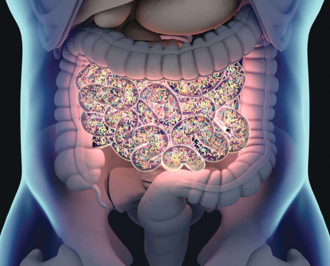
“Maybe we were too bold to say we will go for steroid-free endoscopic remission and response,” said Clara Caenepeel, MD, who was the presenting study investigator. “It’s a very strict endpoint.”
The reasoning for such a strict endpoint, however, was so that the trials’ findings could be compared with some of the other studies that have been done with FMT in UC. Importantly, all those trials have all been positive, making the results of the RESTORE-UC trial at odds with their findings.
“I think in the analysis that we will do now is definitely look at how many steps we went into the right direction,” noted Dr. Caenepeel, who is a doctoral researcher at IBD Leuven (Belgium).
Response to results
Although this is a negative trial, its findings are still important for future work looking at the role of FMT in managing patients with inflammatory bowel disease and identifying the best donor material and ways to deliver it.

While some have suggested it was back to the bench to explore negative results, others such as Michael A. Kamm, MBBS, MD, FRCP, FRACP, congratulated the investigators for undertaking the study, saying that “these studies are very hard to do!”
Dr. Kamm, who is professor of gastroenterology and leads the Kamm Gut Research Group at the University of Melbourne, was part of the Australian team that conducted the FOCUS (Faecal Microbiota Transplantation in Ulcerative Colitis) study. That study used the same primary endpoint of steroid-free endoscopic and clinical remission at 8 weeks but reported positive results – 27% of patients who had FMT versus just 8% of those who had a saline enema as a placebo achieved the endpoint (P = .021). Similarly positive findings have also been reported from five other studies.
“To understand why [RESTORE-UC] study is negative, coming after several positive studies, one needs to explore the differences in study design,” Dr. Kamm observed in an interview. Those differences include how donors were selected, how the FMT was delivered, and how patients were selected.
“All the early studies made no presumption about a favorable donor profile,” Dr. Kamm noted with regard to the selection of donors based on their microbial profile. Moreover “the mode of delivery – sigmoidoscopy without any colonoscopic whole-colon delivery, in contrast to previous studies – as well as patient selection, [with] no information on the anatomical extent of their disease,” could be important.
“There are enough robust positive studies of FMT in ulcerative colitis to believe that this therapy can be effective,” said Dr. Kamm. “Analysis of negative studies like this one should help us to understand what factors are needed to achieve a positive outcome.”
RESTORE-UC trial
“Fecal microbiota transplantation is a new emerging strategy in the treatment of active ulcerative colitis,” Dr. Caenepeel observed during her presentation. At the time the study was started in 2017 there had been four other studies, with “very heterogeneous” designs in terms of the samples used, the placebos given, the delivery of FMT, and the primary endpoints. The idea of superdonor samples also came out of those trials.
So the aim was to try to standardize practice and set up a trial “to examine if we could increase the FMT success rate in our active ulcerative colitis patients by strictly preselecting our donors; by standardized FMT preparation; and a standardized and repeated FMT administration,” Dr. Caenepeel said.
RESTORE-UC was a multicenter, randomized, double-blind, and sham-controlled trial conducted in seven Belgian hospitals. A predefined futility analysis was performed when 66% (n = 72) of the proposed 108 patients had been recruited. Of these, 36 receive autologous FMT and 30 received superdonor FMT.
“We put the emphasis on standardization. This started already with our donor selection,” Dr. Caenepeel said. From a potential 57 healthy donors, 15 were selected and altogether provided more than 500 samples that were then whittled down to the ones that provided the “best” microbial content.
FMT or autologous samples delivered four times – first by sigmoidoscopy and then at weekly intervals by rectal enema. Every patient received the same donor material, Dr. Caenepeel stressed, containing the same enterotype and concentration.
In addition to the primary endpoint of steroid-free endoscopic and clinical remission at Week 8, secondary endpoints included steroid-free clinical remission, steroid-free clinical response, steroid-free endoscopic remission, and steroid-free endoscopic response. Again, however, no significant differences were seen between the two study arms.
Two serious adverse events were seen in the trial, both occurring in the autologous sample group; these were dysuria/constipation and a worsening of colitis that needed surgery.
In discussion, Walter Reinisch, MD, the director of the inflammatory bowel disease study group at the Medical University of Vienna, picked up on why the study may have been negative. He observed that using a steroid-free endoscopic endpoint, where the Mayo score was zero, may have been a factor. A result of 19% at week 8 was not insignificant, he said, observing “if studies from big sponsors would get these results, they would be very happy.”
Perhaps the trials to date have been a little too simplistic by looking at the donor’s microbiota, Dr. Caenepeel. “It goes much further than microbiota,” she said. Thus, future work will perhaps look at the genetics and immunity of those undergoing FMT, she suggested.
Dr. Caenepeel, Dr. Kamm, and Dr. Reinisch had no conflicts of interest to disclose.
The success of fecal microbiota transplantation (FMT) in people with active ulcerative colitis (UC) was not improved by using highly standardized and controlled “superdonor” samples versus control samples, according to results reported at the 17th congress of the European Crohn’s and Colitis Organisation.
Indeed, a similar percentage (10% and 13.9%, respectively; P = .72) of patients achieved combined steroid-free endoscopic and clinical remission at 8 weeks, which was the primary endpoint of the randomized, controlled, RESTORE-UC trial.
“Maybe we were too bold to say we will go for steroid-free endoscopic remission and response,” said Clara Caenepeel, MD, who was the presenting study investigator. “It’s a very strict endpoint.”
The reasoning for such a strict endpoint, however, was so that the trials’ findings could be compared with some of the other studies that have been done with FMT in UC. Importantly, all those trials have all been positive, making the results of the RESTORE-UC trial at odds with their findings.
“I think in the analysis that we will do now is definitely look at how many steps we went into the right direction,” noted Dr. Caenepeel, who is a doctoral researcher at IBD Leuven (Belgium).
Response to results
Although this is a negative trial, its findings are still important for future work looking at the role of FMT in managing patients with inflammatory bowel disease and identifying the best donor material and ways to deliver it.
While some have suggested it was back to the bench to explore negative results, others such as Michael A. Kamm, MBBS, MD, FRCP, FRACP, congratulated the investigators for undertaking the study, saying that “these studies are very hard to do!”
Dr. Kamm, who is professor of gastroenterology and leads the Kamm Gut Research Group at the University of Melbourne, was part of the Australian team that conducted the FOCUS (Faecal Microbiota Transplantation in Ulcerative Colitis) study. That study used the same primary endpoint of steroid-free endoscopic and clinical remission at 8 weeks but reported positive results – 27% of patients who had FMT versus just 8% of those who had a saline enema as a placebo achieved the endpoint (P = .021). Similarly positive findings have also been reported from five other studies.
“To understand why [RESTORE-UC] study is negative, coming after several positive studies, one needs to explore the differences in study design,” Dr. Kamm observed in an interview. Those differences include how donors were selected, how the FMT was delivered, and how patients were selected.
“All the early studies made no presumption about a favorable donor profile,” Dr. Kamm noted with regard to the selection of donors based on their microbial profile. Moreover “the mode of delivery – sigmoidoscopy without any colonoscopic whole-colon delivery, in contrast to previous studies – as well as patient selection, [with] no information on the anatomical extent of their disease,” could be important.
“There are enough robust positive studies of FMT in ulcerative colitis to believe that this therapy can be effective,” said Dr. Kamm. “Analysis of negative studies like this one should help us to understand what factors are needed to achieve a positive outcome.”
RESTORE-UC trial
“Fecal microbiota transplantation is a new emerging strategy in the treatment of active ulcerative colitis,” Dr. Caenepeel observed during her presentation. At the time the study was started in 2017 there had been four other studies, with “very heterogeneous” designs in terms of the samples used, the placebos given, the delivery of FMT, and the primary endpoints. The idea of superdonor samples also came out of those trials.
So the aim was to try to standardize practice and set up a trial “to examine if we could increase the FMT success rate in our active ulcerative colitis patients by strictly preselecting our donors; by standardized FMT preparation; and a standardized and repeated FMT administration,” Dr. Caenepeel said.
RESTORE-UC was a multicenter, randomized, double-blind, and sham-controlled trial conducted in seven Belgian hospitals. A predefined futility analysis was performed when 66% (n = 72) of the proposed 108 patients had been recruited. Of these, 36 receive autologous FMT and 30 received superdonor FMT.
“We put the emphasis on standardization. This started already with our donor selection,” Dr. Caenepeel said. From a potential 57 healthy donors, 15 were selected and altogether provided more than 500 samples that were then whittled down to the ones that provided the “best” microbial content.
FMT or autologous samples delivered four times – first by sigmoidoscopy and then at weekly intervals by rectal enema. Every patient received the same donor material, Dr. Caenepeel stressed, containing the same enterotype and concentration.
In addition to the primary endpoint of steroid-free endoscopic and clinical remission at Week 8, secondary endpoints included steroid-free clinical remission, steroid-free clinical response, steroid-free endoscopic remission, and steroid-free endoscopic response. Again, however, no significant differences were seen between the two study arms.
Two serious adverse events were seen in the trial, both occurring in the autologous sample group; these were dysuria/constipation and a worsening of colitis that needed surgery.
In discussion, Walter Reinisch, MD, the director of the inflammatory bowel disease study group at the Medical University of Vienna, picked up on why the study may have been negative. He observed that using a steroid-free endoscopic endpoint, where the Mayo score was zero, may have been a factor. A result of 19% at week 8 was not insignificant, he said, observing “if studies from big sponsors would get these results, they would be very happy.”
Perhaps the trials to date have been a little too simplistic by looking at the donor’s microbiota, Dr. Caenepeel. “It goes much further than microbiota,” she said. Thus, future work will perhaps look at the genetics and immunity of those undergoing FMT, she suggested.
Dr. Caenepeel, Dr. Kamm, and Dr. Reinisch had no conflicts of interest to disclose.
FROM ECCO 2022
Meta-analysis favors SubQ infliximab biosimilar over vedolizumab in Crohn’s disease
An infliximab biosimilar (infliximab-axxq) delivered via subcutaneous injection was superior to intravenous vedolizumab (Entyvio) for induction and maintenance therapy of Crohn’s disease (CD), according to a systematic review and meta-analysis from French researchers.
In contrast, the same research showed no significant differences in efficacy between these agents for people with ulcerative colitis (UC).
The subcutaneous injection formulation of infliximab-axxq, also known as CT-P13 SC, is approved by the European Medicines Agency for both CD and UC (Remsima SC). This formulation is not yet approved by the U.S. Food and Drug Administration.
“It’s kind of a small revolution, because now we have access, and in France, we are using a lot of subcutaneous infliximab,” Laurent Peyrin-Biroulet, MD, said during a digital oral presentation at ECCO ‘22 Virtual, the European Crohn’s and Colitis Organisation 2022 Congress. He is ECCO president and a gastroenterologist at University Hospital of Nancy in France.
Dr. Peyrin-Biroulet and colleagues compared the efficacy and safety of the two agents in major randomized controlled trials looking at moderate-to-severe CD or UC from January 2010 to April 2021.
Subcutaneous infliximab-axxq data came from NCT02883452, an ongoing phase 1 study started in 2016. The vedolizumab data come from GEMINI I, II, and III; VISIBLE 1 and 2; and VARSITY trials.
Key findings
In CD, induction efficacy was 79% for subcutaneous infliximab-axxq versus a 45% response for vedolizumab, as measured by the Crohn’s Disease Activity Index-70 (CDAI-70).
In addition, the study showed a 62% induction efficacy for subcutaneous infliximab-axxq versus 36% for vedolizumab on the CDAI-100.
The maintenance response on the CDAI-100 was achieved by 64% of the infliximab-axxq patients versus 47% of the vedolizumab patients.
“You can see better efficacy results of subcutaneous infliximab compared to vedolizumab in CD for both induction and maintenance,” Dr. Peyrin-Biroulet said, adding that what is seen here is probably what has been observed.
Similar results for UC
“We know vedolizumab is a good drug in UC, and there was not a difference between UC and CD,” Dr. Peyrin-Biroulet said.
There were no significant differences between subcutaneous infliximab-axxq and vedolizumab in terms of clinical response for induction (77% vs. 69%, respectively) or clinical response for maintenance (62% vs. 63%, respectively).
Similarly, no significant differences emerged in rates of clinical remission or mucosal healing.
Safety so far
Safety profiles for both agents in both indications were similar over 1 year.
However, Dr. Peyrin-Biroulet shared a caveat about that timeline. “It’s a 1-year follow-up, not a 10-year follow-up with 100,000 patients,” he said. “But still, what we see here is a confirmation of what we see in other analyses. There is not a significant [safety] difference between these two drugs.”
The rate of any adverse event in CD was 76% for subcutaneous infliximab-axxq versus 77% for vedolizumab, for example. Rates of any serious adverse event were 9% and 14%, respectively.
In UC studies, the rate of any adverse event was 67% for subcutaneous infliximab-axxq versus 64% for vedolizumab. Rates of serious adverse events were 12% and 11%, respectively.
Rates of discontinuation over 1 year differed between agents. Five percent of subcutaneous infliximab-axxq participants with CD discontinued treatment as because of lack of efficacy compared with 32% with vedolizumab. In UC participants, discontinuation for the same reason was reported for 3% of subcutaneous infliximab-axxq patients versus 15% of vedolizumab.
Although all participants in the subcutaneous infliximab-axxq trial were anti-tumor necrosis factor treatment naive, some in the vedolizumab may have been previously exposed to such therapy, Dr. Peyrin-Biroulet said. “Subgroup analyses would be need to further explore that.”
“It’s interesting and a clinically relevant piece of work,” said Mark Samaan, MD, session moderator and consultant gastroenterologist at Guy’s and St. Thomas’ NHS Foundation Trust in London, who was not involved in the study.
“The results are in keeping with what one might expect from IV infliximab administration. In that sense, [it is] reassuring that the two routes of administration appear to perform in a broadly similar manner,” Dr. Samaan said.
“So long as adequate exposure is achieved, I wouldn’t have expected route of administration to be a significant determinant of efficacy,” he added.
Dr. Peyrin-Biroulet reports receiving personal fees from Galapagos, AbbVie, Janssen, Genetech, Ferring, Tillots, Pharmacosmos, Celltrion, Takeda, Samsung Bioepis, Alma, Sterna, Nestle, Inotrem, Enterome, Allergen, MSD, Roche, Arena, Gilead, Hikma, Amgen, BMS, Vifor, Norgene, Mylan, Lilly, Fresenius Kabi, Opplian Pharma, Sublimity Therapeutics, Applied Molecular Transport, OSE Immunotherapeutics, Enthera, and Theravance. Dr. Samaan reports receiving advisory fees from Janssen, Takeda, Sandoz, Samsung Bioepis, Galapagos, and AbbVie and lecture fees from Bristol-Myers Squibb, Janssen, Takeda, MSD, Falk, AbbVie, and Galapagos.
A version of this article first appeared on Medscape.com.
Two factors linked to higher risk of long COVID in IBD
Two features are significantly associated with a higher risk for developing long COVID symptoms among people with inflammatory bowel disease (IBD), according to a large Danish population study.
People with Crohn’s disease (CD) who experienced adverse acute COVID-19, defined as requiring hospitalization, were nearly three times more likely to report persistent symptoms 12 weeks after acute infection.
“Long-term, persisting symptoms following COVID-19 is a frequently occurring problem, which is probably underappreciated. IBD specialists should therefore be aware of any of these symptoms and actively ask patients whether they have these problems,” lead author Mohamed Attauabi, MD, PhD, said in an interview.
Dr. Attauabi and colleagues also found that people with ulcerative colitis (UC) who discontinued immunosuppressive agents because of COVID-19 were 1.5 times more likely to experience long COVID symptoms, a result that surprised the researchers.
“This has not been shown before and remains to be confirmed,” said Dr. Attauabi, a fellow in the department of gastroenterology at Herlev Hospital at the University of Copenhagen.
Attauabi presented the results as a digital oral presentation at the 17th congress of the European Crohn’s and Colitis Organisation.
A closer look at IBD and COVID-19
Large, hospital-based studies of symptoms consistent with long COVID reveal a high prevalence of fatigue, sleep difficulties, and anxiety at 12 weeks or more post acute infection. However, these were not specific to people with CD or UC, Dr. Attauabi said.
“In patients with IBD, the risk of long-term sequelae of COVID-19 remains to be investigated,” he said.
Dr. Attauabi and colleagues studied 197 people with CD and 319 with UC, all of whom had polymerase chain reaction–confirmed COVID-19. Participants were prospectively enrolled in the population-based Danish IBD-COVID registry from January 28, 2020 to April 1, 2021. At a median of 5.1 months, a subset of 85 people with CD and 137 with UC agreed to report any post-COVID symptoms.
Older age, smoking, IBD disease activity, and presence of comorbidities were not associated with a significantly elevated risk of long COVID.
In a multivariate analysis, hospitalization for COVID-19 among people with CD was significantly associated with long COVID (odds ratio, 2.76; 95% confidence interval, 1.05-3.90; P = .04).
Furthermore, people with UC who stopped taking immunosuppressive agents also had a significantly higher risk (OR, 1.50; 95% CI, 1.07-10.22; P = .01).
“However, IBD medications such as systemic steroids were not associated with this outcome,” Dr. Attauabi said.
Fatigue most common long COVID symptom
Fatigue was the most common long COVID symptom, reported by 37% of patients with CD and 36% with UC.
Anosmia and ageusia were also common, reported by 29% and 28% of patients with CD, and 27% and 19% of those with UC, respectively.
“In our cohort of patients with UC or CD who developed COVID-19, the long-term health effects of COVID-19 did not appear to differ among patients with UC or CD nor according to IBD medications,” Dr. Attauabi said.
That is a “great study,” said session cochair Torsten Kucharzik, MD, PhD, head of internal medicine and gastroenterology at Lueneburg (Germany) Hospital.
When Dr. Kucharzik asked about smoking, Dr. Attauabi responded that they collected information on current and previous smoking, but they chose not to include the data because it was not statistically significant.
Dr. Attauabi has reported no relevant financial relationships. Dr. Kucharzik has reported receiving grants from Takeda and personal fees from companies including MSD/Essex, AbbVie, Falk Foundation, Biogen, Bristol-Myers Squibb, Arena, Celgene, Celltrion, Ferring, Janssen, Galapagos, Olympus, Mundipharma, Takeda, Amgen, Pfizer, Roche, and Vifor Pharma.
A version of this article first appeared on Medscape.com.
FROM ECCO 2022
Handle with care: Managing IBD in older patients
As the saying goes: "Age is a case of mind over matter: If you don't mind, it don't matter."
But for older patients with inflammatory bowel disease (IBD) and the clinicians who treat them, it’s hard to ignore the complications that aging can bring, such as comorbidities, functional limitations, and polypharmacy, said Nana Bernasko, CRNP, DNP, WHNP-BC, a nurse practitioner in the department of gastroenterology at Penn State Milton S. Hershey Medical Center in Hershey, Pa.
“We are seeing a large number of patients in our clinics that are being diagnosed later on in life,” she said in an oral presentation at the annual Crohn’s & Colitis Congress®, a partnership of the Crohn’s & Colitis Foundation and the American Gastroenterological Association.
Between 10% and 30% of all patients with IBD are older than 60, and roughly 10%-15% of patients with IBD are diagnosed after age 60, she said.
The diagnosis of IBD is often delayed in older patients as well, with an estimated 60% of patients initially given an incorrect or incomplete diagnosis that may lead to significant delays in the initiation of appropriate therapy, she said.
Differential diagnoses for IBD in older patients include diverticulitis, ischemic colitis, infectious colitis, and radiation colitis.
Bharati Kochar, MD, MS, from the Crohn’s and Colitis Center at Massachusetts General Hospital in Boston, who was not involved in the presentation, agreed that older adults need special handling.
“The management of IBD in older adults is challenging for a number of reasons, but primarily because until very recently, we have not invested in understanding how IBD should be optimally managed at older ages,” she said in an interview.
“Additionally, like in all fields, older adults with IBD are disproportionately under-represented in clinical trials, meaning that we have less rigorous data guiding the management of older adults,” she added.
Clinical presentations
Older adults tend to differ in clinical presentation, compared with younger adults, Dr. Bernasko said.
For example, among patients with Crohn’s disease, rectal bleeding is a more common symptom among older adults, whereas diarrhea and weight loss are more common among younger adults.
Disease location may also differ, with more senior adults having predominantly colonic disease (L2 according to the Montreal Classification of IBD), compared with more prevalent ileocolonic disease (L3) among their more junior counterparts. And although both generations of patients have inflammatory behavior (B1) at diagnosis, younger patients have more prevalent structuring (B2) and penetrating disease (B3), Dr. Bernasko noted.
Among patients with ulcerative colitis, rectal bleeding, abdominal pain, and extraintestinal manifestations are more common among the younger set, whereas left-sided colitis is more common among older patients. In addition, extensive ulcerative colitis (E3) is more common in younger patients, compared with older patients.
Management considerations
Dr. Kochar noted that “older adults have higher baseline risks for all adverse events – like infections, malignancies, polypharmacy, procedural complications – than younger adults, so any additional risk conferred by treatments seem amplified, but that should not mean that we should avoid effectively treating older adults. It should mean we need to invest in understanding how to best mitigate those risks.”
While younger patients are sometimes on multiple medications prior to starting on IBD therapy, polypharmacy is common among the older set, who may be taking drugs for diabetes, hypertension, prostate disease, and so on.
“There’s just so much going on in terms of their medical background to start off with, so many medications, and then we’re adding more things to it,” Dr. Bernasko said.
She echoed Dr. Kochar in noting that older patients as a subgroup are under-represented in clinical trials, making it difficult to know what treatment approaches may work best for them.
In addition, older patients are at higher risk for malignancies, and for complications from surgery.
Medication adherence in older patients is frequently compromised by memory issues, she added, noting that “I can’t tell you enough how sometimes our older patients forget to take their medications.”
Other challenges for the management of older patients with IBD included psychosocial issues, cognitive decline, and malnutrition.
Medications and adverse events
Dr. Bernasko also discussed specific medications and potential adverse events and drug interactions in older patients.
For example, aminosalicylic acids (5-ASA) are associated with higher risk for nephrotoxicity and pancreatitis in older patients and can interact with thiopurines to cause leukopenia.
Steroids are associated with elevated risk for osteopenia, myopathy, cataracts, glaucoma, diabetes, and hypertension, and can interact with thiazide and loop diuretics to cause hypokalemia.
Methotrexate use in this population is linked to pancytopenia and hepatotoxicity, and it can interact with NSAIDs and multiple antibiotics to cause decreased renal secretion.
Thiopurines in older patients are associated with increased risk for leukopenia, myelosuppression, non-Hodgkin lymphoma, skin cancer, pancreatitis, and hepatotoxicity, and drugs in this class interact with allopurinol and angiotensin-converting enzyme inhibitors to increase risk for myelosuppression. Additionally, warfarin can inhibit the efficacy of thiopurines, and when these drugs are used in combination with tumor necrosis factor (TNF)–alpha inhibitors they can further increase risk of malignancy through immunosuppression.
Cyclosporine is associated with worsening hypertension and renal insufficiency among older patients.
TNF-alpha inhibitors are associated with increased risk for tuberculosis; hepatitis B; and fungal infections, malignant lymphoma, and New York Heart Association class 3 or 4 heart failure.
Ciprofloxacin in older patients with IBD has been linked to tendinopathy and increased risk for Clostridioides difficile infections. Metronidazole increases the likelihood of peripheral neuropathy in these patients.
Colon cancer screening
“When it comes to colon cancer screening, definitely assess the risk prior to doing this,” Dr. Bernasko recommended. “Weigh all the risks and benefits. Why are we doing this for these elderly patients, because there are definitely risks associated with this.”
Older patients with IBD may have difficulty with bowel prep and are at elevated risk, compared with younger patients, for cardiopulmonary complications, perforation, adverse events from sedation, and procedural complications, she cautioned.
“When it comes to our elderly patients, you want to focus on a more personalized approach – not all older people present the same way in terms of comorbidities or medications,” Dr. Bernasko advised in her summary.
Dr. Bernasko and Dr. Kochar reported having no relevant conflicts of interest to disclose. Dr. Kochar is a member of the board of editors for GI & Hepatology News.
This article was updated 2/18/22.
FROM CROHN’S & COLITIS CONGRESS
Malnutrition common in patients with IBD
Malnutrition is common among patients with inflammatory bowel disease (IBD) and is associated with worse outcomes that can prolong hospitalizations and increase patients’ risk for death.
As many as 85% of inpatients with IBD may be malnourished, with the severity of malnutrition affected by disease activity, extent, and duration, said Kelly Issokson, MS, RD, CNSC, clinical nutrition coordinator in the IBD program in the division of gastroenterology at Cedars-Sinai Medical Center, Los Angeles.
“Malnutrition is a severe complication of IBD, and it should not be overlooked,” she said during an oral presentation at the annual Crohn’s & Colitis Congress®, a partnership of the Crohn’s & Colitis Foundation and the American Gastroenterological Association.
In patients with IBD, malabsorption, enteric losses, inadequate intake, and side effects of medical therapy can all lead to malnutrition, which in turn is an independent risk factor for venous thromboembolic events, nonelective surgery, longer hospital stays, and increased mortality.

In addition, malnutrition in IBD increases risk for infection and sepsis, and for perioperative complications, and can more than double the cost of care, compared with adequately nourished IBD patients, she said.
Ms. Issokson cited a definition of malnutrition from the American Society of Parenteral and Enteral Nutrition as “an acute or chronic state of overnutrition or undernutrition with or without inflammatory activity that has led to a change in body composition and diminished function.”
Lab findings of low albumin, low prealbumin, or isolated metrics such as weight loss or change in body mass index do not constitute malnutrition and should not be used to diagnosis it, Ms. Issokson cautioned.
Patients at low risk for malnutrition have no unintentional weight loss, are eating well, have minimal or no dietary restrictions, and no wasting. In contrast, high-risk patients have unintentional weight loss, decreased appetite and/or food intake, restrict multiple foods, or show signs of wasting.
Screening
“Nutrition screening is the first step in diagnosing a patient with malnutrition. This is a process of identifying individuals who may be at nutrition risk and benefit from assessment from a registered dietitian,” Ms. Issokson said.
The Malnutrition Screening Tool is quick, easy to administer, and requires minimal training. It can be used to screen adults for malnutrition regardless of age, medical history, or setting, she said.
The two-item instrument asks, “Have you recently lost weight without trying?” with a “no” scored as 0 and a “yes” scored as 2. The second question is, “Have you been eating poorly because of decreased appetite, with a “no” equal to 0 and a “yes” equal to 1. Patients with a score of 0 or 1 are not at risk, whereas patients with scores of 2 or 3 are deemed to be at risk for malnutrition and require further assessment by a dietitian.
Assessment
Assessment for malnutrition involves a variety of factors, including anthropometric factors such as weight and BMI changes; biochemical markers such as fat-soluble vitamins, water-soluble vitamins, minerals, and urinary sodium; symptoms such as decreased appetite, abdominal pain, cramping or bloating, diarrhea, or urgency or obstructive symptoms; and body composition measures such as handgrip strength, biochemical impedance analysis, skinfold thickness, bone mineral density, and muscle mass.
Other nutritional assessment tools may include 24-hour recall of nutrition intake, diet history, and questions about eating behaviors, food allergies or intolerances, and cultural or religious food preferences.
Assessing food security is also important, especially during the current pandemic, Ms. Issokson emphasized.
“Is your patient running out of food? Do they have money to purchase food? Are they able to go to the grocery store to buy food? This is essential to know when you’re developing a nutrition plan,” she said.
A nutrition-focused physical exam should include assessment of skin manifestation, secondary to malnutrition or malabsorption, such as dry skin, delayed wound healing, stomatitis, scurvy, seborrheic dermatitis, bleeding, and periorificial and acral dermatitis or alopecia.
Diagnosis
Currently available malnutrition criteria have not been validated for use in patients with IBD, and further studies are needed to affirm their applicability to this population, Ms. Issokson said.
The Academy of Nutrition and Dietetics–American Society for Parenteral and Enteral Nutrition (AND-ASPEN) malnutrition criteria require measures of weight loss, energy intake, subcutaneous fat loss, subcutaneous muscle loss, general or local fluid accumulation, and handgrip strength to determine whether a patient is moderately or severely malnourished.
Ms. Issokson said that she finds the European Society for Clinical Nutrition and Metabolism Global Leadership Initiative on Malnutrition (ESPEN GLIM) criteria somewhat easier to use for diagnosis, as they consist of phenotypic and etiologic criteria, with patients who meet at least one of each being considered malnourished.
“When identified, document malnutrition, and of course intervene appropriately by referring to a dietitian providing education and supporting the patient to help them optimize their nutrition and improve their outcomes,” she concluded.
In a discussion following the session, panelist Neha Shah, MPH, RD, CNSC, a dietitian and health education specialist at the University of California, San Francisco, commented on the importance of malnutrition assessment in patients with IBD being considered for surgery.
Patients should be screened for malnutrition, and if they have a positive screen, “should be automatically referred to a registered dietitian specializing in IBD for a nutrition assessment,” she said.
“Certainly, a nutritional assessment, as Kelly has highlighted really well, will encompass an evaluation of various areas of health – patient history, food and nutrition history, changing anthropometrics, alterations in labs – and certainly going into further nutrition history with net food intolerance, intake from each food group, portions, access, support, culture, eating environment, skills in the kitchen, relationship with diet.”
Ms. Issokson is a board member of the Crohn’s & Colitis Foundation and a digital advisory board member of Avant Healthcare. Ms. Shah had no disclosures.
Malnutrition is common among patients with inflammatory bowel disease (IBD) and is associated with worse outcomes that can prolong hospitalizations and increase patients’ risk for death.
As many as 85% of inpatients with IBD may be malnourished, with the severity of malnutrition affected by disease activity, extent, and duration, said Kelly Issokson, MS, RD, CNSC, clinical nutrition coordinator in the IBD program in the division of gastroenterology at Cedars-Sinai Medical Center, Los Angeles.
“Malnutrition is a severe complication of IBD, and it should not be overlooked,” she said during an oral presentation at the annual Crohn’s & Colitis Congress®, a partnership of the Crohn’s & Colitis Foundation and the American Gastroenterological Association.
In patients with IBD, malabsorption, enteric losses, inadequate intake, and side effects of medical therapy can all lead to malnutrition, which in turn is an independent risk factor for venous thromboembolic events, nonelective surgery, longer hospital stays, and increased mortality.
In addition, malnutrition in IBD increases risk for infection and sepsis, and for perioperative complications, and can more than double the cost of care, compared with adequately nourished IBD patients, she said.
Ms. Issokson cited a definition of malnutrition from the American Society of Parenteral and Enteral Nutrition as “an acute or chronic state of overnutrition or undernutrition with or without inflammatory activity that has led to a change in body composition and diminished function.”
Lab findings of low albumin, low prealbumin, or isolated metrics such as weight loss or change in body mass index do not constitute malnutrition and should not be used to diagnosis it, Ms. Issokson cautioned.
Patients at low risk for malnutrition have no unintentional weight loss, are eating well, have minimal or no dietary restrictions, and no wasting. In contrast, high-risk patients have unintentional weight loss, decreased appetite and/or food intake, restrict multiple foods, or show signs of wasting.
Screening
“Nutrition screening is the first step in diagnosing a patient with malnutrition. This is a process of identifying individuals who may be at nutrition risk and benefit from assessment from a registered dietitian,” Ms. Issokson said.
The Malnutrition Screening Tool is quick, easy to administer, and requires minimal training. It can be used to screen adults for malnutrition regardless of age, medical history, or setting, she said.
The two-item instrument asks, “Have you recently lost weight without trying?” with a “no” scored as 0 and a “yes” scored as 2. The second question is, “Have you been eating poorly because of decreased appetite, with a “no” equal to 0 and a “yes” equal to 1. Patients with a score of 0 or 1 are not at risk, whereas patients with scores of 2 or 3 are deemed to be at risk for malnutrition and require further assessment by a dietitian.
Assessment
Assessment for malnutrition involves a variety of factors, including anthropometric factors such as weight and BMI changes; biochemical markers such as fat-soluble vitamins, water-soluble vitamins, minerals, and urinary sodium; symptoms such as decreased appetite, abdominal pain, cramping or bloating, diarrhea, or urgency or obstructive symptoms; and body composition measures such as handgrip strength, biochemical impedance analysis, skinfold thickness, bone mineral density, and muscle mass.
Other nutritional assessment tools may include 24-hour recall of nutrition intake, diet history, and questions about eating behaviors, food allergies or intolerances, and cultural or religious food preferences.
Assessing food security is also important, especially during the current pandemic, Ms. Issokson emphasized.
“Is your patient running out of food? Do they have money to purchase food? Are they able to go to the grocery store to buy food? This is essential to know when you’re developing a nutrition plan,” she said.
A nutrition-focused physical exam should include assessment of skin manifestation, secondary to malnutrition or malabsorption, such as dry skin, delayed wound healing, stomatitis, scurvy, seborrheic dermatitis, bleeding, and periorificial and acral dermatitis or alopecia.
Diagnosis
Currently available malnutrition criteria have not been validated for use in patients with IBD, and further studies are needed to affirm their applicability to this population, Ms. Issokson said.
The Academy of Nutrition and Dietetics–American Society for Parenteral and Enteral Nutrition (AND-ASPEN) malnutrition criteria require measures of weight loss, energy intake, subcutaneous fat loss, subcutaneous muscle loss, general or local fluid accumulation, and handgrip strength to determine whether a patient is moderately or severely malnourished.
Ms. Issokson said that she finds the European Society for Clinical Nutrition and Metabolism Global Leadership Initiative on Malnutrition (ESPEN GLIM) criteria somewhat easier to use for diagnosis, as they consist of phenotypic and etiologic criteria, with patients who meet at least one of each being considered malnourished.
“When identified, document malnutrition, and of course intervene appropriately by referring to a dietitian providing education and supporting the patient to help them optimize their nutrition and improve their outcomes,” she concluded.
In a discussion following the session, panelist Neha Shah, MPH, RD, CNSC, a dietitian and health education specialist at the University of California, San Francisco, commented on the importance of malnutrition assessment in patients with IBD being considered for surgery.
Patients should be screened for malnutrition, and if they have a positive screen, “should be automatically referred to a registered dietitian specializing in IBD for a nutrition assessment,” she said.
“Certainly, a nutritional assessment, as Kelly has highlighted really well, will encompass an evaluation of various areas of health – patient history, food and nutrition history, changing anthropometrics, alterations in labs – and certainly going into further nutrition history with net food intolerance, intake from each food group, portions, access, support, culture, eating environment, skills in the kitchen, relationship with diet.”
Ms. Issokson is a board member of the Crohn’s & Colitis Foundation and a digital advisory board member of Avant Healthcare. Ms. Shah had no disclosures.
FROM THE CROHN’S & COLITIS CONGRESS
Breakthrough COVID-19 milder in vaccinated patients with IBD
Vaccination against SARS-CoV-2 appears to protect people with inflammatory bowel disease (IBD) from the more serious consequences of breakthrough COVID-19 infections, but results may vary by which vaccine was received, results of a small study suggest.
In a study of patients with IBD who had completed a primary vaccine series but went on to develop COVID-19, there were trends toward worse outcomes for patients who received a non-mRNA vaccine, older patients, and those who were on combination therapy rather than monotherapy, reported Emily Spiera, a medical student at the Icahn School of Medicine at Mount Sinai, New York.
“Overall, we saw that vaccinated patients who subsequently developed COVID-19 had low rates of hospitalization, severe COVID, and death,” she said in an oral abstract at the annual Crohn’s & Colitis Congress®, a partnership of the Crohn’s & Colitis Foundation and the American Gastroenterological Association.
The study was conducted before the highly infectious Omicron variant of SARS-CoV-2 became dominant, however, and the sample size of 88 patients, combined with a low number of study events, was too small for statistical significance to emerge for most measures, Ms. Spiera acknowledged.
Nonetheless, the findings support the protective benefit of vaccines in this population, said Freddy Caldera, DO, associate professor of gastroenterology at the University of Wisconsin–Madison, who was not involved in the study.
“In my mind, when we think about COVID vaccines, the whole goal is to prevent severe disease,” he said.

Dr. Caldera and colleagues conducted an earlier study of humoral immunogenicity of mRNA COVID-19 vaccines in 122 patients with IBD and 60 healthy controls, and found that all controls and 97% of patients with IBD developed antibodies, although antibody concentrations were lower in patients with IBD, compared with controls (P < .001). Those who received the mRNA-1273 (Moderna) COVID-19 had significantly higher antibody concentrations than those who received the Pfizer-BNT vaccine series (P < .001).
They also found that patients on immune-modifying therapy had lower antibody concentrations, compared with those who were not on such therapy, or those who received aminosalicylates or vedolizumab (Entyvio; P = .003).
The protective effect of vaccines in this population became even more apparent after patients received an additional vaccine dose.
“We actually have a study in preprint of what happens after a third dose, where everyone made antibodies,” he said. “What we tell patients is that vaccines work.”
SECURE-IBD data
The investigators at Mount Sinai, the University of North Carolina at Chapel Hill, and Tel Aviv University analyzed data from the Surveillance Epidemiology of Coronavirus Under Research Exclusion in Inflammatory Bowel Disease (SECURE-IBD) database, an international web-based registry that includes reports from 74 countries, with data reported by 48 U.S. states.
The study sample consisted of patients enrolled from Dec. 12, 2020, to Oct. 1, 2021, who had completed a primary vaccination series with either mRNA vaccines (Pfizer or Moderna) adenoviral vector-based vaccines (AstraZeneca, Sputnik, CanSino, or Janssen/Johnson & Johnson), or an inactivated SARS-CoV-2 vaccine (Sinovac).
Of 2,477 patients with COVID-19 infections reported to SECURE-IBD, 160 reported being vaccinated. Of this group, 53 were excluded because they were only partially vaccinated, and 19 were excluded because of missing data on either vaccine type, number of doses, or COVID-19 outcomes, leaving 88 patients with completed primary vaccination series at the time of COVID-19 infection.
The median patient age was 40.1 years. Nearly two-thirds of the patients had a diagnosis of Crohn’s disease, and slightly more than one-third has a diagnosis of ulcerative colitis. The patients came from 18 countries, with 45.3% of the sample in the United States.
A total of 58% of patients were on biologic monotherapy, with either a tumor necrosis factor antagonist, integrin antagonist, or anti–interleukin-12/13. In addition, 3.4% were on immunomodulator monotherapy, 21.6% were on combination therapy, and 5.7% were receiving corticosteroids.
Lower severity
COVID-19 severity was numerically but not significantly lower among the 88 vaccinated patients, with a rate of 5.7%, compared with 9.3% among 2,317 patients with COVID-19 infections in the database who were not vaccinated.
COVID-19 severity defined as a composite of ICU admission, need for mechanical ventilation and/or death was actually slightly higher among the vaccinated patients, with a rate of 3.4% versus 1.9% for nonvaccinated patients, but this difference was not statistically significant.
There was 1 death among vaccinated patients (1.1%) versus 29 among the unvaccinated (1.2%).
There were trends toward fewer hospitalizations and less-frequent severe COVID-19 infection among patients who received a mRNA vaccine, compared with other vaccine types, but again these differences did not reach statistical significance.
As noted before, there was a higher frequency of severe COVID-19 among patients on combination therapy than on monotherapy, but this difference too was not statistically significant.
As seen with COVID-19 in the general population older patients tended to have worse outcomes, with a mean age of 53 for patients requiring hospitalization, compared with 39 years for patients who stayed out of the hospital (P = .04), and a mean age of 59 among patients with severe COVID-19 infections, compared with 39 for patients with moderate or mild infections (P = .03).
Ms. Spiera described the case of the single vaccinated patient who died. The 63-year-old woman had moderately active Crohn’s disease treated with corticosteroids, adalimumab (Humira) and azathioprine at the time of COVID-19 infection. She had received the AstraZeneca adenoviral-based vaccine more than 30 days prior to infection. She was hospitalized and intubated, and died from gastrointestinal bleeding.
Ms. Spiera noted that, although the sample size was small, and only patients known to have COVID-19 were included, it is one of the largest cohorts to date of vaccinated patients with IBD who developed COVID-19. She said that the study supports prior studies showing that combination therapy and tumor necrosis factor antagonists may result in reduced immunity, and that mRNA vaccines may offer better protection against severe illness in this population.
The study was supported by a Digestive Disease Research Foundation Fellowship. Ms. Spiera and Dr. Caldera reported no relevant disclosures.
Vaccination against SARS-CoV-2 appears to protect people with inflammatory bowel disease (IBD) from the more serious consequences of breakthrough COVID-19 infections, but results may vary by which vaccine was received, results of a small study suggest.
In a study of patients with IBD who had completed a primary vaccine series but went on to develop COVID-19, there were trends toward worse outcomes for patients who received a non-mRNA vaccine, older patients, and those who were on combination therapy rather than monotherapy, reported Emily Spiera, a medical student at the Icahn School of Medicine at Mount Sinai, New York.
“Overall, we saw that vaccinated patients who subsequently developed COVID-19 had low rates of hospitalization, severe COVID, and death,” she said in an oral abstract at the annual Crohn’s & Colitis Congress®, a partnership of the Crohn’s & Colitis Foundation and the American Gastroenterological Association.
The study was conducted before the highly infectious Omicron variant of SARS-CoV-2 became dominant, however, and the sample size of 88 patients, combined with a low number of study events, was too small for statistical significance to emerge for most measures, Ms. Spiera acknowledged.
Nonetheless, the findings support the protective benefit of vaccines in this population, said Freddy Caldera, DO, associate professor of gastroenterology at the University of Wisconsin–Madison, who was not involved in the study.
“In my mind, when we think about COVID vaccines, the whole goal is to prevent severe disease,” he said.
Dr. Caldera and colleagues conducted an earlier study of humoral immunogenicity of mRNA COVID-19 vaccines in 122 patients with IBD and 60 healthy controls, and found that all controls and 97% of patients with IBD developed antibodies, although antibody concentrations were lower in patients with IBD, compared with controls (P < .001). Those who received the mRNA-1273 (Moderna) COVID-19 had significantly higher antibody concentrations than those who received the Pfizer-BNT vaccine series (P < .001).
They also found that patients on immune-modifying therapy had lower antibody concentrations, compared with those who were not on such therapy, or those who received aminosalicylates or vedolizumab (Entyvio; P = .003).
The protective effect of vaccines in this population became even more apparent after patients received an additional vaccine dose.
“We actually have a study in preprint of what happens after a third dose, where everyone made antibodies,” he said. “What we tell patients is that vaccines work.”
SECURE-IBD data
The investigators at Mount Sinai, the University of North Carolina at Chapel Hill, and Tel Aviv University analyzed data from the Surveillance Epidemiology of Coronavirus Under Research Exclusion in Inflammatory Bowel Disease (SECURE-IBD) database, an international web-based registry that includes reports from 74 countries, with data reported by 48 U.S. states.
The study sample consisted of patients enrolled from Dec. 12, 2020, to Oct. 1, 2021, who had completed a primary vaccination series with either mRNA vaccines (Pfizer or Moderna) adenoviral vector-based vaccines (AstraZeneca, Sputnik, CanSino, or Janssen/Johnson & Johnson), or an inactivated SARS-CoV-2 vaccine (Sinovac).
Of 2,477 patients with COVID-19 infections reported to SECURE-IBD, 160 reported being vaccinated. Of this group, 53 were excluded because they were only partially vaccinated, and 19 were excluded because of missing data on either vaccine type, number of doses, or COVID-19 outcomes, leaving 88 patients with completed primary vaccination series at the time of COVID-19 infection.
The median patient age was 40.1 years. Nearly two-thirds of the patients had a diagnosis of Crohn’s disease, and slightly more than one-third has a diagnosis of ulcerative colitis. The patients came from 18 countries, with 45.3% of the sample in the United States.
A total of 58% of patients were on biologic monotherapy, with either a tumor necrosis factor antagonist, integrin antagonist, or anti–interleukin-12/13. In addition, 3.4% were on immunomodulator monotherapy, 21.6% were on combination therapy, and 5.7% were receiving corticosteroids.
Lower severity
COVID-19 severity was numerically but not significantly lower among the 88 vaccinated patients, with a rate of 5.7%, compared with 9.3% among 2,317 patients with COVID-19 infections in the database who were not vaccinated.
COVID-19 severity defined as a composite of ICU admission, need for mechanical ventilation and/or death was actually slightly higher among the vaccinated patients, with a rate of 3.4% versus 1.9% for nonvaccinated patients, but this difference was not statistically significant.
There was 1 death among vaccinated patients (1.1%) versus 29 among the unvaccinated (1.2%).
There were trends toward fewer hospitalizations and less-frequent severe COVID-19 infection among patients who received a mRNA vaccine, compared with other vaccine types, but again these differences did not reach statistical significance.
As noted before, there was a higher frequency of severe COVID-19 among patients on combination therapy than on monotherapy, but this difference too was not statistically significant.
As seen with COVID-19 in the general population older patients tended to have worse outcomes, with a mean age of 53 for patients requiring hospitalization, compared with 39 years for patients who stayed out of the hospital (P = .04), and a mean age of 59 among patients with severe COVID-19 infections, compared with 39 for patients with moderate or mild infections (P = .03).
Ms. Spiera described the case of the single vaccinated patient who died. The 63-year-old woman had moderately active Crohn’s disease treated with corticosteroids, adalimumab (Humira) and azathioprine at the time of COVID-19 infection. She had received the AstraZeneca adenoviral-based vaccine more than 30 days prior to infection. She was hospitalized and intubated, and died from gastrointestinal bleeding.
Ms. Spiera noted that, although the sample size was small, and only patients known to have COVID-19 were included, it is one of the largest cohorts to date of vaccinated patients with IBD who developed COVID-19. She said that the study supports prior studies showing that combination therapy and tumor necrosis factor antagonists may result in reduced immunity, and that mRNA vaccines may offer better protection against severe illness in this population.
The study was supported by a Digestive Disease Research Foundation Fellowship. Ms. Spiera and Dr. Caldera reported no relevant disclosures.
FROM THE CROHN’S & COLITIS CONGRESS
A deep dive on tofacitinib’s mode of action
A new study has revealed potential cell-specific effects of the human Janus kinase (JAK) inhibitor tofacitinib, including possible targets – such as intestinal inflammation – for future research and even for increasing the drug’s effects.
The work used both mice and human cell models to explore the drug’s effect in inflammatory bowel disease (IBD). The mouse models suggested that the drug’s pharmacokinetics may be affected by intestinal inflammation. The human cell models seem to identify equilibrative nucleoside transporters as the likely route of cellular uptake of tofacitinib; this mechanism appears to be upregulated during inflammation and could present a therapeutic target to bolster the drug’s effects.
“We identify intestinal inflammation as a decisive modulator of the systemic pharmacokinetics of tofacitinib in mice, which needs to be studied and confirmed in humans. Finally, we decipher an important membrane transport mechanism that regulates cellular uptake of tofacitinib into activated immune cells, suggesting a model that explains a preferred uptake of tofacitinib into activated immune cells and a potential starting point to interfere with and channel such an uptake,” wrote the authors, led by Bernhard Texler and Andreas Zollner, both with the Christian Doppler Laboratory for Mucosal Immunology at the Johannes Kepler University in Linz, Austria, who published the results in Cellular and Molecular Gastroenterology and Hepatology.
IBD-related inflammation likely involves multiple cytokine pathways. The JAK-signal transducers and activator of transcription (JAK-STAT) pathway is downstream to more than 50 cytokines and growth factors, so disruption of their activity by JAK-STAT inhibitors like tofacitinib could counter the effects of more than one cytokine at a time.
Tofacitinib received FDA approval for the treatment of ulcerative colitis in 2018, but the details of its mechanism of action against intestinal inflammation remain poorly understood. For example, despite its efficacy against UC, the drug doesn’t work for Crohn’s disease patients. That may be because the drug affects specific cell populations involved only in UC pathogenesis.
To better understand the drug’s pharmacokinetics, the researchers examined the effects of tofacitinib in cells isolated from human peripheral blood, as well as an experimental mouse model of colitis.
The drug inhibited proliferation of both naïve and memory cytotoxic and helper T cells. At higher concentrations, it had strong effects on innate immune system cells, including monocytes, macrophages, and human intestinal epithelial organoids. It promotes the anti-inflammatory M2 phenotype among monocytes and macrophages. The drug also inhibited the pro-inflammatory M1 phenotype. The researchers observed similar effects in the mouse model of colitis.
The investigators also linked equilibrative nucleoside transporters (ENTs) with uptake of tofacitinib, specifically as a mediating role. These membrane proteins transport nucleosides, nucleobases, and therapeutic analogs like tofacitinib, which mimics the nucleotide adenosine triphosphate (ATP). Targeted inhibitors could potentially influence this process.
The researchers created three-dimensional, in vitro colonic organoids using intestinal epithelial cells from UC patients and healthy human controls. In this model, TNF-alpha can lead to production of pro-inflammatory cytokines, but tofacitinib blocked this effect. That result suggests that intestinal epithelial cells are a previously unidentified tofacitinib target.
Although a large amount of work has been done on the pharmacokinetics of therapeutic antibodies used to treat IBD, the authors point out that little is known about tofacitinib. In a mouse model, the serum concentration of the drug increased after exposure to dextran sulfate sodium (DSS), which triggers an IBD-like condition, and the spike was higher during more intense inflammation. The finding was surprising, considering that therapeutic antibodies typically get eliminated through feces during inflammation. Mice treated with DSS versus control had similar levels of tofacitinib in both urine and the feces, suggesting that inflammation may somehow inhibit the enzymes that metabolize the drug.
The researchers also noted that uptake of tofacitinib into leukocytes increased following stimulation with lipopolysaccharide. Given its structural similarity to ATP, the researchers propose that tofacitinib may enter the cells through adenosine cell membrane transporters ENT1 and ENT2, and some evidence even suggested that the pathway may be strengthened in activated immune cells.
The study received funding from: the Christian Doppler Research Association; the Austrian Federal Ministry of Science, Research, and Economy; and the National Foundation for Research, Technology, and Development. One author is receiving research support from AbbVie and Takeda under the framework of the Christian Doppler Research Society, but the remaining authors have no relevant conflicts of interest.
Growing understanding of underlying immunopathogenic mechanisms of inflammatory bowel diseases (IBD) have led to the development of targeted therapies that have considerably improved patient outcomes. However, insights into their respective effector mechanisms are still scarce.

Pharmacokinetics of biological therapies with assessment of serum drug levels have since been an indispensable part of the optimal management of IBD patients. The presented findings on the pharmacokinetics of tofacitinib during inflammation both on a systemic and on a cellular level might have comparable potential therapeutic consequences. Therapeutic modulation of the responsible membrane transport mechanism for the cellular uptake of tofacitinib might lead to enhanced therapeutic efficacy in the future. Further research in humans is needed to confirm the presented findings.
Raja Narayana Atreya, MD is a professor of medicine, Heisenberg Professor of Translational Immunology in IBD and head of the IBD Unit and Clinical Study Centre at the Erlangen University Hospital, Friedrich-Alexander University of Erlangen-Nürnberg, Erlangen, Germany. He has no conflicts.
Growing understanding of underlying immunopathogenic mechanisms of inflammatory bowel diseases (IBD) have led to the development of targeted therapies that have considerably improved patient outcomes. However, insights into their respective effector mechanisms are still scarce.
Pharmacokinetics of biological therapies with assessment of serum drug levels have since been an indispensable part of the optimal management of IBD patients. The presented findings on the pharmacokinetics of tofacitinib during inflammation both on a systemic and on a cellular level might have comparable potential therapeutic consequences. Therapeutic modulation of the responsible membrane transport mechanism for the cellular uptake of tofacitinib might lead to enhanced therapeutic efficacy in the future. Further research in humans is needed to confirm the presented findings.
Raja Narayana Atreya, MD is a professor of medicine, Heisenberg Professor of Translational Immunology in IBD and head of the IBD Unit and Clinical Study Centre at the Erlangen University Hospital, Friedrich-Alexander University of Erlangen-Nürnberg, Erlangen, Germany. He has no conflicts.
A new study has revealed potential cell-specific effects of the human Janus kinase (JAK) inhibitor tofacitinib, including possible targets – such as intestinal inflammation – for future research and even for increasing the drug’s effects.
The work used both mice and human cell models to explore the drug’s effect in inflammatory bowel disease (IBD). The mouse models suggested that the drug’s pharmacokinetics may be affected by intestinal inflammation. The human cell models seem to identify equilibrative nucleoside transporters as the likely route of cellular uptake of tofacitinib; this mechanism appears to be upregulated during inflammation and could present a therapeutic target to bolster the drug’s effects.
“We identify intestinal inflammation as a decisive modulator of the systemic pharmacokinetics of tofacitinib in mice, which needs to be studied and confirmed in humans. Finally, we decipher an important membrane transport mechanism that regulates cellular uptake of tofacitinib into activated immune cells, suggesting a model that explains a preferred uptake of tofacitinib into activated immune cells and a potential starting point to interfere with and channel such an uptake,” wrote the authors, led by Bernhard Texler and Andreas Zollner, both with the Christian Doppler Laboratory for Mucosal Immunology at the Johannes Kepler University in Linz, Austria, who published the results in Cellular and Molecular Gastroenterology and Hepatology.
IBD-related inflammation likely involves multiple cytokine pathways. The JAK-signal transducers and activator of transcription (JAK-STAT) pathway is downstream to more than 50 cytokines and growth factors, so disruption of their activity by JAK-STAT inhibitors like tofacitinib could counter the effects of more than one cytokine at a time.
Tofacitinib received FDA approval for the treatment of ulcerative colitis in 2018, but the details of its mechanism of action against intestinal inflammation remain poorly understood. For example, despite its efficacy against UC, the drug doesn’t work for Crohn’s disease patients. That may be because the drug affects specific cell populations involved only in UC pathogenesis.
To better understand the drug’s pharmacokinetics, the researchers examined the effects of tofacitinib in cells isolated from human peripheral blood, as well as an experimental mouse model of colitis.
The drug inhibited proliferation of both naïve and memory cytotoxic and helper T cells. At higher concentrations, it had strong effects on innate immune system cells, including monocytes, macrophages, and human intestinal epithelial organoids. It promotes the anti-inflammatory M2 phenotype among monocytes and macrophages. The drug also inhibited the pro-inflammatory M1 phenotype. The researchers observed similar effects in the mouse model of colitis.
The investigators also linked equilibrative nucleoside transporters (ENTs) with uptake of tofacitinib, specifically as a mediating role. These membrane proteins transport nucleosides, nucleobases, and therapeutic analogs like tofacitinib, which mimics the nucleotide adenosine triphosphate (ATP). Targeted inhibitors could potentially influence this process.
The researchers created three-dimensional, in vitro colonic organoids using intestinal epithelial cells from UC patients and healthy human controls. In this model, TNF-alpha can lead to production of pro-inflammatory cytokines, but tofacitinib blocked this effect. That result suggests that intestinal epithelial cells are a previously unidentified tofacitinib target.
Although a large amount of work has been done on the pharmacokinetics of therapeutic antibodies used to treat IBD, the authors point out that little is known about tofacitinib. In a mouse model, the serum concentration of the drug increased after exposure to dextran sulfate sodium (DSS), which triggers an IBD-like condition, and the spike was higher during more intense inflammation. The finding was surprising, considering that therapeutic antibodies typically get eliminated through feces during inflammation. Mice treated with DSS versus control had similar levels of tofacitinib in both urine and the feces, suggesting that inflammation may somehow inhibit the enzymes that metabolize the drug.
The researchers also noted that uptake of tofacitinib into leukocytes increased following stimulation with lipopolysaccharide. Given its structural similarity to ATP, the researchers propose that tofacitinib may enter the cells through adenosine cell membrane transporters ENT1 and ENT2, and some evidence even suggested that the pathway may be strengthened in activated immune cells.
The study received funding from: the Christian Doppler Research Association; the Austrian Federal Ministry of Science, Research, and Economy; and the National Foundation for Research, Technology, and Development. One author is receiving research support from AbbVie and Takeda under the framework of the Christian Doppler Research Society, but the remaining authors have no relevant conflicts of interest.
REPORTING FROM CELLULAR AND MOLECULAR GASTROENTEROLOGY AND HEPATOLOGY
C. difficile: New vancomycin-resistant strains raise concerns
Samples from patients in the United States and Kenya show an increasing emergence of previously undetected vancomycin-resistant strains of Clostridioides difficile, sparking concern as recurrences in the treatment of C. difficile infection (CDI) continue to rise.
“Our results may help explain a decreasing effectiveness of antibiotic-based therapy in C. difficile infection, since a significant proportion of patients harboring strains with reduced susceptibility to vancomycin may not respond to treatment,” reported the authors in research published recently in Clinical Infectious Diseases.
The spread of the resistant strains “has serious public health implications, underscoring an urgent need for a comprehensive analysis of the circulating strains to help inform clinical decisions,” they added.
Commenting on the findings, Cornelius J. Clancy, MD, professor of medicine at the University of Pittsburgh, and chief of infectious diseases at the Veterans Affairs Pittsburgh Healthcare System, echoed the concern.
“The casual belief has been that [C. difficile] strains at most centers can be assumed to be vancomycin susceptible,” he told this news organization. “This study shows that this assumption can no longer be taken as a given.”
Dr. Clancy, who was not involved with this research, noted that “based on this study, there might be need for the Infectious Diseases Society of America and other organizations to offer guidance on generating good, quality surveillance data for C. difficile resistance.”
With C. difficile showing the ability to resist multiple antibiotics, drugs in the armamentarium to treat the infection have declined in recent years, and recurrences with the infection are reported in up to 25% of cases.
Oral vancomycin is recommended as the antibiotic of choice by the IDSA and the Society for Healthcare Epidemiology of America for severe as well as nonsevere cases of CDI, and although there are reports of nine vancomycin-resistant gene clusters, most involve Enterococcus.
To take a closer look at the prevalence of vancomycin-resistant C. difficile strains, first author Charles Darkoh, PhD, with the Center for Infectious Diseases at the University of Texas Health Science Center, Houston, and colleagues analyzed stool samples from patients with CDI, including 438 patients in Houston, taken between 2012 and 2017, and 98 in Nairobi, Kenya, taken in 2017.
They found that, among samples from patients in Houston, over the time period, 26% showed vancomycin nonsusceptible C. difficile isolates and 29% had isolates that were metronidazole resistant.
And among samples from the Nairobi patients, 67% harbored vancomycin-resistant isolates and 85% had isolates resistant to metronidazole.
Of note, the proportion of samples containing vancomycin-resistant C. difficile in the Houston patients showed a marked increase over time, from «complete absence» in 2012 to approximately 35% in 2017, the authors reported.
“These nonsusceptibility rates significantly exceeded prior reports from other studies conducted in the United States and Europe from 2011 to 2014, suggesting a lower percentage of resistance to both metronidazole and vancomycin,” the authors wrote.
Further experiments on mouse models infected with one of the vancomycin-resistant isolates showed that treatment with vancomycin failed to eradicate the infection, and 5-day survival was significantly lower after vancomycin treatment in those mice (25%) versus those infected with strains known to be vancomycin sensitive (50%).
Unrecognized genetic strains
Whole-genome sequencing of 10 of the resistant isolates showed no matches with gene clusters that have been previously recognized as being vancomycin resistant, suggesting the emergence of new clusters.
“Together, these results suggest unknown genetic elements associated with vancomycin nonsusceptibility in isolates circulating in the patient population,” the authors wrote.
Dr. Darkoh told this news organization that the research team is currently working to further investigate the patterns and mechanisms.
“We are currently working on a follow-up study for the next 5 years to find out how widespread this is,” he said. “We want to make sure it’s not necessarily just occurring in the settings we studied, and we also need to establish the mechanism of resistance.”
Further commenting on the results, Dr. Clancy noted that “the extent of resistance caught many in the field a bit off guard, as they are higher than previously reported.”
“The data are also concerning because most centers do not routinely test C. difficile for drug susceptibility.”
Dr. Clancy noted that “another immediately pressing need is to understand mechanisms of resistance. It was quite striking that vancomycin-resistant strains in this study did not carry vanA genes, pointing to previously unrecognized mechanisms of resistance.”
“As is often the case, antibiotic overuse was likely a factor in the resistances, with overtesting often leading to overtreatment of C. difficile,” Dr. Clancy said. “The situation may have been compounded by failure to appreciate how entrenched C. difficile resistance may be at certain hospitals, since widespread susceptibility testing is generally not routinely performed.”
As alternative treatments, Dr. Clancy pointed to the recent IDSA update, which included a stronger endorsement of fidaxomicin.
“Of course, there is also the need to assure that data on resistance to agents like fidaxomicin are generated going forward,” he noted.
The study was supported by was supported by National Institutes of Health, the National Institute of Allergy and Infectious Diseases, the Texas Medical Center Digestive Diseases Center, and the University of Texas Health Science Center. Dr. Darkoh has disclosed no relevant financial relationships. One coauthor received grant support from Merck, Entasis Pharmaceuticals, and MeMed Diagnostics. Dr. Clancy disclosed advisory board, consulting and/or research relationships with Merck, Qpex Biopharma, Shionogi, Astellas, Cidara, Scynexis, and Needham & Associates.
Help your patients understand their C. difficile diagnosis by sharing patient education from the AGA GI Patient Center: www.gastro.org/Cdiff.
A version of this article first appeared on Medscape.com.
FROM CLINICAL INFECTIOUS DISEASES