User login
ALL therapies grow, so do the complexities of choosing the order of treatments
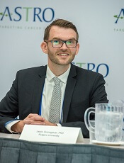
SAN FRANCISCO – A growing number of immunotherapy options for adults with acute lymphocytic leukemia (ALL) – rituximab, inotuzumab ozogamicin, blinatumomab and chimeric antigen receptor (CAR) T-cell therapy – have improved remission rates, but their collective effects on patient outcomes remain to be seen, David Maloney, MD, PhD, said at the National Comprehensive Cancer Network Annual Congress: Hematologic Malignancies.
The main challenge for the field is deciding when and how to use a variety of therapies, he said. “How are we going to put these together? What’s the order?” he asked. “Are we going to be able to decrease the need for allogeneic stem cell transplant? And, obviously, that’s the goal.”
About 30%-50% of adults with ALL exhibit CD20-positive cells, making them potentially treatable with rituximab. Data show a better event-free survival rate and a reduced relapse rate when rituximab is added to standard chemotherapy as compared with standard chemotherapy alone, Dr. Maloney of the clinical research division at the Fred Hutchinson Cancer Research Center, Seattle, noted (N Engl J Med. 2016 Sep 15;375[11]:1044-53). But the improvement was only “modest,” he said.
The anti-CD22 antibody inotuzumab ozogamicin has produced complete remission in 81% of relapsed or refractory ALL patients, compared with those getting standard therapy (N Engl J Med. 2016 Aug 25;375:740-53). Dr. Maloney said it seems well tolerated, but there is concern about an increase in veno-occlusive disease in patients who have undergone or will undergo an allogeneic stem cell transplant.
Blinatumomab produces moderate response rates and minimal residual disease–negative remissions, but delivery of the drug is “cumbersome,” requiring a 4-week continuous infusion, he said. The drug seems to be more effective in those with a lower burden of disease, he noted.
CAR T-cell therapy has produced MRD-negative complete responses in 94% of patients, based on results from a clinical trial at Fred Hutchinson. And using the chemotherapy drug fludarabine in combination with this therapy “dramatically” boosts the peak number of the CAR T cells and how long they persist, Dr. Maloney said. Still, CAR T-cell therapy is a work-intensive treatment requiring cells harvested from the patient, and the procedure often brings on cytokine-release syndrome and neurotoxicity, though both adverse events are typically reversible, he said.
It may be that using fewer CAR T cells can reduce toxicity without compromising treatment response, he said.
Questions remain over whether to transplant patients who are in remission after CAR T-cell therapy. “This is a hot debate,” he said. The decision will likely depend on their prior therapy, whether they’ve had a prior transplant, and the how robust the CAR T-cell expansion has been, he said.
Dr. Maloney reports financial relationships with Celgene, Gilead Sciences, Kite Pharma, and Roche.
EXPERT ANALYSIS FROM THE NCCN ANNUAL CONGRESS: HEMATOLOGIC MALIGNANCIES
Primary care may be inadequate for cancer survivors

Primary care may not meet the healthcare needs of cancer survivors in the US, according to research published in JAMA Internal Medicine.
Researchers examined 12 advanced primary care practices selected from a national registry of “workforce innovators” and found that none of these practices had a comprehensive survivorship care program in place.
In addition, there were 3 main barriers to survivorship care—not treating cancer survivors as a distinct population, limitations of electronic health records, and a lack of information and guidance for clinicians.
“This is troubling because these are highly innovative practices that have a national reputation,” said study author Benjamin Crabtree, PhD, of Rutgers Robert Wood Johnson Medical School in New Brunswick, New Jersey.
Dr Crabtree and his colleagues evaluated survivorship care* at the 12 practices, which were based in Colorado, Illinois, Maine, New York, Pennsylvania, and Washington.
Over nearly 2 years, the team spent 10 to 12 days observing each of the practices and interviewing clinicians and administrators.
In this way, the researchers identified 3 main barriers to integrating survivorship care into primary medicine.
Barrier 1
The first barrier was that clinicians did not treat cancer survivors as a distinct population or clinical category.
“There is no diagnosis code for ‘cancer survivor’ that can be entered into the medical record, which is important if you want physicians to pay attention,” Dr Crabtree said.
Some of the clinicians interviewed said their care was comprehensive enough to address the needs of all patients. Other clinicians did not understand what survivorship care entails.
Barrier 2
The second barrier was that electronic health record systems didn’t support survivorship care.
Clinicians reported an inability to identify patients with a history of cancer. Even if a patient’s cancer history was included in his or her record, it might take searching through multiple screens to find the information.
In addition, medical records were sometimes lost as patients changed clinicians over the years, which left it up to patients to report their cancer histories.
Barrier 3
The third barrier was that clinicians did not receive adequate information or guidance for follow-up care of cancer survivors.
Although some of the practices received cancer-related information about their patients, it was considered “inadequate” or “not actionable.”
Clinicians expressed concerns about their knowledge gaps in cancer care and the need to monitor changing information in oncology.
“There is nothing in the residency curriculum about cancer survivorship,” Dr Crabtree said. “There is also nothing in Continuing Medical Education courses. It’s just not there.”
Dr Crabtree and his colleagues believe these barriers must be addressed so that comprehensive cancer survivorship services can move to the forefront of primary care. ![]()
* Survivorship care includes checking for cancer recurrence, monitoring long-term effects of radiation and chemotherapy, and assessing a patient’s psychological well-being.
Newer blood cancer drugs may not improve OS, QOL

A study of cancer drugs approved by the European Commission from 2009 to 2013 showed that few hematology drugs were known to provide a benefit in overall survival (OS) or quality of life (QOL) over existing treatments.
Of 12 drugs approved for 17 hematology indications, 3 drugs had been shown to provide a benefit in OS (for 3 indications) at the time of approval.
None of the other hematology drugs were known to provide an OS benefit even after a median follow-up of 5.4 years.
Two hematology drugs were shown to provide a benefit in QOL (for 2 indications) after approval, but none of the drugs were known to provide a QOL benefit at the time of approval.
These findings were published in The BMJ alongside a related editorial, feature article, and patient commentary.
All cancer drugs
Researchers analyzed reports on all cancer drug approvals by the European Commission from 2009 to 2013.
There were 48 drugs approved for 68 cancer indications during this period. Fifty-one of the indications were for solid tumor malignancies, and 17 were for hematologic malignancies.
For 24 indications (35%), research had demonstrated a significant improvement in OS at the time of the drugs’ approval. For 3 indications, an improvement in OS was demonstrated after approval.
There was a known improvement in QOL for 7 of the indications (10%) at the time of approval and for 5 indications after approval.
The median follow-up was 5.4 years (range, 3.3 years to 8.1 years).
Overall, there was a significant improvement in OS or QOL during the study period for 51% of the indications (35/68). For the other half (49%, n=33), it wasn’t clear if the drugs provide any benefits in OS or QOL.
All cancer trials
The 68 approvals of cancer drugs were supported by 72 clinical trials.
Sixty approvals (88%) were supported by at least 1 randomized, controlled trial. Eight approvals (12%) were based on a single-arm study. This included 6 of 10 conditional marketing authorizations and 2 of 58 regular marketing authorizations.
Eighteen of the approvals (26%) were supported by a pivotal study powered to evaluate OS as the primary endpoint. And 37 of the approvals (54%) had a supporting pivotal trial evaluating QOL, but results were not reported for 2 of these trials.
Hematology trials and drugs
Of the 12 drugs approved for 17 hematology indications, 4 were regular approvals, 5 were conditional approvals, and 8 had orphan drug designation.
The approvals were supported by data from 18 trials—13 randomized and 5 single-arm trials.
The study drug was compared to an active comparator in 9 of the trials. The drug was evaluated as an add-on treatment in 4 trials. And the drug was not compared to anything in 5 trials (the single-arm trials).
OS was the primary endpoint in 1 of the trials, and 17 trials had OS or QOL as a secondary endpoint.
There were 3 drugs that had demonstrated an OS benefit at the time of approval but no QOL benefit at any time:
- Decitabine used for first-line treatment of acute myeloid leukemia in adults 65 and older who are ineligible for chemotherapy
- Pomalidomide in combination with dexamethasone as third-line therapy for relapsed/refractory multiple myeloma (MM)
- Rituximab plus chemotherapy for first-line treatment of chronic lymphocytic leukemia (CLL).
There were 2 drugs that had demonstrated a QOL benefit, only after approval, but they were not known to provide an OS benefit at any time:
- Nilotinib as a treatment for adults with newly diagnosed, chronic phase, Ph+ chronic myeloid leukemia (CML)
- Ofatumumab for CLL that is refractory to fludarabine and alemtuzumab
For the remaining drugs, there was no evidence of an OS or QOL benefit at any time during the period studied. The drugs included:
- Bortezomib given alone or in combination with doxorubicin or dexamethasone as second-line therapy for MM patients ineligible for hematopoietic stem cell transplant (HSCT)
- Bortezomib plus dexamethasone with or without thalidomide as first-line therapy in MM patients eligible for HSCT
- Bosutinib as second- or third-line treatment of Ph+ CML (any phase)
- Brentuximab vedotin for relapsed or refractory systemic anaplastic large-cell lymphoma
- Brentuximab vedotin for relapsed or refractory, CD30+ Hodgkin lymphoma after autologous HSCT or as third-line treatment for patients ineligible for autologous HSCT
- Dasatinib for first-line treatment of chronic phase, Ph+ CML
- Pixantrone for multiply relapsed or refractory B-cell non-Hodgkin lymphoma
- Ponatinib for patients with Ph+ acute lymphoblastic leukemia who are ineligible for imatinib or have disease that is resistant or intolerant to dasatinib or characterized by T315I mutation
- Ponatinib for patients with any phase of CML who are ineligible for imatinib or have disease that is resistant or intolerant to dasatinib/nilotinib or characterized by T315I mutation
- Rituximab as maintenance after induction for patients with follicular lymphoma
- Rituximab plus chemotherapy for relapsed or refractory CLL
- Temsirolimus for relapsed or refractory mantle cell lymphoma.
Sperm banking may be underused by young cancer patients

New research suggests sperm banking may be underutilized by adolescent and young adult males with cancer who are at risk of infertility.
However, the study also showed that patients were more likely to attempt sperm banking if they were physically mature, met with fertility specialists, or their parents recommended sperm banking.
These findings were published in the Journal of Clinical Oncology.
“Research has found that the majority of males who survive childhood cancer desire biological children,” said study author James Klosky, PhD, of St. Jude Children’s Research Hospital in Memphis, Tennessee.
“Fertility preservation is also associated with a variety of benefits for survivors, including increased optimism about the future. While sperm banking is not for everyone, it is an effective method for preserving male fertility. Yet this study shows that sperm banking remains underutilized by at-risk patients with cancer.”
Dr Klosky and his colleagues surveyed 146 young males with cancer who were at risk of infertility. The researchers also surveyed 144 parents or guardians and 52 oncologists and other healthcare providers.
The patients’ mean age was 16.49 (range, 13.0-21.99). Diagnoses included leukemia and lymphoma (56.2%), solid tumor malignancies (37.7%), and brain tumors (6.2%).
Slightly more than half of the patients (53.4%, n=78) attempted sperm banking prior to starting treatment. Sixty-two, or 82.1%, of those who attempted sperm banking were successful.
In all, 43.8% of the patients successfully banked sperm.
Of the 68 patients who did not attempt sperm banking, 29 reported discussing the option with their families but deciding against it. Twenty-six patients indicated they did not believe sperm banking was necessary, and 9 patients were unsure what it was.
There were several factors that influenced the likelihood of patients making sperm collection attempts as well as successfully banking sperm.
In a multivariable analysis, the following factors were associated with an increased likelihood of attempting to bank sperm:
- Meeting with a fertility specialist (odds ratio[OR]=29.96; 95% CI, 2.48 to 361.41; P=0.007)
- Parent recommending banking (OR=12.30; 95% CI, 2.01 to 75.94; P=0.007)
- Higher Tanner stage (OR=5.42; 95% CI, 1.75 to 16.78; P=0.003).
In another multivariable analysis, successful sperm banking was associated with:
- Patient history of masturbation (OR=5.99; 95% CI, 1.25 to 28.50; P=0.025)
- Higher self-efficacy for banking coordination (OR=1.23; 95% CI, 1.05 to 1.45; P=0.012)
- Medical team member recommending banking (OR=4.26; 95% CI, 1.45 to 12.43; P=0.008)
- Parent recommending banking (OR=4.62; 95% CI, 1.46 to 14.73; P=0.010).
“These results highlight factors that providers can target to empower adolescents to actively participate in their own healthcare,” Dr Klosky said. “These decisions, which are typically made at the time of diagnosis, have high potential to affect their lives as survivors.” ![]()
Team discovers oncogenic driver of T-ALL

Preclinical research suggests the TOX protein is an oncogenic driver of T-cell acute lymphoblastic leukemia (T-ALL).
Results indicate that TOX may be expressed in as many as 95% of human T-ALL cases, and the protein is required for the cancer’s growth and persistence.
“A major role for TOX in T-ALL is to elicit defects in DNA repair, leading to genetic changes that drive normal cells into cancer,” said study author David Langenau, PhD, of Massachusetts General Hospital in Boston.
“TOX then continues to be expressed within leukemic cells and is required for continued tumor growth. That means that, if we can successfully target TOX with small molecules in the future, the 95% of T-ALL patients whose tumors express TOX would have new treatment options for this aggressive leukemia.”
Dr Langenau and his colleagues described this new role for TOX in Cancer Discovery.
The team noted that T-ALL has several molecular subtypes, many of which are driven by common oncogenes such as MYC and NOTCH. However, evidence has suggested the cancer’s initiation is likely driven by aberrations in DNA repair.
To identify genes that might help drive T-ALL, the researchers performed a transgenic screen in zebrafish.
The team found that TOX collaborates with known oncogene pathways to transform T-cell precursors into leukemia cells by altering DNA repair and then expanding the population of transformed cells.
In human T-ALL cells, TOX was shown to suppress non-homologous end joining (NHEJ) repair, a pathway required for repairing double-strand DNA breaks that, when disrupted, is known to cause errant DNA repair and genomic instability.
Nearly all of the human T-ALL samples the researchers tested were found to express TOX. And TOX proved essential for the proliferation and survival of T-ALL.
Dr Langenau explained that TOX is known to have important roles in the development and maturation of several types of immune cells, yet its roles in leukemia initiation and genomic instability were not described until this work.
TOX belongs to a group of proteins known to regulate the configuration or expression of genes by binding to DNA molecules, yet its mechanism in T-ALL—blocking NHEJ repair by binding to DNA repair proteins rather than directly to DNA—was totally unexpected.
The researchers believe that, in addition to better understanding how TOX regulates the continued growth of T-ALL, it will be important to determine whether related proteins have similar molecular functions in other cancers. ![]()
How Abl ‘shape-shifts’ in drug-resistant CML

Researchers say they have determined how the structure of Abl kinase regulates its activity, enabling the enzyme to switch itself on and off.
The team believes these findings will pave the way to new treatment strategies that can overcome drug resistance in chronic myeloid leukemia (CML) and other malignancies.
Charalampos Kalodimos, PhD, of St Jude Children’s Research Hospital in Memphis, Tennessee, and his colleagues described this research in Nature Structural & Molecular Biology.
The researchers sought to understand how Abl manages to switch itself on and off by altering its shape. Abl controls this switching through allosteric regulation, in which a part of the molecule distant from its kinase domain somehow inhibits or activates Abl.
“We knew we had these 2 functional states, but we had no idea about the conditions under which Abl switched from one to another,” Dr Kalodimos said.
“We also didn’t understand how external molecules that regulate Abl acted on these 2 states. Nor did we understand how mutations that confer drug resistance affected the states.”
To investigate, the researchers used NMR spectroscopy to view Abl’s structure and watch the kinase change. The team explored how the region of Abl called the allosteric regulatory module interacted with the kinase domain to control it.
The research revealed that, in its shape-shifting, Abl was precisely balanced between its inhibition and activation states.
“We saw this very fast ‘breathing’ motion of several thousand times a second, in which the molecule goes on and off, on and off,” Dr Kalodimos said. “This motion is important because it allows other molecules that regulate Abl to adjust its activity one way or the other in a graded manner—like turning a rheostat up or down.”
Such regulation would involve pushing the Abl molecule toward either the inhibited or activated state, Dr Kalodimos said.
Newfound activator region
The researchers also discovered new details about how Abl’s structure affects its activation state. For example, the team’s experiments revealed a previously unknown activator region within Abl.
The researchers noted that the Abl regulatory module consists of 5 regions:
- An unstructured N-terminal region called the cap (residues 1–80)
- The SH3 domain (residues 85–138)
- A short linker called the connectorSH3/2 (residues 139–152), which links the SH3 and SH2 domains
- The SH2 domain (residues 153–237)
- A linker (linkerSH2–KD; residues 238–250) that connects SH2 to the kinase domain (residues 255–534).
The previously unknown activator region the researchers identified is part of the cap region comprising residues 14 to 20 (capPxxP), which carries a PxxP sequence motif, a preferred binding site of the SH3 domain.
The team found that capPxxP is an SH3-binding site that can compete with and displace the linkerSH2–KD from the SH3 domain, thereby destabilizing the inhibiting state.
The researchers said they believe the recently reported A19V drug-resistance mutation exerts its function by promoting the activated state of Abl by means of capPxxP.
Implications for treatment
The researchers also analyzed mutations in Bcr-Abl that allow it to become resistant to imatinib. The drug has proven effective in treating CML by plugging into the kinase domain of the over-activated Abl enzyme and shutting it down. However, in many patients, a mutation in the gene that produces Abl renders it drug-resistant.
While many of the mutations block imatinib from plugging into the kinase domain, others appear to interfere with the allosteric regulation. In effect, they may “warp” the enzyme to keep it activated.
In analyzing the structure of these allosteric mutants, Dr Kalodimos and his colleagues discovered the mutants altered Abl’s shape to activate it and did not interfere with how imatinib plugs into the kinase domain.
This finding points the way to new treatments to overcome such resistance, according to Dr Kalodimos.
“There is now a new generation of drugs that bind to the allosteric pocket to inhibit its activity,” he said. “These could be combined with [imatinib] to overcome allosteric mutations to shift Abl into an inhibited state.”
Dr Kalodimos said that treatment strategy could also be applied to other forms of leukemia that have uncontrolled Bcr-Abl activity. And this new basic understanding of Abl regulation will yield insight into similar enzymes in which allosteric regulation controls a kinase domain. ![]()
Doc advocates depression screening for cancer patients
SAN DIEGO—New research suggests a need for mental health screening among cancer patients.
The study revealed a 40% rate of depression among patients treated at an urban cancer center over a 3-year period.
Three-quarters of the depressed patients were previously undiagnosed.
Female patients and those who were unable to work due to disability were more likely to be depressed.
Jason Domogauer, PhD, of Rutgers New Jersey Medical School in Newark, New Jersey, presented these findings at ASTRO’s 59th Annual Meeting.
“Depression is widely recognized as an underdiagnosed disorder, particularly among older adults and cancer patients,” Dr Domogauer said. “Our findings point to a clear need for action, including depression screening during initial and continuing patient visits, initiation of mental health treatments for identified patients, and increased collaboration with mental health providers in cancer treatment centers. These efforts are particularly important for patients in urban centers, those who are female, and those who are unable to work because of their disease.”
Dr Domogauer and his colleagues studied 400 cancer patients who received treatment at Rutgers New Jersey Medical School/University Hospital Cancer Center between 2013 and 2016.
The average patient age was 55 (range, 20-86), and 53% of patients were female. Forty-eight percent of patients were African-American, 29% were non-Hispanic white, and 16% were Hispanic.
Nearly equal numbers of patients reported being able to work (49%) or unable to work due to disability (51%). Most patients (85%) received radiation as part of their cancer treatment.
The researchers assessed depression in the patients using a minimum score of 16 on the Center for Epidemiologic Studies Depression Scale.
In this way, 40% of the patients were diagnosed with depression. In 75% of these patients, depression was previously undiagnosed. This means roughly 30% of the overall patient population suffered from undiagnosed and untreated depression.
Depression was more common among females than males—47% and 32%, respectively (odds ratio [OR]=1.9, P=0.007).
Depression was also more likely among patients who were unable to work due to disability—48%, compared to 33% of those able to work (OR=1.9, P=0.005).
Depression prevalence did not differ significantly among racial/ethnic groups.
When the researchers looked specifically at patients who were previously not diagnosed with depression, the effects of being female or unable to work persisted.
In this subgroup, depression was more common among women than men—43% and 29%, respectively (OR=1.9, P=0.02)—and patients with disability compared to able patients—43% and 31%, respectively (OR=1.7, P=0.03). ![]()
Drugs could improve treatment of CML

Preclinical research suggests 2 drugs already approved for use in the US may improve upon tyrosine kinase inhibitor (TKI) therapy in patients with chronic myeloid leukemia (CML).
The drugs are prostaglandin E1 (PGE1), which is used to treat erectile dysfunction, and misoprostol, which is used to prevent stomach ulcers.
Researchers found that each of these drugs could suppress leukemic stem cells (LSCs) and enhance the activity of imatinib in mice with CML.
Hai-Hui (Howard) Xue, MD, PhD, of University of Iowa in Iowa City, and his colleagues reported these findings in Cell Stem Cell.
“A successful treatment [for CML] is expected to kill the bulk leukemia cells and, at the same time, get rid of the leukemic stem cells,” Dr Xue said. “Potentially, that could lead to a cure.”
Therefore, Dr Xue and his colleagues set out to find drugs that could eradicate LSCs.
The researchers had previously shown that CML LSCs are “strongly dependent” on 2 transcription factors—Tcf1 and Lef1—for self-renewal, whereas normal hematopoietic stem and progenitor cells are not.
With their current research, the team found that Tcf1/Lef1 deficiency “at least partly impairs the transcriptional program” that maintains LSCs in mice and humans with CML.
So the researchers used connectivity maps to identify molecules that could replicate Tcf1/Lef1 deficiency. This screen revealed PGE1.
The team found that PGE1 inhibited the activity and self-renewal of CML LSCs. And the combination of PGE1 and imatinib could reduce leukemia growth in mouse models of CML.
When the mice received no treatment or imatinib alone, LSCs persisted. However, PGE1 enhanced the efficacy of imatinib, and mice that received this combination saw their LSCs “greatly diminished.”
The researchers then transplanted LSCs from these mice into secondary hosts and monitored their survival without administering additional treatment.
Mice that received PGE1-pretreated LSCs lived significantly longer (P<0.001) than mice that received imatinib-pretreated LSCs. And mice that received LSCs pretreated with PGE1 and imatinib lived significantly longer than mice that received PGE1-pretreated LSCs (P=0.039).
Investigating how PGE1 works to suppress LSCs, the researchers found the effect relies on a critical interaction between PGE1 and its receptor, EP4.
So the team tested misoprostol, which also interacts with EP4, in mice with CML.
The researchers found that misoprostol alone diminished LSCs, and the combination of misoprostol and imatinib “exhibited stronger effects.”
In addition, mice that received LSCs from animals previously treated with misoprostol survived longer and had a reduction in leukemia burden compared to mice that received untreated LSCs.
“We would like to be able to test these compounds in a clinical trial,” Dr Xue said. “If we could show that the combination of TKI with PGE1 or misoprostol can eliminate both the bulk tumor cells and the stem cells that keep the tumor going, that could potentially eliminate the cancer to the point where a patient would no longer need to depend on TKI.” ![]()
Preclinical research suggests 2 drugs already approved for use in the US may improve upon tyrosine kinase inhibitor (TKI) therapy in patients with chronic myeloid leukemia (CML).
The drugs are prostaglandin E1 (PGE1), which is used to treat erectile dysfunction, and misoprostol, which is used to prevent stomach ulcers.
Researchers found that each of these drugs could suppress leukemic stem cells (LSCs) and enhance the activity of imatinib in mice with CML.
Hai-Hui (Howard) Xue, MD, PhD, of University of Iowa in Iowa City, and his colleagues reported these findings in Cell Stem Cell.
“A successful treatment [for CML] is expected to kill the bulk leukemia cells and, at the same time, get rid of the leukemic stem cells,” Dr Xue said. “Potentially, that could lead to a cure.”
Therefore, Dr Xue and his colleagues set out to find drugs that could eradicate LSCs.
The researchers had previously shown that CML LSCs are “strongly dependent” on 2 transcription factors—Tcf1 and Lef1—for self-renewal, whereas normal hematopoietic stem and progenitor cells are not.
With their current research, the team found that Tcf1/Lef1 deficiency “at least partly impairs the transcriptional program” that maintains LSCs in mice and humans with CML.
So the researchers used connectivity maps to identify molecules that could replicate Tcf1/Lef1 deficiency. This screen revealed PGE1.
The team found that PGE1 inhibited the activity and self-renewal of CML LSCs. And the combination of PGE1 and imatinib could reduce leukemia growth in mouse models of CML.
When the mice received no treatment or imatinib alone, LSCs persisted. However, PGE1 enhanced the efficacy of imatinib, and mice that received this combination saw their LSCs “greatly diminished.”
The researchers then transplanted LSCs from these mice into secondary hosts and monitored their survival without administering additional treatment.
Mice that received PGE1-pretreated LSCs lived significantly longer (P<0.001) than mice that received imatinib-pretreated LSCs. And mice that received LSCs pretreated with PGE1 and imatinib lived significantly longer than mice that received PGE1-pretreated LSCs (P=0.039).
Investigating how PGE1 works to suppress LSCs, the researchers found the effect relies on a critical interaction between PGE1 and its receptor, EP4.
So the team tested misoprostol, which also interacts with EP4, in mice with CML.
The researchers found that misoprostol alone diminished LSCs, and the combination of misoprostol and imatinib “exhibited stronger effects.”
In addition, mice that received LSCs from animals previously treated with misoprostol survived longer and had a reduction in leukemia burden compared to mice that received untreated LSCs.
“We would like to be able to test these compounds in a clinical trial,” Dr Xue said. “If we could show that the combination of TKI with PGE1 or misoprostol can eliminate both the bulk tumor cells and the stem cells that keep the tumor going, that could potentially eliminate the cancer to the point where a patient would no longer need to depend on TKI.”
Mutations impact outcomes in AML, MDS
Researchers say they have identified genetic mutations that can significantly affect treatment outcomes in patients with acute myeloid leukemia (AML) and myelodysplastic syndromes (MDS).
The findings come from a clinical trial in which the team examined whether combining vorinostat with azacitidine could improve survival in patients with AML and MDS.
The results showed no additional benefit with the combination, when compared to azacitidine alone.
However, researchers did find that patients had significantly shorter survival times if they had mutations in CDKN2A, IDH1, or TP53.
“This important trial . . . has rapidly answered the important question of whether combining azacitidine with vorinostat improves outcomes for people with AML and MDS and emphasizes the need for further studies with new drug partners for azacitidine,” said Charles Craddock, DPhil, of the Queen Elizabeth Hospital in Birmingham, UK.
“Importantly, the linked molecular studies have shed new light on which people will benefit most from azacitidine. Furthermore, discovering that the CDKN2A gene mutation affects treatment response may be hugely valuable in helping doctors to design new treatment combinations in the future.”
Dr Craddock and his colleagues reported their discoveries in Clinical Cancer Research.
Previous, smaller trials had suggested that adding vorinostat to treatment with azacitidine could improve outcomes for patients with AML and MDS.
To test this idea, Dr Craddock and his colleagues enrolled 259 patients in the current trial. Most of these patients (n=217) had AML—111 were newly diagnosed, 73 had relapsed AML, and 33 had refractory disease.
The remaining 42 patients had MDS—36 were newly diagnosed, 5 had relapsed MDS, and 1 had refractory disease.
Half of patients (n=130) received azacitidine and vorinostat, and the other half received azacitidine alone (n=129).
In both arms, azacitidine was given at 75 mg/m2 on a 5-2-2 schedule, beginning on day 1 of a 28-day cycle for up to 6 cycles. In the combination arm, patients also received vorinostat at 300 mg twice daily for 7 consecutive days, beginning on day 3 of each cycle.
Results
The combination did not significantly improve response rates or survival times.
The overall response rate was 41% in the azacitidine arm and 42% in the combination arm (odds ratio [OR]=1.05, P=0.84).
The rate of compete response (CR)/CR with incomplete count recovery/marrow CR was 22% in the azacitidine arm and 26% in the combination arm (OR=0.82, P=0.49).
The median overall survival (OS) was 9.6 months in the azacitidine arm and 11.0 months in the combination arm (hazard ratio[HR]=1.15, P=0.32).
Impact of mutations
In a multivariable analysis adjusted for all clinical variables, mutations in NPM1 were associated with reduced overall response (OR=8.6, P=0.012).
In another multivariate analysis, mutations in CDKN2A, IDH1, and TP53 were associated with decreased OS. The HRs were 10.0 (P<0.001), 3.6 (P=0.001), and 4.7 (P<0.001), respectively.
The median OS was 4.5 months in patients with CDKN2A mutations and 11.0 months in patients without them.
The median OS was 7.6 months in patients with TP53 mutations and 11.3 months in patients without them.
And the median OS was 5.6 months in patients with IDH1 mutations and 11.1 months in patients without them.
The researchers believe that testing patients newly diagnosed with AML and MDS for CDKN2A, IDH1, and TP53 mutations could help doctors tailor treatment for patients who are less likely to do well.
The team also said the information gleaned from this trial will guide the choice of new drug partners with the potential to increase azacitidine’s clinical activity.
Representation in cancer clinical trials

ATLANTA—New research suggests some racial/ethnic minority groups are underrepresented in clinical trials for cancer patients in the US.
African-American and Hispanic patients were underrepresented in the trials studied, while Asian and non-Hispanic white patients were not.
Patients belonging to other racial/ethnic groups were not studied in detail.
The research also showed that elderly patients were less likely than other age groups to enroll in a cancer trial.
However, the percentage of elderly patients in the trials studied (36%) was more than double the percentage of elderly individuals in the US population (15.2%).
This research was presented at the 10th AACR Conference on The Science of Cancer Health Disparities in Racial/Ethnic Minorities and the Medically Underserved (abstract A26).
“Clinical trials are crucial in studying the effectiveness of new drugs and ultimately bringing them to the market to benefit patients,” said Narjust Duma, MD, of the Mayo Clinic in Rochester, Minnesota.
“However, many clinical trials lack appropriate representation of certain patient populations. As a result, the findings of a clinical trial might not be generalizable to all patients.”
Dr Duma and her colleagues analyzed enrollment data from all cancer therapeutic trials reported as completed on clinicaltrials.gov from 2003 to 2016. These trials included 55,689 subjects, and the racial/ethnic breakdown of the group was as follows:
- Non-Hispanic white—83%
- African-American—6%
- Asian—5.3%
- Hispanic—2.6%
- “Other”—2.4%.
According to the US Census Bureau, as of July 1, 2016, the estimated total US population was 323,127,516. The racial/ethnic breakdown of that population is as follows:
- White alone (excluding Hispanics/Latinos)—61.3%
- Hispanic/Latino*—17.8%
- Black/African-American alone—13.3%
- Asian alone—5.7%
- American Indian/Alaska Native—1.3%
- Native Hawaiian/Other Pacific Islander—0.2%
- Two or more races—2.6%.
Dr Duma and her colleagues said their study suggests African-American and Hispanic representation in cancer trials has declined in recent years, when compared to historical data from 1996 to 2002.
In the 1996-2002 period, African-Americans represented 9.2% of patients in cancer trials (vs 6% in 2003-2016), and Hispanics represented 3.1% (vs 2.6% in 2003-2016).
On the other hand, the recruitment of Asians in cancer trials has more than doubled, from 2% in the historical data to 5.3% in the current data.
The current study also showed that elderly patients (age 65 and older) represented 36% of the subjects enrolled in cancer trials. In comparison, 15.2% of the total US population is 65 or older.
Previous research suggested the elderly are often underrepresented in clinical trials, despite the fact that most cancer cases are diagnosed in individuals age 65 and older, according to the National Cancer Institute’s Surveillance, Epidemiology and End Results database.
Dr Duma said the increasing use of genetic information in clinical trials may be decreasing the numbers of ethnic minorities and elderly patients. In recent years, researchers have sought to study drugs that treat cancers by targeting certain mutations. In order to identify the patients who are most likely to respond to the drugs, many trials now require molecular testing of tumors.
“This is leading to significant advances,” Dr Duma said. “However, it is vastly more expensive to run these trials, often leaving a limited budget to recruit patients or do outreach to the elderly or minorities.”
“Also, this type of testing can only be conducted at the major cancer centers. The mid-sized, regional hospitals are excluded because they don’t have the capacity, and, sadly, this leaves us farther away from these populations.”
Dr Duma added that cultural biases may also make minorities less likely to enroll in clinical trials. Previous research has indicated that members of certain minority groups may be less likely to trust healthcare providers.
Language barriers may also be a factor for minority patients, and the elderly may be dissuaded by difficulty in traveling to and from major cancer centers, Dr Duma noted.
She identified a few potential ways to narrow the gap of participation in clinical trials:
- Increase clinical trial partnerships between major cancer centers and satellite hospitals. Dr Duma suggested that patients could be enrolled at their local hospital and undergo treatment there, while data could be sent to the partnering cancer center.
- Targeted interventions, such as Spanish interpreters, could be used to help enroll minority patients in clinical trials.
- Healthcare providers should be mindful of the need to enroll more patients from underrepresented populations and should be willing to discuss risks and benefits with patients.
Dr Duma said the main limitation of this study is that race and ethnicity are generally self-reported, which could lead to some inconsistencies in data.
*The US Census Bureau notes that Hispanics may be of any race, so they are also included in applicable race categories.