User login
Cryotherapy can reduce signs of CIPN

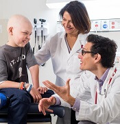
A new study suggests cryotherapy can reduce symptoms of chemotherapy-induced peripheral neuropathy (CIPN).
Researchers found that having chemotherapy patients wear frozen gloves and socks for 90-minute periods significantly reduced the incidence of CIPN symptoms.
Hiroshi Ishiguro, MD, PhD, of International University of Health and Welfare Hospital in Tochigi, Japan, and colleagues reported these findings in the Journal of the National Cancer Institute.
The researchers prospectively evaluated the efficacy of cryotherapy for preventing CIPN. Breast cancer patients treated weekly with paclitaxel (80 mg/m2 for 1 hour) wore frozen gloves and socks on one side of their bodies for 90 minutes, including the entire duration of drug infusion.
The researchers then compared symptoms on the treated sides with those on the untreated sides.
The primary endpoint was CIPN incidence assessed by changes in tactile sensitivity from a pretreatment baseline. The researchers also assessed subjective symptoms, as reported in the Patient Neuropathy Questionnaire, and patients' manual dexterity.
Among the 40 patients studied, 4 did not reach the cumulative dose due to the occurrence of pneumonia, severe fatigue, liver dysfunction, and macular edema. Of the 36 remaining patients, none dropped out due to cold intolerance.
The incidence of objective and subjective signs of CIPN was clinically and statistically significantly lower on the intervention side than on the control side for most measurements, which includes (among other measures):
- Hand tactile sensitivity—27.8% and 80.6%, respectively (odds ratio[OR]= 20.00, P<0.001)
- Foot tactile sensitivity—25.0% and 63.9%, respectively (OR=infinite, P<0.001)
- Hand warm sense—8.8% and 32.4%, respectively (OR=9.00, P=0.02)
- Foot warm sense—33.4% and 57.6%, respectively (OR=5.00, P=0.04)
- Hand cold sense—2.8% and 13.9%, respectively (OR=infinite, P=0.13)
- Foot cold sense—12.6% and 18.8%, respectively (OR=2.00, P=0.69)
- Severe CIPN in the hand according to the Patient Neuropathy Questionnaire—2.8% and 41.7%, respectively (OR=infinite, P<0.001)
- Severe CIPN in the foot according to the Patient Neuropathy Questionnaire—2.8% and 36.1%, respectively (OR=infinite, P<0.001).
Natural selection opportunities tied to cancer rates

Countries with the lowest opportunities for natural selection have higher cancer rates than countries with the highest opportunities for natural selection, according to a study published in Evolutionary Applications.
Researchers said this is because modern medicine is enabling people to survive cancers, and their genetic backgrounds are passing from one generation to the next.
The team said the rate of some cancers has doubled and even quadrupled over the past 100 to 150 years, and human evolution has moved away from “survival of the fittest.”
“Modern medicine has enabled the human species to live much longer than would otherwise be expected in the natural world,” said study author Maciej Henneberg, PhD, DSc, of the University of Adelaide in South Australia.
“Besides the obvious benefits that modern medicine gives, it also brings with it an unexpected side-effect—allowing genetic material to be passed from one generation to the next that predisposes people to have poor health, such as type 1 diabetes or cancer.”
“Because of the quality of our healthcare in western society, we have almost removed natural selection as the ‘janitor of the gene pool.’ Unfortunately, the accumulation of genetic mutations over time and across multiple generations is like a delayed death sentence.”
Country comparison
The researchers studied global cancer data from the World Health Organization as well as other health and socioeconomic data from the United Nations and the World Bank of 173 countries. The team compared the top 10 countries with the highest opportunities for natural selection to the 10 countries with the lowest opportunities for natural selection.
“We looked at countries that offered the greatest opportunity to survive cancer compared with those that didn’t,” said study author Wenpeng You, a PhD student at the University of Adelaide. “This does not only take into account factors such as socioeconomic status, urbanization, and quality of medical services but also low mortality and fertility rates, which are the 2 distinguishing features in the ‘better’ world.”
“Countries with low mortality rates may allow more people with cancer genetic background to reproduce and pass cancer genes/mutations to the next generation. Meanwhile, low fertility rates in these countries may not be able to have diverse biological variations to provide the opportunity for selecting a naturally fit population—for example, people without or with less cancer genetic background. Low mortality rate and low fertility rate in the ‘better’ world may have formed a self-reinforcing cycle which has accumulated cancer genetic background at a greater rate than previously thought.”
Based on the researchers’ analysis, the 20 countries are:
| Lowest opportunities for natural selection | Highest opportunities for natural selection |
| Iceland | Burkina Faso |
| Singapore | Chad |
| Japan | Central African Republic |
| Switzerland | Afghanistan |
| Sweden | Somalia |
| Luxembourg | Sierra Leone |
| Germany | Democratic Republic of the Congo |
| Italy | Guinea-Bissau |
| Cyprus | Burundi |
| Andorra | Cameroon |
Cancer incidence
The researchers found the rates of most cancers were higher in the 10 countries with the lowest opportunities for natural selection. The incidence of all cancers was 2.326 times higher in the low-opportunity countries than the high-opportunity ones.
The increased incidences of hematologic malignancies were as follows:
- Non-Hodgkin lymphoma—2.019 times higher in the low-opportunity countries
- Hodgkin lymphoma—3.314 times higher in the low-opportunity countries
- Leukemia—3.574 times higher in the low-opportunity countries
- Multiple myeloma—4.257 times higher in the low-opportunity countries .
Dr Henneberg said that, having removed natural selection as the “janitor of the gene pool,” our modern society is faced with a controversial issue.
“It may be that the only way humankind can be rid of cancer once and for all is through genetic engineering—to repair our genes and take cancer out of the equation,” he said. ![]()
Study supports prophylaxis in kids with ALL
Results of an observational study support targeted antibacterial prophylaxis in children undergoing induction therapy for acute lymphoblastic leukemia (ALL).
Prophylaxis effectively prevented febrile neutropenia and systemic infection in the children studied.
Prophylaxis with the drug levofloxacin reduced the use of treatment antibiotics and the incidence of Clostridium difficile infection.
“This research provides the first major evidence supporting targeted use of antibacterial prophylaxis for at-risk pediatric ALL patients, particularly use of the broad-spectrum antibiotic levofloxacin,” said study author Joshua Wolf, MD, of St. Jude Children’s Research Hospital in Memphis, Tennessee.
“Prophylactic antibiotic therapy with levofloxacin is routine for at-risk adult ALL patients, but it has remained controversial in children. Until this study, evidence supporting the safety and efficacy of prophylactic antibiotic therapy in children with ALL has been sparse.”
Dr Wolf and his colleagues described their study in Clinical Infectious Diseases.
The study included 344 patients newly diagnosed with ALL who were enrolled in the St. Jude Total XVI clinical trial (NCT00549848). Patients were enrolled from 2007 to 2016.
Until July 2014, the patients received prophylactic antibiotic therapy at the discretion of their physicians. Patients typically received cefepime, ciprofloxacin, or vancomycin plus cefepime or ciprofloxacin. And prophylaxis was typically started at the onset of neutropenia after chemotherapy.
Beginning in August 2014, hospital treatment guidelines changed to recommend prophylactic levofloxacin during induction for ALL patients who develop neutropenia expected to last at least 7 days.
Dr Wolf and his colleagues used the change to compare infection rates and other questions in the following patient groups.
| Patient characteristics | No prophylaxis (n=173) | Levofloxacin prophylaxis (n=69) | Other prophylaxis (n=102) |
| Median age in years (range) | 5.8 (3-11.9) | 6.8 (3.9-11.1) | 7 (3.6-11.9) |
| B-ALL | 83% | 78% | 79% |
| Low-risk ALL | 51% | 54% | 50% |
| Standard-risk ALL | 47% | 41% | 42% |
| High-risk ALL | 2% | 6% | 8% |
| Median duration of neutropenia in days (range) | 17 (11-24) | 18 (12-23) | 20 (17-25) |
| Median duration of profound neutropenia in days (range) | 6 (2-13) | 7 (4-12) | 11 (5-16) |
Results
Researchers reported that patients with neutropenia who received any prophylactic therapy were far less likely than those who did not to develop fever, documented or likely infections, or bloodstream infections.
In a multivariate analysis, the adjusted odds ratios in patients who received prophylaxis, compared to those who did not, were as follows.
- Febrile neutropenia—0.23, P<0.001
- Febrile neutropenia with clinically documented infection—0.30, P=0.002
- Febrile neutropenia with microbiologically documented infection—0.25, P<0.001
- Clinically documented infection—0.54, P=0.02
- Microbiologically documented infection—0.40, P<0.001
- Bloodstream infection—0.30, P=0.008
- C difficile infection—0.38, P=0.04
- Likely bacterial infection—0.26, P<0.001
- Any enterocolitis—0.44, P=0.03.
Analysis also revealed that patients who received levofloxacin had a greater reduction in C difficile infection than patients who received other prophylaxis. The adjusted odds ratio was 0.04 (P<0.001).
However, there was no significant difference between the prophylaxis groups when it came to other infections.
Patients who received levofloxacin prophylaxis had significantly less exposure to other antibiotics than patients who received other prophylaxis or no prophylaxis.
This included exposure to cefepime/ceftazidime (P<0.001 for both comparisons), vancomycin (P<0.001 for both), meropenem (P<0.001 for both), and aminoglycosides (P=0.002 for no prophylaxis, P=0.04 for other prophylaxis).
The reduction in exposure to other antibiotics may partly explain why C difficile infections declined in levofloxacin-treated patients, Dr Wolf said.
He also noted that antibiotic resistance did not significantly increase in this study, despite the greater use of levofloxacin to prevent infections.
“We are cautiously optimistic that any impact of levofloxacin on antibacterial resistance will be balanced by the reduction in use of other antibiotics,” Dr Wolf said, “but long-term monitoring of antibiotic resistance patterns in young ALL patients will be needed to prove this.” ![]()
NCCN completes resource on radiation therapy

The National Comprehensive Cancer Network® (NCCN) has announced the release of the newly completed NCCN Radiation Therapy Compendium™.
This resource includes information designed to support clinical decision-making regarding the use of radiation therapy in cancer patients.
The content is based on the NCCN Clinical Practice Guidelines in Oncology and includes information from the 41 guidelines that reference radiation therapy.
“By compiling every recommendation for radiation therapy in one place, we’ve made it significantly easier for specialists . . . to stay up-to-date on the very latest recommendations, regardless of how many different cancer types they treat,” said Robert W. Carlson, MD, chief executive officer of NCCN.
“This targeted content provides radiation oncologists with the specific, cutting-edge information they need, without forcing them to sift through any extraneous information. It’s part of our ongoing effort to always provide the most pertinent data on emerging treatment practices in the clearest, most efficient way possible.”
The NCCN Radiation Therapy Compendium includes a full complement of radiation therapy recommendations found in the current NCCN guidelines, including specific treatment modalities such as 2D/3D conformal external beam radiation therapy, intensity modulated radiation therapy, intra-operative radiation therapy, stereotactic radiosurgery/stereotactic body radiotherapy/stereotactic ablative body radiotherapy, image-guided radiation therapy, low dose-rate/high dose-rate brachytherapy, radioisotope, and particle therapy.
NCCN first announced the launch of the Radiation Therapy Compendium in March at the NCCN Annual Conference: Improving the Quality, Effectiveness, and Efficiency of Cancer Care.
At the time, the NCCN released a preliminary version of the compendium featuring 24 cancer types. The newly completed version now contains all 41 disease sites that are currently being treated using radiation therapy.
The compendium will be updated on a continual basis in conjunction with the library of clinical guidelines.
For more information and to access the NCCN Radiation Therapy Compendium, visit NCCN.org/RTCompendium. The compendium is available free-of-charge through March 2018. ![]()
Minimal residual disease measures not yet impactful for AML patients
SAN FRANCISCO – Routine testing for minimal residual disease is probably not of value in acute myeloid leukemia, as there is no evidence that changing treatment based on MRD status currently makes a difference in patient outcomes, experts said at the annual congress on Hematologic Malignancies held by the National Comprehensive Cancer Network.
“If we find minimal residual disease, we don’t always have a better therapy to offer our patients,” Jessica Altman, MD, associate professor of hematology and oncology at the Northwestern University Feinberg School of Medicine, said.
Beyond that therapeutic reality, there are no clear guidelines and standards for MRD testing. The optimal timing for MRD testing and a standard threshold for an MRD classification are not yet established, she said.
“Having MRD is bad, not having it is better,” Richard Stone, MD, PhD, clinical director of the adult leukemia program at the Dana-Farber Cancer Institute, said. The problem in AML, he said, is, “So?” There is no reliable “MRD eraser” in AML, he said. Until then, there is not much point in knowing whether a patient is MRD positive or not.
A recent survey conducted by researchers at Moffitt Cancer Center, Tampa, addressed MRD testing at 13 major cancer centers. While most centers reported that they test for MRD, many physicians said that they are unsure about what to do with the results.
A 2013 study by the HOVON group found that patients who were in complete remission but MRD positive after their first course of therapy, subsequently became MRD negative after their second course of therapy. But the second regimen would not have been different based on knowledge of MRD status, according to the HOVON/SAKK AML 42A study (J Clin Oncol. 2013; 31:3889-97).
The AML community is awaiting guidelines on MRD use from the NCCN and other groups, Dr. Altman said. An option for using NPM1 mutations to assess MRD should be available soon, and could be an improvement on existing options (N Engl J Med 2016; 374:422-33).
Given the treatment limitations, knowing about MRD status can have a negative mental toll on patients, Dr. Stone said. “I would not underplay the psychological burden.” Nevertheless, MRD should be measured in clinical trials, and it could be a valuable surrogate marker by which to compare drug efficacy.
One of the biggest hopes is that MRD status could eventually be useful in determining the need for allogeneic stem cell transplant in patients deemed intermediate risk, Dr. Altman said. “I think we are finally on the brink of this being actionable.”
Dr. Altman reports financial relationships with Astellas, Bristol-Myers Squibb, Celgene, Janssen, Novartis, and Syros. Dr. Stone reports financial relationships with AbbVie, Actinium, Agios, Amgen and many other companies.
EXPERT ANALYSIS AT NCCN HEMATOLOGIC MALIGNANCIES CONGRESS
Predicting neurotoxicity after CAR T-cell therapy

Researchers say they have identified potential biomarkers that may be used to help identify patients at an increased risk of neurotoxicity after chimeric antigen receptor (CAR) T-cell therapy.
The team also created an algorithm intended to identify patients whose symptoms were most likely to be life-threatening.
The researchers discovered the biomarkers and developed the algorithm based on data from a trial of JCAR014, an anti-CD19 CAR T-cell therapy, in patients with B-cell malignancies.
Cameron J. Turtle, MBBS, PhD, of Fred Hutchinson Cancer Research Center in Seattle, Washington, and his colleagues described this research in Cancer Discovery.
“It’s essential that we understand the potential side effects of CAR T therapies” Dr Turtle said. “While use of these cell therapies is likely to dramatically increase because they’ve been so effective in patients with resistant or refractory B-cell malignancies, there is still much to learn.”
Dr Turtle and his colleagues sought to provide a detailed clinical, radiological, and pathological characterization of neurotoxicity arising from anti-CD19 CAR T-cell therapy.
So the team analyzed data from a phase 1/2 trial of 133 adults with relapsed and/or refractory CD19+ B-cell acute lymphoblastic leukemia, non-Hodgkin lymphoma, or chronic lymphocytic leukemia.
The patients received lymphodepleting chemotherapy followed by an infusion of JCAR014.
Neurotoxicity
Within 28 days of treatment, 53 patients (40%) developed grade 1 or higher neurologic adverse events (AEs), 28 patients (21%) had grade 3 or higher neurotoxicity, and 4 patients (3%) developed fatal neurotoxicity.
Of the 53 patients with any neurologic AE, 48 (91%) also had cytokine release syndrome (CRS). All neurologic AEs in the 5 patients who did not have CRS were mild (grade 1) and transient.
Neurologic AEs included delirium with preserved alertness (66%), headache (55%), language disturbance (34%), decreased level of consciousness (25%), seizures (8%), and macroscopic intracranial hemorrhage (2%).
For most patients, neurotoxicity resolved by day 28 after CAR T-cell infusion. The exceptions were 1 patient in whom a grade 1 neurologic AE resolved 2 months after CAR T-cell infusion and the 4 patients who died of neurotoxicity.
The 4 neurotoxicity-related deaths were due to:
- Acute cerebral edema (n=2)
- Multifocal brainstem hemorrhage and edema associated with disseminated intravascular coagulation (n=1)
- Cortical laminar necrosis with a persistent minimally conscious state until death (n=1).
Potential biomarkers
In a univariate analysis, neurotoxicity was significantly more frequent in patients who:
- Had CRS (P<0.0001)
- Received a high CAR T-cell dose (P<0.0001)
- Had pre-existing neurologic comorbidities at baseline (P=0.0059).
In a multivariable analysis (which did not include CRS as a variable), patients had an increased risk of neurotoxicity if they:
- Had pre-existing neurologic comorbidities (P=0.0023)
- Received cyclophosphamide and fludarabine lymphodepletion (P=0.0259)
- Received a higher CAR T-cell dose (P=0.0009)
- Had a higher burden of malignant CD19+ B cells in the bone marrow (P=0.0165).
The researchers noted that patients who developed grade 3 or higher neurotoxicity had more severe CRS (P<0.0001).
“It appears that cytokine release syndrome is probably necessary for most cases of severe neurotoxicity, but, in terms of what triggers a person with cytokine release syndrome to get neurotoxicity, that’s something we need to investigate further,” said study author Kevin Hay, MD, of Fred Hutchinson Cancer Research Center.
Dr Hay and his colleagues also found that patients with severe neurotoxicity exhibited evidence of endothelial activation, which could contribute to manifestations such as capillary leak, disseminated intravascular coagulation, and disruption of the blood-brain barrier.
Algorithm
The researchers developed a predictive classification tree algorithm to identify patients who have an increased risk of severe neurotoxicity.
The algorithm suggests patients who meet the following criteria in the first 36 hours after CAR T-cell infusion have a high risk of grade 4-5 neurotoxicity:
- Fever of 38.9°C or greater
- Serum levels of IL6 at 16 pg/mL or higher
- Serum levels of MCP1 at 1343.5 pg/mL or higher.
This algorithm predicted severe neurotoxicity with 100% sensitivity and 94% specificity. Eight patients were misclassified, 1 of whom did not subsequently develop grade 2-3 neurotoxicity and/or grade 2 or higher CRS.
Funding
This research was funded by Juno Therapeutics Inc. (the company developing JCAR014), the National Cancer Institute, Life Science Discovery Fund, the Bezos family, the University of British Columbia Clinical Investigator Program, and via institutional funds from Bloodworks Northwest.
Dr Turtle receives research funding from Juno Therapeutics, holds patents licensed by Juno, and has pending patent applications that could be licensed by nonprofit institutions and for-profit companies, including Juno.
The Fred Hutchinson Cancer Research Center has a financial interest in Juno and receives licensing and other payments from the company. ![]()
FDA grants drug fast track designation for rel/ref AML

The US Food and Drug Administration (FDA) has granted fast track designation to gilteritinib for the treatment of adults with FLT3 mutation-positive relapsed or refractory acute myeloid leukemia (AML).
Gilteritinib is a compound that has demonstrated inhibitory activity against FLT3 internal tandem duplication (ITD) and FLT3 tyrosine kinase domain, 2 mutations that are seen in approximately one-third of patients with AML.
Gilteritinib has also demonstrated inhibition of AXL, which is reported to be associated with therapeutic resistance.
Astellas Pharma Inc. is currently investigating gilteritinib in phase 3 trials of AML patients.
Results from a phase 1/2 study of gilteritinib in AML were presented at the 2017 ASCO Annual Meeting.
The goal of the study was to determine the tolerability and antileukemic activity of once-daily gilteritinib in a FLT3-ITD-enriched, relapsed/refractory AML population.
Researchers said the drug exhibited potent FLT3 inhibition at doses greater than 80 mg/day. In patients who received such doses, the greatest overall response rate was 52%, and the longest median overall survival was 31 weeks.
The maximum tolerated dose of gilteritinib was 300 mg/day. Dose-limiting toxicities included diarrhea and liver function abnormalities.
About fast track designation
The FDA’s fast track program is designed to facilitate the development and expedite the review of products intended to treat or prevent serious or life-threatening conditions and address unmet medical need.
Through the fast track program, a product may be eligible for priority review. In addition, the company developing the product may be allowed to submit sections of the new drug application or biologics license application on a rolling basis as data become available.
Fast track designation also provides the company with opportunities for more frequent meetings and written communications with the FDA.
About orphan designation
Gilteritinib also has orphan drug designation for the treatment of AML.
The FDA grants orphan designation to products intended to treat, diagnose, or prevent diseases/disorders that affect fewer than 200,000 people in the US.
The designation provides incentives for sponsors to develop products for rare diseases. This may include tax credits toward the cost of clinical trials, prescription drug user fee waivers, and 7 years of market exclusivity if the product is approved. ![]()
AML trial placed on full clinical hold

The US Food and Drug Administration (FDA) has placed a full clinical hold on a phase 1/2 trial of SEL24, a dual PIM/FLT3 kinase inhibitor, in patients with relapsed/refractory acute myeloid leukemia (AML).
The hold is due to a fatal cerebral adverse event that is considered possibly related to SEL24.
The clinical hold means no new patients will be enrolled in the trial and enrolled patients will not receive SEL24 until the hold is lifted.
Selvita S.A., the company developing SEL24, received a clinical hold letter from the FDA on October 6 and said it plans to work with the agency to have the hold lifted.
As part of this process, Selvita will provide the FDA with additional data and analysis on patients treated with SEL24 as well as a proposed protocol amendment.
The trial began in the first quarter of 2017. The study is designed to determine the maximum tolerated dose and recommended dose of SEL24 in patients with relapsed and refractory AML. The study began with a 25 mg daily dose, which was then escalated following cohort reviews.
One AML patient started treatment with a 150 mg dose of SEL24 as the third patient in this dose cohort and received 4 doses of the drug. This patient developed a life-threatening, grade 4 venous thrombus in the brain with subsequent intracerebral hemorrhage, which required hospitalization.
The patient died in hospice 4 days later due to the cerebral event. The patient’s death was subsequently evaluated as possibly related to SEL24.
A safety report and a review of data by the trial’s data monitoring committee were submitted to the FDA. The agency then placed a clinical hold on the trial and requested more safety data on patients who have received SEL24, as well as specific protocol changes and additional guidance to the study staff.
Selvita said it plans to comply with the requests and provide additional information to the agency and clinical trial centers, in collaboration with the Menarini Group, its global development partner for SEL24.
The FDA has 30 days from the receipt of Selvita’s response to let the company know whether the clinical hold is lifted. ![]()
The US Food and Drug Administration (FDA) has placed a full clinical hold on a phase 1/2 trial of SEL24, a dual PIM/FLT3 kinase inhibitor, in patients with relapsed/refractory acute myeloid leukemia (AML).
The hold is due to a fatal cerebral adverse event that is considered possibly related to SEL24.
The clinical hold means no new patients will be enrolled in the trial and enrolled patients will not receive SEL24 until the hold is lifted.
Selvita S.A., the company developing SEL24, received a clinical hold letter from the FDA on October 6 and said it plans to work with the agency to have the hold lifted.
As part of this process, Selvita will provide the FDA with additional data and analysis on patients treated with SEL24 as well as a proposed protocol amendment.
The trial began in the first quarter of 2017. The study is designed to determine the maximum tolerated dose and recommended dose of SEL24 in patients with relapsed and refractory AML. The study began with a 25 mg daily dose, which was then escalated following cohort reviews.
One AML patient started treatment with a 150 mg dose of SEL24 as the third patient in this dose cohort and received 4 doses of the drug. This patient developed a life-threatening, grade 4 venous thrombus in the brain with subsequent intracerebral hemorrhage, which required hospitalization.
The patient died in hospice 4 days later due to the cerebral event. The patient’s death was subsequently evaluated as possibly related to SEL24.
A safety report and a review of data by the trial’s data monitoring committee were submitted to the FDA. The agency then placed a clinical hold on the trial and requested more safety data on patients who have received SEL24, as well as specific protocol changes and additional guidance to the study staff.
Selvita said it plans to comply with the requests and provide additional information to the agency and clinical trial centers, in collaboration with the Menarini Group, its global development partner for SEL24.
The FDA has 30 days from the receipt of Selvita’s response to let the company know whether the clinical hold is lifted.
FDA rejects pegfilgrastim biosimilar

The US Food and Drug Administration (FDA) has issued a complete response letter saying the agency cannot approve MYL-1401H, a proposed biosimilar of pegfilgrastim (Neulasta).
Biocon and Mylan are seeking approval of MYL-1401H to reduce the duration of neutropenia and the incidence of febrile neutropenia in adults receiving chemotherapy to treat non-myeloid malignancies.
Biocon and Mylan filed the biologics license application for MYL-1401H in February.
The FDA had planned to issue a decision on the application by October 9.
Biocon and Mylan said the FDA’s complete response letter relates to a pending update to the application. The update involves chemistry manufacturing and control data from facility requalification activities after recent plant modifications.
The complete response letter did not raise any questions on the biosimilarity of MYL-1401H, pharmacokinetic/pharmacodynamic data, clinical data, or immunogenicity. (Results of a phase 3 study presented at ESMO 2016 Congress suggested MYL-1401H is equivalent to Neulasta.)
Biocon and Mylan said they do not expect the complete response letter for MYL-1401H to impact the commercial launch timing of the drug in the US. The companies said they are committed to working with the FDA to resolve the issues outlined in the letter.
FDA grants drug orphan designation for AML

The US Food and Drug Administration (FDA) has granted orphan drug designation to PCM-075 for the treatment of patients with acute myeloid leukemia (AML).
PCM-075 is an oral adenosine triphosphate competitive inhibitor of the serine/threonine Polo-like kinase 1 (PLK1) enzyme, which appears to be overexpressed in several hematologic and solid tumor malignancies.
PCM-075 is being developed by Trovagene, Inc.
The company is initiating a phase 1b/2 trial of PCM-075 in combination with standard care (low-dose cytarabine or decitabine) in patients with AML (NCT03303339).
Trovagene has already completed a phase 1 dose-escalation study of PCM-075 in patients with advanced metastatic solid tumor malignancies. This study was recently published in Investigational New Drugs.
According to Trovagene, preclinical studies have shown that PCM-075 synergizes with more than 10 drugs used to treat hematologic and solid tumor malignancies, including FLT3 and HDAC inhibitors, taxanes, and cytotoxins.
Trovagene believes the combination of PCM-075 with other compounds has the potential for improved clinical efficacy in AML, non-Hodgkin lymphoma, castration-resistant prostate cancer, triple-negative breast cancer, and adrenocortical carcinoma.
About orphan designation
The FDA grants orphan designation to products intended to treat, diagnose, or prevent diseases/disorders that affect fewer than 200,000 people in the US.
The designation provides incentives for sponsors to develop products for rare diseases. This may include tax credits toward the cost of clinical trials, prescription drug user fee waivers, and 7 years of market exclusivity if the product is approved.