User login
HVPG predicts clinical benefit after sustained virologic response
BOSTON – For patients with hepatitis C virus infection who achieve sustained virologic response to interferon-free therapy, changes in hepatic venous pressure gradient (HVPG) predict clinical benefit, according to investigators.

This finding will allow investigators to use HVPG as a surrogate endpoint for etiologic therapies, which could accelerate future research, reported lead author Mattias Mandorfer, MD, PhD, of the Medical University of Vienna and colleagues.
“Sustained virologic response to interferon-free therapies ameliorates portal hypertension,” Dr. Mandorfer said during a presentation at the annual meeting of the American Association for the Study of Liver Diseases. “[Previous research has shown that] nearly two-thirds of patients with pretreatment clinically significant portal hypertension had an HVPG decrease above or equal to 10%, which denotes a clinically meaningful change according to current recommendations. However, evidence is limited to studies evaluating the impact of HVPG response to nonselective beta-blockers, and nonselective beta-blockers have a completely different mode of action than etiological therapies. Accordingly, it is unclear whether a decrease in HVPG after the cure of hepatitis C translates into the same clinical benefit.”
To find out, the investigators enrolled 90 patients with hepatitis C virus who had an elevated HVPG of 6 mm Hg or higher prior to sustained virologic response. Before and after interferon-free therapy, patients underwent paired HVPG measurement. In addition, to evaluate noninvasive methods of HVPG assessment, transient elastography and von Willebrand factor to platelet count ratio testing were performed.
Analysis showed that HVPG measurements after, but not before, interferon-free therapy predicted liver decompensation. Specifically, HVPG was associated with an 18% increased risk of hepatic decompensation per mm Hg. After 3 years, 40.1% of patients with posttherapy HVPG measurements of 16 mm Hg or more developed hepatic decompensation, an event that occurred in none of the patients with a posttherapy HVPG of 9 mm Hg or less. Among patients who had a baseline HVPG of 10 mm Hg or more, which is considered a clinically significant level of portal hypertension, a decrease in HVPG of least 10% after therapy was associated with a similar level of protection against decompensation, compared with those who had no such decrease (2.5% vs. 31.8%).
While the two noninvasive methods (transient elastography and von Willebrand factor to platelet count ratio) were able to detect clinically significant portal hypertension (at least 10 mm Hg), they were not accurate enough to detect the protective 10% drop in HVPG.
“These results support the concept of applying HVPG as a surrogate endpoint for interventions that primarily aim at decreasing intrahepatic resistance (e.g., etiological therapies),” the investigators concluded in their abstract.
Jaime Bosch, MD, PhD, of the University of Barcelona provided some expert insight into the findings.
“The significance of the work is very important,” Dr. Bosch said in a public comment. “This provides, for the first time, firm evidence that HVPG can be taken as a surrogate endpoint ... for studies involving portal hypertension and cirrhosis in general.”
In an interview, Dr. Bosch elaborated on this statement. “The problem is, it takes a long time to get rid of cirrhosis [after sustained virologic response], and meanwhile, as long as portal hypertension remains, there is a risk for decompensation, so the patients cannot be said to be cured. They are cured of the infection, of the consequences of the infection, but it may take 10 years or more [to resolve cirrhosis], so the patient needs clinical surveillance and treatment after curing the cause of the disease.
“An academic consequence of these findings is that they’ve proved that decreasing HVPG by means of achieving sustained virologic response is followed by an improvement in prognosis. ... And when you can influence prognosis, and the influence in prognosis is reflected by a measurement independent from the way that we achieve this effect on the measurement, it means that this measurement is robust and now has to be used as a surrogate marker of resolution of cirrhosis.”
The study was funded by the Medical Scientific Fund of the city of Vienna. The investigators disclosed relationships with AbbVie, Bristol-Myers Squibb, Gilead, and others.
SOURCE: Mandorfer M et al. The Liver Meeting 2019, Abstract 146.
BOSTON – For patients with hepatitis C virus infection who achieve sustained virologic response to interferon-free therapy, changes in hepatic venous pressure gradient (HVPG) predict clinical benefit, according to investigators.
This finding will allow investigators to use HVPG as a surrogate endpoint for etiologic therapies, which could accelerate future research, reported lead author Mattias Mandorfer, MD, PhD, of the Medical University of Vienna and colleagues.
“Sustained virologic response to interferon-free therapies ameliorates portal hypertension,” Dr. Mandorfer said during a presentation at the annual meeting of the American Association for the Study of Liver Diseases. “[Previous research has shown that] nearly two-thirds of patients with pretreatment clinically significant portal hypertension had an HVPG decrease above or equal to 10%, which denotes a clinically meaningful change according to current recommendations. However, evidence is limited to studies evaluating the impact of HVPG response to nonselective beta-blockers, and nonselective beta-blockers have a completely different mode of action than etiological therapies. Accordingly, it is unclear whether a decrease in HVPG after the cure of hepatitis C translates into the same clinical benefit.”
To find out, the investigators enrolled 90 patients with hepatitis C virus who had an elevated HVPG of 6 mm Hg or higher prior to sustained virologic response. Before and after interferon-free therapy, patients underwent paired HVPG measurement. In addition, to evaluate noninvasive methods of HVPG assessment, transient elastography and von Willebrand factor to platelet count ratio testing were performed.
Analysis showed that HVPG measurements after, but not before, interferon-free therapy predicted liver decompensation. Specifically, HVPG was associated with an 18% increased risk of hepatic decompensation per mm Hg. After 3 years, 40.1% of patients with posttherapy HVPG measurements of 16 mm Hg or more developed hepatic decompensation, an event that occurred in none of the patients with a posttherapy HVPG of 9 mm Hg or less. Among patients who had a baseline HVPG of 10 mm Hg or more, which is considered a clinically significant level of portal hypertension, a decrease in HVPG of least 10% after therapy was associated with a similar level of protection against decompensation, compared with those who had no such decrease (2.5% vs. 31.8%).
While the two noninvasive methods (transient elastography and von Willebrand factor to platelet count ratio) were able to detect clinically significant portal hypertension (at least 10 mm Hg), they were not accurate enough to detect the protective 10% drop in HVPG.
“These results support the concept of applying HVPG as a surrogate endpoint for interventions that primarily aim at decreasing intrahepatic resistance (e.g., etiological therapies),” the investigators concluded in their abstract.
Jaime Bosch, MD, PhD, of the University of Barcelona provided some expert insight into the findings.
“The significance of the work is very important,” Dr. Bosch said in a public comment. “This provides, for the first time, firm evidence that HVPG can be taken as a surrogate endpoint ... for studies involving portal hypertension and cirrhosis in general.”
In an interview, Dr. Bosch elaborated on this statement. “The problem is, it takes a long time to get rid of cirrhosis [after sustained virologic response], and meanwhile, as long as portal hypertension remains, there is a risk for decompensation, so the patients cannot be said to be cured. They are cured of the infection, of the consequences of the infection, but it may take 10 years or more [to resolve cirrhosis], so the patient needs clinical surveillance and treatment after curing the cause of the disease.
“An academic consequence of these findings is that they’ve proved that decreasing HVPG by means of achieving sustained virologic response is followed by an improvement in prognosis. ... And when you can influence prognosis, and the influence in prognosis is reflected by a measurement independent from the way that we achieve this effect on the measurement, it means that this measurement is robust and now has to be used as a surrogate marker of resolution of cirrhosis.”
The study was funded by the Medical Scientific Fund of the city of Vienna. The investigators disclosed relationships with AbbVie, Bristol-Myers Squibb, Gilead, and others.
SOURCE: Mandorfer M et al. The Liver Meeting 2019, Abstract 146.
REPORTING FROM THE LIVER MEETING 2019
Pediatric study characterizes recurrent PSC
BOSTON – Children who have recurrence of primary sclerosing cholangitis after liver transplant tend to be younger and have more rapidly progressive disease, based on an international retrospective analysis.
Within 5 years of transplant, the probability of primary sclerosing cholangitis (PSC) recurrence in pediatric patients is 26%, reported lead author Mercedes Martinez, MD, of Columbia University, New York, and colleagues.
“The aim of our study was to identify risk factors for primary sclerosing cholangitis recurrence following transplant,” Dr. Martinez said during a presentation at the annual meeting of the American Association for the Study of Liver Diseases. This may be the largest pediatric study evaluating recurrent PSC to date, she added.
The investigators drew data from 35 centers around the world via the Pediatric PSC Consortium database. Recurrence was defined by cholestatic biochemistry with nonanastomotic biliary strictures and beading of bile ducts on cholangiography. Recurrences caused by hepatic artery thrombosis or chronic rejection were excluded, as were any cases that recurred within 6 months of transplant.
The final analysis included 149 patients with a median age at diagnosis and liver transplant of 12 years and 15.4 years, respectively. Of these, 31 patients had recurrence after a median of 3.3 years. A closer look at the data showed that recurrence was linked with younger median age at time of transplant (13.2 vs. 16.2 years). In cases of recurrence, PSC was generally more aggressive prior to transplant, with a shorter interval between diagnosis and transplant (1.6 vs. 4.1 years), higher total bilirubin (7.8 vs. 3.8 mg/dL), and higher ALT (118 vs. 62 U/L). Furthermore, almost half of the patients (45%) who had recurrence also had pretransplant autoimmune hepatitis overlap, compared with approximately one-quarter of the patients (27%) who did not have recurrence, although this trend was not statistically significant (P = .06).
Recurrent PSC was also associated with poorer outcomes; almost half of those with recurrence (48%) were relisted for liver transplant, developed portal hypertension, or died within 2 years of diagnosis. Mean rejection rates were higher in recurrent versus nonrecurrent cases (3 vs. 1); recurrent cases also had shorter time until rejection (3 vs. 6 months) and greater prevalence of rejection that was refractory to steroids (23% vs. 12%). Moreover, a significantly greater proportion of patients with recurrence had Epstein-Barr viremia (41% vs. 21%).
Dr. Martinez noted that ongoing therapy involving mammalian target of rapamycin inhibition was associated with lower rates of recurrence and suggested that this deserves further investigation; however, owing to small population size, she urged a cautious interpretation of this finding.
“We have to do prospective research,” Dr. Martinez said, emphasizing that tissue immunophenotyping was needed, as a better understanding of underlying immune processes and disease subtypes may open doors to more effective therapies.
The investigators disclosed relationships with Gilead, Merck, Novartis, and others.
SOURCE: Martinez M et al. The Liver Meeting 2019, Abstract 44.
REPORTING FROM THE LIVER MEETING 2019
Bezafibrate beats placebo in pruritus of chronic cholestasis: The FITCH trial
BOSTON – Bezafibrate was superior to placebo for ameliorating pruritus in patients with chronic cholestatic liver diseases, investigators reported at the annual meeting of the American Association for the Study of Liver Diseases.
Improvements in itch were reported by four times as many patients treated with the peroxisome proliferator-activated receptor (PPAR) agonist, compared with those treated with placebo, according to results of the FITCH (Fibrates for cholestatic ITCH) trial.
That finding from FITCH is very encouraging for patients with this “vexing” clinical issue, which can be highly distressing and is a common feature of cholestatic liver diseases, said Michael R. Charlton, MBBS, FRCP, director of the Transplant Institute and hepatology chief at the University of Chicago.
“It’s generally a misery-making condition,” Dr. Charlton said in a podium discussion of the FITCH study results at the Liver Meeting 2019. “I had a patient tell me that they felt like the subject of Edvard Munch’s ‘Scream’ painting.”
As of this meeting, bezafibrate should be considered superior to placebo for treatment of pruritus in cholangiopathies and should be “considered as first-line treatment” for pruritus in primary sclerosing cholangitis (PSC) and primary biliary cholangitis (PBC), added Dr. Charlton, who was not involved in the study.
Investigators in FITCH recruited a total of 74 patients – all enrolled in the Netherlands or Spain – with cholestasis-induced pruritus who reported itch with an intensity of least 5 out of 10 on a visual analogue scale (VAS). Of the 70 patients who completed the trial, 44 had PSC, 24 had PBC, and 2 had secondary sclerosing cholangitis. Patients were randomly allocated to receive bezafibrate 400 mg once daily or placebo for 21 days.
The hypothesis was that PPAR agonist treatment would relieve itch by alleviating hepatobiliary inflammation and reducing formation of a biliary itch factor, according to the investigators, led by Elsemieke de Vries, MD, of the department of gastroenterology and hepatology, Amsterdam University Medical Centers.
“Guideline-approved pharmacological strategies show limited efficacy and can provoke serious side effects,” Dr. de Vries and coauthors said in the published abstract on the study.
The primary study endpoint, a 50% reduction in pruritus VAS score, was achieved in 45% of patients in the bezafibrate treatment arm (17 of 38 patients) versus just 11% in the placebo arm (4 of 36 patients; P = .003), according to updated results presented at the meeting.
The mean VAS score, comparable at baseline, was significantly lower in the bezafibrate group vs. the placebo group at day 21 (P < .001), the results showed.
Authors of the FITCH study reported disclosures related to Intercept Pharmaceuticals, Gilead, Takeda, Tillotts, Pliant, and Dr. Falk GmbH.
SOURCE: de Vries E et al. The Liver Meeting 2019. Abstract 13.
REPORTING FROM THE LIVER MEETING 2019
Key clinical point: Bezafibrate was superior to placebo for improving pruritus in chronic cholestatic liver diseases.
Major finding: A 50% reduction in pruritus visual analogue scale (VAS) score was achieved in 45% of patients in the bezafibrate treatment arm versus 11% in the placebo arm (P = .003).
Study details: Report on the randomized, placebo-controlled FITCH trial including 74 patients with cholestasis-induced pruritus.
Disclosures: Authors of the FITCH study reported disclosures related to Intercept Pharmaceuticals, Gilead, Takeda, Tillotts, Pliant, and Dr. Falk GmbH.
Source: de Vries E et al. The Liver Meeting 2019. Abstract 13.
Over half of NASH patients have improvement in liver fibrosis after bariatric surgery
BOSTON – For almost half of patients with nonalcoholic steatohepatitis (NASH), bariatric surgery does not resolve severe fibrosis, even after significant weight loss and resolution of multiple metabolic comorbidities, according to investigators.
Still, bariatric surgery was highly effective at improving liver histology in patients without severe fibrosis, reported lead author Raluca Pais, MD, of Sorbonne University, Paris, and colleagues.
“There are many targeted agents for NASH at this time, but their response rate is limited to less than 30%,” Dr. Pais said in a presentation at the annual meeting of the American Association for the Study of Liver Diseases. “In very selective patients, bariatric surgery is a very attractive therapeutic option, as it promotes massive weight loss and sustained improvement in metabolic comorbidities, which are concomitant with very high rates of hepatic histological improvement in most but not all patients.” Despite the known benefits of bariatric surgery, histologic outcomes have been minimally studied, Dr. Pais said, which prompted the present trial.
The investigators began by analyzing data from 868 patients with NASH who underwent bariatric surgery with perioperative liver biopsy between 2004 and 2014. Of these patients, 181 had advanced NASH, a diagnosis that was subclassified by severe fibrosis (F3 or F4) or high activity (steatosis, activity, and fibrosis score of 3-4 with F0-2). Out of the 181 patients with advanced NASH, 65 consented to follow-up liver biopsy, which was conducted a mean of 6 years after surgery. Among these patients, 53 had undergone gastric bypass surgery, while 12 had undergone sleeve gastrectomy.
Almost one-third (29%) of the 65 patients who underwent bariatric surgery had normal livers at follow-up biopsy. Among the 35 patients who had severe fibrosis at baseline, slightly more than half (54%) had resolution of severe fibrosis. In contrast, resolution of high activity occurred in almost all affected patients (97%).
While the findings highlighted some of the benefits associated with bariatric surgery, Dr. Pais emphasized the other side of the coin; many patients did not have resolution of severe fibrosis, even years after surgery. Specifically, 45% of patients had persistent severe fibrosis at follow-up biopsy, despite improvements in comorbidities. On average, these patients lost 23% of their baseline body weight, and two-thirds of the group achieved normal ALT and resolution of NASH. Many also had improvements in insulin resistance and nonalcoholic fatty liver disease activity scores. These findings suggest that changes in severe fibrosis occur independently of many other improvements, Dr. Pais said.
Although multiple comorbidities were not correlated with changes in severe fibrosis, several other predictors were identified. Compared with patients who had resolution of severe fibrosis, nonresponders were typically older (56 vs. 49 years), more often had persistent diabetes (79% vs. 50%), and generally had shorter time between surgery and follow-up biopsy (4.2 vs. 7.5 years). Compared with nonresponders, responders were more likely to have undergone gastric bypass surgery (100% vs. 69%), suggesting that this procedure was more effective at resolving severe fibrosis than sleeve gastrectomy.
During the question-and-answer session following the presentation, multiple conference attendees suggested that the title of the study, “Persistence of severe liver fibrosis despite substantial weight loss with bariatric surgery,” was unnecessarily negative, when in fact the study offered strong support for bariatric surgery.
“This is wonderful data that shows surgery is very effective,” one attendee said.
“This is really positive data,” said another. “I mean, you’ve got reversal of advanced fibrosis in half of the population by the time you follow up for several years, so I would say this is really, very positive data.”
The investigators disclosed relationships with Allergan, Boehringer Ingelheim, Novo Nordisk, and others.
*This story was updated on November 15, 2019.
SOURCE: Pais R et al. The Liver Meeting 2019, Abstract 66.
REPORTING FROM THE LIVER MEETING 2019
Cilofexor passes phase 2 for primary biliary cholangitis
BOSTON – Cilofexor, a nonsteroidal farnesoid X receptor (FXR) agonist, can improve disease biomarkers in patients with primary biliary cholangitis (PBC), based on results of a phase 2 trial.
Compared with placebo, patients treated with cilofexor had significant reductions in serum alkaline phosphatase (ALP), gamma-glutamyltransferase (GGT), C-reactive protein (CRP), and primary bile acids, reported lead author Kris V. Kowdley, MD, of Swedish Medical Center in Seattle, and colleagues.
Dr. Kowdley, who presented findings at the annual meeting of the American Association for the Study of Liver Diseases, began by offering some context for the trial.
“There’s a strong rationale for FXR agonist therapy in PBC,” he said. “FXR is the key regulator of bile acid homeostasis, and FXR agonists have shown favorable effects on fibrosis, inflammatory activity, bile acid export and synthesis, as well as possibly effects on the microbiome and downstream in the gut.” He went on to explain that cilofexor may benefit patients with PBC, primary sclerosing cholangitis, or nonalcoholic steatohepatitis (NASH), noting preclinical data that have demonstrated reductions in bile acids, inflammation, fibrosis, and portal pressure.
The present trial involved 71 patients with PBC who lacked cirrhosis and had a serum ALP level that was at least 1.67 times greater than the upper limit of normal, and an elevated serum total bilirubin that was less than 2 times the upper limit of normal. Patients were randomized to receive either cilofexor 30 mg, cilofexor 100 mg, or placebo, once daily for 12 weeks. Stratification was based on use of ursodeoxycholic acid, which was stable for at least the preceding year. Safety and efficacy were evaluated, with the latter based on liver biochemistry, serum C4, bile acids, and serum fibrosis markers.
Across the entire population, baseline median serum bilirubin was 0.6 mg/dL and median serum ALP was 286 U/L. After 12 weeks, compared with placebo, patients treated with cilofexor, particularly those who received the 100-mg dose, showed significant improvements across multiple measures of liver health. Specifically, patients in the 100-mg group achieved median reductions in ALP (–13.8%; P = .005), GGT (–47.7%; P less than .001), CRP (–33.6%; P = .03), and primary bile acids (–30.5%; P = .008). These patients also exhibited trends toward reduced aspartate aminotransferase and aminoterminal propeptide of type III procollagen; Dr. Kowdley attributed the lack of statistical significance to insufficient population size.
Highlighting magnitude of ALP improvement, Dr. Kowdley noted that reductions in ALP greater than 25% were observed in 17% and 18% of patients in the 100-mg and 30-mg cilofexor groups, respectively, versus 0% of patients in the placebo group.
Although the 100-mg dose of cilofexor appeared more effective, the higher dose did come with some trade-offs in tolerability; grade 2 or 3 pruritus was more common in patients treated with the higher dose than in those who received the 30-mg dose (39% vs. 10%). As such, 7% of patients in the 100-mg group discontinued therapy because of the pruritus, compared with no patients in the 30-mg or placebo group.
Responding to a question from a conference attendee, Dr. Kowdley said that ALP reductions to below the 1.67-fold threshold were achieved by 9% and 14% of patients who received the 30-mg dose and 100-mg dose of cilofexor, respectively.
“We believe these data support further evaluation of cilofexor for the treatment of cholestatic liver disorders,” Dr. Kowdley concluded.
The study was funded by Gilead. The investigators disclosed additional relationships with Allergan, Novartis, GlaxoSmithKline, and others.
SOURCE: Kowdley KV et al. The Liver Meeting 2019. Abstract 45.
REPORTING FROM THE LIVER MEETING 2019
Direct-acting antiviral treatment linked to lower mortality in patients with HCC history
BOSTON – For patients with a complete response following treatment for hepatitis C virus (HCV)–related hepatocellular carcinoma (HCC), treatment with direct-acting antiviral therapy was linked to significantly reduced mortality compared with no such treatment, according to results of a large cohort study.

The mortality benefit associated with direct-acting antiviral (DAA) therapy was consistent across most subgroups, suggesting that the association was driven by achieving sustained virological response (SVR), according to Amit G. Singal, MD, associate professor of medicine at UT Southwestern Medical Center, Dallas.
Those results suggest that DAA therapy in patients with a history of HCC is not only safe, but is also beneficial, Dr. Singal said at the annual meeting of the American Association for the Study of Liver Diseases.
“To be slightly controversial, I think that this changes the paradigm in this subgroup of patients from ‘can be treated’ for their hepatitis C to ‘should be treated,’ ” he concluded in an oral presentation of the results.
While DAA treatment is proven to reduce risk of incident HCC in patients with cirrhosis, the risk-benefit ratio is “less clear” in patients with a history of HCC following complete response, according to Dr. Singal.
Moreover, concerns were raised about the safety of DAA therapy in patients with an HCC history, after early data suggested a potentially higher recurrence risk, he added.
In the current multicenter, retrospective North American cohort study, Dr. Singal and colleagues reviewed data for 797 patients with HCV–related HCC who achieved complete response following ablation, resection, radiotherapy, transarterial chemoembolization, or transarterial radioembolization.
Treatment with DAA therapy was associated with improved overall survival, according to results of multivariable analysis, with a hazard ratio of 0.54 (95% confidence interval, 0.33-0.90). Median time from HCC complete response to death was 25.7 months for the DAA treatment group, versus 11.5 months for the untreated group.
The association between DAA treatment and death was apparently driven by SVR, as reduced mortality was seen in the DAA-treated patients who did achieve SVR, but not in those without SVR, Dr. Singal reported.
While these findings together suggest that DAA treatment is linked to reduced mortality after HCC complete response, prospective studies are needed to confirm this association, Dr. Singal said.
Dr. Singal reported disclosures related to AbbVie, Bayer, BMS, Eisai, Exact Sciences, Exelixis, Genentech, Gilead FOCUS, Glycotest, GRAIL, Merck, Roche, TARGET Pharmasolutions, and Wako Diagnostics.
SOURCE: Singal AG et al. The Liver Meeting 2019, Abstract 199.
BOSTON – For patients with a complete response following treatment for hepatitis C virus (HCV)–related hepatocellular carcinoma (HCC), treatment with direct-acting antiviral therapy was linked to significantly reduced mortality compared with no such treatment, according to results of a large cohort study.
The mortality benefit associated with direct-acting antiviral (DAA) therapy was consistent across most subgroups, suggesting that the association was driven by achieving sustained virological response (SVR), according to Amit G. Singal, MD, associate professor of medicine at UT Southwestern Medical Center, Dallas.
Those results suggest that DAA therapy in patients with a history of HCC is not only safe, but is also beneficial, Dr. Singal said at the annual meeting of the American Association for the Study of Liver Diseases.
“To be slightly controversial, I think that this changes the paradigm in this subgroup of patients from ‘can be treated’ for their hepatitis C to ‘should be treated,’ ” he concluded in an oral presentation of the results.
While DAA treatment is proven to reduce risk of incident HCC in patients with cirrhosis, the risk-benefit ratio is “less clear” in patients with a history of HCC following complete response, according to Dr. Singal.
Moreover, concerns were raised about the safety of DAA therapy in patients with an HCC history, after early data suggested a potentially higher recurrence risk, he added.
In the current multicenter, retrospective North American cohort study, Dr. Singal and colleagues reviewed data for 797 patients with HCV–related HCC who achieved complete response following ablation, resection, radiotherapy, transarterial chemoembolization, or transarterial radioembolization.
Treatment with DAA therapy was associated with improved overall survival, according to results of multivariable analysis, with a hazard ratio of 0.54 (95% confidence interval, 0.33-0.90). Median time from HCC complete response to death was 25.7 months for the DAA treatment group, versus 11.5 months for the untreated group.
The association between DAA treatment and death was apparently driven by SVR, as reduced mortality was seen in the DAA-treated patients who did achieve SVR, but not in those without SVR, Dr. Singal reported.
While these findings together suggest that DAA treatment is linked to reduced mortality after HCC complete response, prospective studies are needed to confirm this association, Dr. Singal said.
Dr. Singal reported disclosures related to AbbVie, Bayer, BMS, Eisai, Exact Sciences, Exelixis, Genentech, Gilead FOCUS, Glycotest, GRAIL, Merck, Roche, TARGET Pharmasolutions, and Wako Diagnostics.
SOURCE: Singal AG et al. The Liver Meeting 2019, Abstract 199.
REPORTING FROM THE LIVER MEETING 2019
Short-course DAA therapy may prevent hepatitis transmission in transplant patients
BOSTON – A short course of results of a recent study show.
The regimen, given right before transplantation and for 7 days afterward, reduced the cost of direct-acting antiviral (DAA) therapy and allowed patients to complete hepatitis C virus (HCV) therapy before hospital discharge, according to authors of the study, which was presented at the annual meeting of the American Association for the Study of Liver Diseases.
If confirmed in subsequent studies, this regimen could become the standard of care for donor-positive, recipient-negative transplantation, said lead study author Jordan J. Feld, MD, R. Phelan Chair in translational liver disease research at the University of Toronto and research director at the Toronto Centre for Liver Disease.
“Transplant recipients are understandably nervous about accepting organs from people with HCV infection,” said Dr. Feld in a press release. “This very short therapy allows them to leave hospital free of HCV, which is a huge benefit. Not only is it cheaper and likely safer, but the patients really prefer not having to worry about HCV with all of the other challenges after a transplant.”
Results of this study come at a time when the proportion of overdose death organ donors is on the rise, from just 1% in 2000 to 15% in 2016, according to Dr. Feld. Overdose deaths account for the largest percentage of HCV-infected donors, most of whom are young and often otherwise healthy, he added.
Recipients of HCV-infected organs can be cured after transplant as a number of studies have previously shown. However, preventing transmission would be better than cure, Dr. Feld said, in part because of issues with drug-drug interactions, potential for relapse, and issues with procuring the drugs after transplant.
Accordingly, Dr. Feld and colleagues sought to evaluate “preemptive” treatment with DAA therapy combined with ezetimibe, which they said has been shown to inhibit HCV entry blockers. The recipients, who were listed for heart, lung, kidney, or kidney-pancreas transplant, were given glecaprevir/pibrentasvir plus ezetimibe starting 6-12 hours prior to transplantation, and then daily for 7 days.
The median age was 36 years for the 16 donors reported, and 61 years for the 25 recipients. Most recipients (12 patients) had a lung transplant, while 8 had a heart transplant, 4 had a kidney transplant, and 1 had a kidney-pancreas transplant.
There were no virologic failures, according to the investigators, with sustained virologic response (SVR) after 6 weeks in 7 patients, and SVR after 12 weeks in the remaining 18. Three recipients did have detectable HCV RNA, though all cleared and had SVR at 6 weeks in one case, and SVR at 12 weeks in the other two, according to the investigators’ report.
Of 22 serious adverse events noted in the study, 1 was considered treatment related, according to the report, and there were 2 deaths among lung transplant patients, caused by sepsis in 1 case to sepsis and subarachnoid hemorrhage in another.
It’s not clear whether ezetimibe is needed in this short-duration regimen, but in any case, it is well tolerated and inexpensive, and so there is “minimal downside” to include it, Dr. Feld and coinvestigators wrote in their report.
Dr. Feld reported disclosures related to Abbvie, Abbott, Enanta Pharmaceuticals, Gilead, Janssen, Merck, and Roche.
SOURCE: Feld JJ et al. The Liver Meeting 2019, Abstract 38.
REPORTING FROM THE LIVER MEETING 2019
Study spotlights severe NAFLD in lean patients
BOSTON – For patients with nonalcoholic fatty liver disease (NAFLD), leaner may really be meaner, according to a recent French study involving more than 100,000 individuals of the general population.
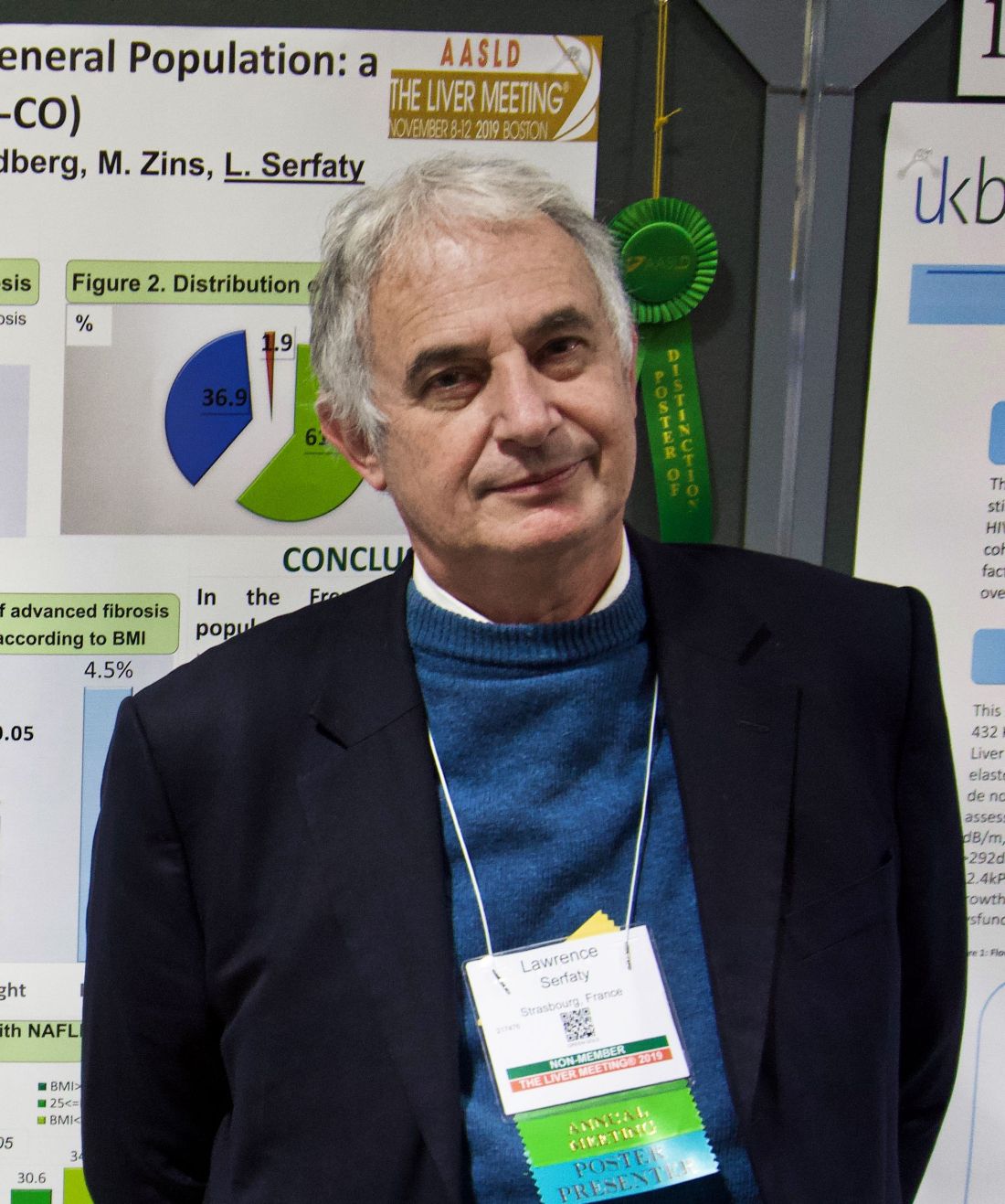
NAFLD was uncommon among participants with normal bodyweight, but when NAFLD was present, lean patients had almost twice the risk of advanced fibrosis as that of overweight and obese patients, reported principal author Lawrence Serfaty, MD, chief of the department of hepatology at Strasbourg (France) University, who conducted the project with colleagues at the French public research organization, Inserm.
“Normally, NAFLD and [nonalcoholic steatohepatitis (NASH)] are part of metabolic syndrome,” Dr. Serfaty said in an interview at the annual meeting of the American Association for the Study of Liver Diseases. “But there are some patients with no metabolic risk factors who are lean and who may have NAFLD.”
To determine the prevalence and characteristics of these patients, the investigators drew data from the CONSTANCES cohort, which is composed of 118,664 members of the general public in France. After excluding those who withdrew consent, had a history of other liver diseases, or reported an excess of alcohol consumption (more than 20 g/day), the analyzed dataset included 102,344 subjects. Among these participants, NAFLD and advanced fibrosis were diagnosed with the Fatty Liver Index (FLI) and the Forns Index (FI), respectively, in which an FLI score greater than 60 indicated NAFLD and an FI score greater than 6.9 indicated advanced fibrosis.
Participants were sorted into three weight categories by body mass index: lean (BMI less than 25 kg/m2), overweight (BMI 25-30), or obese (BMI more than 30). In addition, a variety of other health measures were recorded, including presence of metabolic risk factors, such as diabetes or metabolic syndrome, and elevated alanine transaminase (ALT).
Analysis showed that the prevalence of NAFLD in the general population was 18.2%. Of those diagnosed with NAFLD, 2.6% had advanced fibrosis. As expected, Dr. Serfaty said, NAFLD was much less common among those who were of normal bodyweight, with lean patients accounting for only 1.9% of NAFLD diagnoses. However, when NAFLD was diagnosed in lean patients, it was more often severe. Among lean patients with NAFLD, 4.5% had advanced fibrosis, compared with 2.4% of overweight patients and 2.3% of obese patients.
Dr. Serfaty noted that lean patients with NAFLD tended to have fewer metabolic risk factors, but most had at least one. Lifestyle factors were likely to blame, he said, because lean patients with NAFLD were relatively heavy users of tobacco and alcohol, compared with obese or overweight patients. Cardiovascular disease was also more common among lean patients with NAFLD.
“There are probably other factors [that are different, such as] genetic factors, the microbiome, and maybe the immune system,” Dr. Serfaty said.
While drivers of NAFLD among lean patients remain to be clarified, Dr. Serfaty highlighted the importance of recognizing this unique patient population, and if encountered, not discounting the severity of disease based on a lack of other metabolic risk factors.
“It is very important to know that these patients exist because it is very difficult to identify these patients,” Dr. Serfaty said. “If you have a lean patient with NAFLD, normally you [might] say, it’s not very severe for this patient because he’s lean; but no, [that’s not correct], because maybe he has more advanced fibrosis. So you have to go through and check with other noninvasive markers to be sure that this patient does not have advanced fibrosis.”
Concerning the difficulty of identifying such patients in the first place, Dr. Serfaty suggested that elevated ALT may be the most reliable red flag because this laboratory abnormality occurred in more than half of the lean patients diagnosed with NAFLD, despite no excessive alcohol consumption or prior hepatitis. The investigators disclosed relationships with Gilead, AbbVie, Echosens, and others.
SOURCE: Serfaty L et al. The Liver Meeting 2019, Abstract 1188.
BOSTON – For patients with nonalcoholic fatty liver disease (NAFLD), leaner may really be meaner, according to a recent French study involving more than 100,000 individuals of the general population.
NAFLD was uncommon among participants with normal bodyweight, but when NAFLD was present, lean patients had almost twice the risk of advanced fibrosis as that of overweight and obese patients, reported principal author Lawrence Serfaty, MD, chief of the department of hepatology at Strasbourg (France) University, who conducted the project with colleagues at the French public research organization, Inserm.
“Normally, NAFLD and [nonalcoholic steatohepatitis (NASH)] are part of metabolic syndrome,” Dr. Serfaty said in an interview at the annual meeting of the American Association for the Study of Liver Diseases. “But there are some patients with no metabolic risk factors who are lean and who may have NAFLD.”
To determine the prevalence and characteristics of these patients, the investigators drew data from the CONSTANCES cohort, which is composed of 118,664 members of the general public in France. After excluding those who withdrew consent, had a history of other liver diseases, or reported an excess of alcohol consumption (more than 20 g/day), the analyzed dataset included 102,344 subjects. Among these participants, NAFLD and advanced fibrosis were diagnosed with the Fatty Liver Index (FLI) and the Forns Index (FI), respectively, in which an FLI score greater than 60 indicated NAFLD and an FI score greater than 6.9 indicated advanced fibrosis.
Participants were sorted into three weight categories by body mass index: lean (BMI less than 25 kg/m2), overweight (BMI 25-30), or obese (BMI more than 30). In addition, a variety of other health measures were recorded, including presence of metabolic risk factors, such as diabetes or metabolic syndrome, and elevated alanine transaminase (ALT).
Analysis showed that the prevalence of NAFLD in the general population was 18.2%. Of those diagnosed with NAFLD, 2.6% had advanced fibrosis. As expected, Dr. Serfaty said, NAFLD was much less common among those who were of normal bodyweight, with lean patients accounting for only 1.9% of NAFLD diagnoses. However, when NAFLD was diagnosed in lean patients, it was more often severe. Among lean patients with NAFLD, 4.5% had advanced fibrosis, compared with 2.4% of overweight patients and 2.3% of obese patients.
Dr. Serfaty noted that lean patients with NAFLD tended to have fewer metabolic risk factors, but most had at least one. Lifestyle factors were likely to blame, he said, because lean patients with NAFLD were relatively heavy users of tobacco and alcohol, compared with obese or overweight patients. Cardiovascular disease was also more common among lean patients with NAFLD.
“There are probably other factors [that are different, such as] genetic factors, the microbiome, and maybe the immune system,” Dr. Serfaty said.
While drivers of NAFLD among lean patients remain to be clarified, Dr. Serfaty highlighted the importance of recognizing this unique patient population, and if encountered, not discounting the severity of disease based on a lack of other metabolic risk factors.
“It is very important to know that these patients exist because it is very difficult to identify these patients,” Dr. Serfaty said. “If you have a lean patient with NAFLD, normally you [might] say, it’s not very severe for this patient because he’s lean; but no, [that’s not correct], because maybe he has more advanced fibrosis. So you have to go through and check with other noninvasive markers to be sure that this patient does not have advanced fibrosis.”
Concerning the difficulty of identifying such patients in the first place, Dr. Serfaty suggested that elevated ALT may be the most reliable red flag because this laboratory abnormality occurred in more than half of the lean patients diagnosed with NAFLD, despite no excessive alcohol consumption or prior hepatitis. The investigators disclosed relationships with Gilead, AbbVie, Echosens, and others.
SOURCE: Serfaty L et al. The Liver Meeting 2019, Abstract 1188.
REPORTING FROM THE LIVER MEETING 2019
Level of hepatitis B core–related antigen is risk factor for hepatocellular carcinoma
A high level of hepatitis B core–related antigen (HBcrAg) was a complementary risk factor for hepatocellular carcinoma, according to the results of a retrospective cohort study of more than 2,600 noncirrhotic adults with untreated hepatitis B virus (HBV) infection with a median of 16 years of follow-up.
SOURCE: AMERICAN GASTROENTEROLOGICAL ASSOCIATION
“Patients with an intermediate viral load and high levels of HBcrAg had a risk for hepatocellular carcinoma that did not differ significantly from that of patients with a high viral load. [An] HBcrAg of 10 KU/mL may serve as a novel biomarker for the management of patients with intermediate viral load in our clinical practice,” wrote Tai-Chung Tseng, MD, PhD, of National Taiwan University Hospital in Taipei and associates in Gastroenterology.
Deciding whether to start antiviral therapy is controversial for some patients with HBV infection. Typically, monitoring without treatment is recommended for patients who have both low hepatitis B surface antigen levels (less than 1,000 IU/mL) and low levels of HBV DNA (less than 2,000 IU/mL), and early antiviral therapy is recommended for patients who have high levels of HBV DNA (20,000 IU/mL or more). However, there is no clear evidence that early antiviral therapy benefits patients who have intermediate levels of HBV DNA (2,000-19,999 IU/mL) and are negative for hepatitis B e antigen. Biomarkers for risk-stratifying these patients also are lacking, the researchers noted.
Therefore, they studied a cohort of 2,666 adults who had tested positive for hepatitis B surface antigen and were followed at National Taiwan University Hospital from 1985 through 2000. No patient had cirrhosis at baseline. In all, 209 patients developed hepatocellular carcinoma, yielding an incidence rate of 4.91 cases per 1,000 person-years.
Hepatitis B core–related antigen level remained an independent risk factor for hepatocellular carcinoma after accounting for age, sex, serum alanine aminotransferase (ALT) level, FIB-4 index, hepatitis B e antigen status, hepatitis B genotype (B, C, or undetermined), and HBV DNA level. Compared with patients whose HBcrAg level was less than 10 KU, a level of 10-99 KU/mL was associated with a nearly threefold increase in risk for hepatocellular carcinoma (HR, 2.93; 95% CI, 1.67-4.80), and this risk rose even further as HBcrAg levels increased.
In the subgroup of patients who tested negative for hepatitis B e antigen, had an intermediate HBV DNA load (2,000-19,999 IU/mL), and had a normal baseline ALT level (less than 40 U/L), a high HBcrAg level (10 KU/mL or more) was tied to a nearly fivefold greater risk for hepatocellular carcinoma (HR, 4.89; 95% CI, 2.18-10.93). This approximated the risk that is observed with high viral load (20,000 IU/mL), the researchers noted. In contrast, a low HBcrAg level was associated with a risk similar to that of minimal risk carriers (annual incidence rate, 0.10%; 95% CI, 0.04%-0.24%).
“To the best of our knowledge, this is the first study to report HBcrAg level as an independent viral biomarker to stratify hepatocellular risks in a large number of patients with intermediate viral load,” the researchers commented. Among the study limitations, 412 patients received antiviral therapy during follow-up. “This is a retrospective cohort study including Asian HBV patients with genotype B or C infection,” the investigators added. “It is unclear whether this finding could be extrapolated to populations with other HBV genotype infections. Nonetheless, we had a sound cohort, as several HBsAg-related clinical findings based on our cohort have already been validated by other prospective cohort studies, implying that our data were unlikely to be biased by the study design.”
Funders included National Taiwan University Hospital, the Ministry of Science and Technology, Executive Yuan in Taiwan, and National Health Research Institutes. The researchers reported having no conflicts of interest.
SOURCE: Tseng T-C et al. Gastroenterology. 2019 Aug 27. doi: 10.1053/j.gastro.2019.08.028.
FROM GASTROENTEROLOGY

Daily aspirin might cut risk of fibrosis progression
Taking daily aspirin may help keep nonalcoholic fatty liver disease (NAFLD) from progressing to liver fibrosis and nonalcoholic steatohepatitis (NASH), suggest the results of a prospective study of 361 adults.
Previously, preclinical evidence had linked aspirin to fibrogenesis prevention in fatty liver disease, but this is the first report of a prospective study to do so. Daily aspirin use “was associated with less severe histologic features of NAFLD (nonalcoholic fatty liver disease) at study enrollment and with significantly lower risk for advanced fibrosis over time in a duration-dependent manner,” wrote Tracey G. Simon, MD, MPH, and her associates. Their report is in Clinical Gastroenterology and Hepatology.
The study comprised 361 adults with biopsy-confirmed NAFLD who were enrolled in the Massachusetts General Hospital NAFLD Repository between 2006 and 2015. At baseline, 151 individuals were already on daily aspirin, usually to reduce the primary (54%) or secondary (30%) risk of cardiovascular disease. Median duration of aspirin use was 2.5 years. After a median 7.4 years of follow-up (which was similar between aspirin users and nonusers), daily aspirin use was associated with significantly lower odds of NASH (adjusted odds ratio, 0.68; 95% confidence interval, 0.37-0.89) and fibrosis (aOR, 0.54; 95% CI, 0.31-0.82).
The researchers did not find a similar protective effect for nonsteroidal anti-inflammatory drugs (NSAIDs) other than aspirin (adjusted hazard ratio for advanced fibrosis, 0.93; 95% CI, 0.81–1.05). This might be because of differences between how aspirin and nonaspirin NSAIDs affect COX isoforms – aspirin does so irreversibly, while other NSAIDs have a reversible effect, they added. “Nonaspirin NSAIDs also disrupt the intestinal barrier, increasing delivery of proinflammatory cytokines to the liver,” they wrote. “Finally, aspirin uniquely modulates bioactive lipids by stimulating the biosynthesis of pro-resolving mediators and inhibiting proinflammatory lipids, which in turn may prevent progressive liver damage.”
In this study, a single blinded hepatopathologist interpreted baseline liver biopsy specimens, and patients were followed every 3-6 months with clinical examinations and serial calculations of FIB-4, NFS, and APRI scores. All patients were followed for at least a year. Patients were classified as users of nonaspirin NSAIDs if they reported using an NSAID besides aspirin at least twice weekly, or if they had been prescribed drugs such as ibuprofen, naproxen, ketoprofen, diclofenac, or indomethacin.
In a longitudinal analysis of the 317 patients who had early-stage (F0-2) fibrosis at baseline, 86 developed new-onset advanced fibrosis over a median of 3,692 person-years, the researchers said. In all, 26 individuals developed hepatic decompensation and 18 patients died, including eight from liver-related causes. Importantly, the link between aspirin and decreased risk of fibrosis progression seemed to depend on duration of use (adjusted P trend = .026), with the greatest benefit seen with 4 years or more of use (aHR, 0.50; 95% CI, 0.35-0.73). Although subgroup analyses were limited by lack of power, daily aspirin use was associated with a 36% lower odds of incident advanced fibrosis among the 72 study participants who had paired biopsy samples, even after accounting for the effect of age, sex, baseline fibrosis stage, and time between biopsies (aOR, 0.64; 95% CI, 0.50-0.80).
“Our findings add to the growing literature supporting the potential hepatoprotective effects of aspirin in NAFLD,” the researchers concluded. “Research to uncover the mechanisms by which aspirin might prevent fibrogenesis could help develop urgently needed antifibrotic therapies for NAFLD.”
Funders included the National Institutes of Health and the AASLD Foundation. The investigators reported having no conflicts of interest.
SOURCE: Simon TG et al. Clin Gastroenterol Hepatol. 2019 May 8.
Slowing, preventing, or reversing fibrogenesis in patients with NAFLD remains an unmet need. Lifestyle interventions are beneficial to this population but challenging because of concerns with adherence and sustainability, thus, favoring pharmacologic interventions.
The study by Simon et al. provides initial prospective evidence of the role of aspirin in reducing progression of fibrosis. In a thoughtful design, authors showed both cross-sectional and longitudinal associations of reduced risk for progressed fibrosis among aspirin users, all with biological coherence and while accounting for various confounding factors. Although the accuracy of blood-based noninvasive assessment of liver fibrosis (by FIB-4, NFS, and APRI) to determine progression of fibrosis in NAFLD has moderate accuracy at its best, the relatively high FIB-4 cutoff value used by the authors and their sensitivity analyses (including liver biopsy and combinations of blood-based markers combined endpoints) bring certainty to their results.
However, before we can start prescribing aspirin to halt progression of fibrosis in NAFLD, larger and adequately powered studies are needed. Caution with the use of aspirin as prophylaxis for atherosclerotic cardiovascular disease (ASCVD) is now advised, based on results from large clinical trials (i.e., ASCEND). NAFLD patients represent a particular population with both a high ASCVD risk and a high risk for gastrointestinal bleeding, and it is unclear what the number needed to treat or to harm would be without confirmatory studies.
Andres Duarte-Rojo, MD, PhD, is associate professor of medicine, division of gastroenterology, hepatology, and nutrition at the University of Pittsburgh Medical Center, and Pittsburgh Liver Research Center. He received research support from Echosens, USA.
Taking daily aspirin may help keep nonalcoholic fatty liver disease (NAFLD) from progressing to liver fibrosis and nonalcoholic steatohepatitis (NASH), suggest the results of a prospective study of 361 adults.
Previously, preclinical evidence had linked aspirin to fibrogenesis prevention in fatty liver disease, but this is the first report of a prospective study to do so. Daily aspirin use “was associated with less severe histologic features of NAFLD (nonalcoholic fatty liver disease) at study enrollment and with significantly lower risk for advanced fibrosis over time in a duration-dependent manner,” wrote Tracey G. Simon, MD, MPH, and her associates. Their report is in Clinical Gastroenterology and Hepatology.
The study comprised 361 adults with biopsy-confirmed NAFLD who were enrolled in the Massachusetts General Hospital NAFLD Repository between 2006 and 2015. At baseline, 151 individuals were already on daily aspirin, usually to reduce the primary (54%) or secondary (30%) risk of cardiovascular disease. Median duration of aspirin use was 2.5 years. After a median 7.4 years of follow-up (which was similar between aspirin users and nonusers), daily aspirin use was associated with significantly lower odds of NASH (adjusted odds ratio, 0.68; 95% confidence interval, 0.37-0.89) and fibrosis (aOR, 0.54; 95% CI, 0.31-0.82).
The researchers did not find a similar protective effect for nonsteroidal anti-inflammatory drugs (NSAIDs) other than aspirin (adjusted hazard ratio for advanced fibrosis, 0.93; 95% CI, 0.81–1.05). This might be because of differences between how aspirin and nonaspirin NSAIDs affect COX isoforms – aspirin does so irreversibly, while other NSAIDs have a reversible effect, they added. “Nonaspirin NSAIDs also disrupt the intestinal barrier, increasing delivery of proinflammatory cytokines to the liver,” they wrote. “Finally, aspirin uniquely modulates bioactive lipids by stimulating the biosynthesis of pro-resolving mediators and inhibiting proinflammatory lipids, which in turn may prevent progressive liver damage.”
In this study, a single blinded hepatopathologist interpreted baseline liver biopsy specimens, and patients were followed every 3-6 months with clinical examinations and serial calculations of FIB-4, NFS, and APRI scores. All patients were followed for at least a year. Patients were classified as users of nonaspirin NSAIDs if they reported using an NSAID besides aspirin at least twice weekly, or if they had been prescribed drugs such as ibuprofen, naproxen, ketoprofen, diclofenac, or indomethacin.
In a longitudinal analysis of the 317 patients who had early-stage (F0-2) fibrosis at baseline, 86 developed new-onset advanced fibrosis over a median of 3,692 person-years, the researchers said. In all, 26 individuals developed hepatic decompensation and 18 patients died, including eight from liver-related causes. Importantly, the link between aspirin and decreased risk of fibrosis progression seemed to depend on duration of use (adjusted P trend = .026), with the greatest benefit seen with 4 years or more of use (aHR, 0.50; 95% CI, 0.35-0.73). Although subgroup analyses were limited by lack of power, daily aspirin use was associated with a 36% lower odds of incident advanced fibrosis among the 72 study participants who had paired biopsy samples, even after accounting for the effect of age, sex, baseline fibrosis stage, and time between biopsies (aOR, 0.64; 95% CI, 0.50-0.80).
“Our findings add to the growing literature supporting the potential hepatoprotective effects of aspirin in NAFLD,” the researchers concluded. “Research to uncover the mechanisms by which aspirin might prevent fibrogenesis could help develop urgently needed antifibrotic therapies for NAFLD.”
Funders included the National Institutes of Health and the AASLD Foundation. The investigators reported having no conflicts of interest.
SOURCE: Simon TG et al. Clin Gastroenterol Hepatol. 2019 May 8.
FROM CLINICAL GASTROENTEROLOGY AND HEPATOLOGY