User login
VRD pretransplant induction deepens responses in myeloma
Pretransplant induction therapy with subcutaneous bortezomib, lenalidomide, and dexamethasone (VRD) deepened responses in patients with newly diagnosed multiple myeloma, according to an interim analysis of a phase 3 study.
Overall, the regimen was well tolerated, with a minimal number of patients discontinuing treatment because of treatment-emergent adverse events.
The ongoing, open-label, randomized, phase 3 study is designed to compare two transplant-conditioning regimens – intravenous busulfan plus melphalan versus melphalan – in patients who received VRD induction and consolidation, wrote Laura Rosiñol, MD, PhD, of the August Pi i Sunyer Biomedical Research Institute in Barcelona, and colleagues. The findings were published in Blood.
The PETHEMA/GEM2012 study included 458 patients with newly diagnosed multiple myeloma who were eligible for autologous stem cell transplantation. Study patients were previously untreated and aged younger than 65 years.
All patients received VRD induction, which consisted of subcutaneous bortezomib 1.3 mg/m2 on days 1, 4, 8, and 11 of each cycle; lenalidomide 25 mg/day on days 1-21; and dexamethasone 40 mg on days 1-4 and 9-12 at 4-week intervals for six cycles. Posttransplant consolidation consisted of two cycles of VRD.
The researchers conducted a grouped-response analysis of three different treatment phases: induction, transplant, and consolidation.
After analysis, the researchers found that responses deepened over the duration of treatment. In patients who started the sixth induction cycle, the response rates were 55.6%, 63.8%, 68.3%, and 70.4% after cycles 3, 4, 5, and post induction, respectively.
After six cycles of induction, the complete response rate was 33.4%, with a rate of undetectable minimal residual disease of 28.8%, which further increased at transplant (42.1%), and consolidation (45.2%).
With respect to safety, the most frequently reported grade 3 or higher treatment-emergent adverse events were neutropenia (12.9%) and infection (9.2%). The rate of grade 2 or higher peripheral neuropathy throughout induction was 17.0%, with lower rates of grade 3 (3.7%) and 4 (0.2%) toxicities.
“The regimen [used in the present study] has the highest lenalidomide and dexamethasone dose intensity per cycle and a lower bortezomib dose intensity per cycle than the 21-day regimens, which may offer high activity with low levels of toxicity, thereby enabling delivery of all planned induction cycles,” the researchers wrote, adding that “these results confirm that VRD is an effective pretransplant induction regimen and may be considered a new standard of care.”
The study was supported by Celgene, Janssen, Pierre Fabré, and the Instituto de Salud Carlos III. The authors reported financial affiliations with Celgene, Janssen, and several other companies.
SOURCE: Rosiñol L et al. Blood. 2019 Sep 4. doi: 10.1182/blood.2019000241.
FROM BLOOD
Identifying Progression-free Survival in Veterans with Diffuse Large B-Cell Lymphoma Using Electronic Health Care Records
Purpose: To establish a gold-standard methodology for accurately extracting progression-free survival (PFS) following Diffuse Large B-Cell Lymphoma (DLBCL) treatment using real-world electronic healthcare record (EHR) data.
Background: Randomized controlled trials using response evaluation criteria have long served as the gold standard for assessing response to therapy and PFS. However, characteristics of participants in clinical trials do not reflect the overall patient population, and formal response evaluation criteria are not used in realworld contexts. Furthermore, real-world data are often unstructured, preventing accurate comparison of PFS using structured clinical trial data versus real-world data, and existing approaches define PFS inconsistently. Despite the importance of assessing PFS in patients outside of controlled clinical trials, no goldstandard method for collecting and validating PFS from real-world evidence has been established.
Methods: Clinicians, programmers, and data scientists collaborated to develop an R Shiny10 application using Veterans Affairs Corporate Data Warehouse data from the EHR of 352 DLBCL patients. The application takes unstructured data such as clinical notes and facilitates the capture, annotation, and tagging of key words or phrases indicative of progression, thus allowing accurate determination of the date of first identification of progression by a treating clinician.
Data Analysis: In order to refine data-collection techniques and evaluate whether the application can enable calculation of real-world PFS, we conducted an adaptive and iterative process of reviewing EHR documents and capturing and annotating data until a consistent schema and methodology was established. In order to validate annotation schema and methodology, annotations of 50 patient records were performed by 2 annotators and assessed for concordance.
Results: We produced an R Shiny application that can capture, annotate, and transform unstructured EHR data into structured data—specifically, treatment lines, cycles, and response criteria with corresponding dates—ready for analysis of PFS. An annotation schema for capturing real-world data was also developed. Mapping of common phrases used by clinicians in real-world practice to response criteria resulted in a dictionary of these phrases.
Implications: These efforts show that it is possible to convert EHR context reliably into analyzable data such as PFS. Further attempts will be made to establish a gold-standard methodology.
Angioimmunoblastic T-cell Lymphoma: Patient Characteristics and Survival Outcomes
Background: Angioimmunoblastic T-cell lymphoma (AITL) is a rare and aggressive malignancy, representing approximately 18.5% of Peripheral T-cell lymphomas. The clinical presentation varies greatly, and literature describing this disease is limited. In this study we utilize the National Cancer Database (NCDB) to describe the patient characteristics, demographics, and overall survival of AITL.
Methods: The NCDB for non-Hodgkin’s lymphoma was used to identify 3,708 patients diagnosed with AITL between 2004 and 2016. We examined demographic information including age, race, gender, stage, and treatment modality. Kaplan-Meier analysis was used to analyze overall survival and compare survival by patient age, stage, and year of diagnosis. Bivariate and multivariate analysis was used to obtain hazard ratios and assess the association of patient characteristics and treatment methods with survival.
Results: The majority of AITL patients were white 87.8%, males 53%, with an average age of 67 years. 90% of patients received treatment, with 77.4% receiving chemotherapy, 10.8% receiving hematopoietic transplant, and 2.6% receiving radiation. B symptoms were present in 57% of patients. 43% were diagnosed with stage III disease and 43.5% at stage IV.
Median survival was 22 months (CI 19.7-24.4), with a 5- and 10-year survival of 30% and 22%. Median survival for patients aged 35-50 was 74 months, 50-65 was 37.7 months, 65-80 was 20.5 months, and for patients aged > 80 years old median survival was 6.4 months. Patients with stage I disease survived 35.1 months whereas those with stage IV disease survived 17.2 months. On multivariate analysis black race, increasing age, charleson-deyo comorbidity score, and stage at diagnoses were signi cantly associated with increased mortality risk. All forms of treatment, including chemotherapy, hematopoietic transplant, and radiation were all associated with improved survival.
Discussion: Our study found that the majority of AITL patients present with late stage disease, often with B symptoms, and have poor prognosis. We found a five-year survival of 30% for this malignancy when all stages of disease were combined. Knowledge of the patient characteristics, treatment modalities, and overall survival in AITL can serve to enhance the care of providers who encounter this uncommon diagnosis.
Rituximab, bendamustine look better than chemo alone in MCL
In older patients with newly diagnosed mantle cell lymphoma (MCL), first-line therapy with rituximab- and bendamustine-based regimens significantly reduced 1-year mortality rates versus chemotherapy alone, according to a retrospective analysis.
“This study evaluated the comparative effectiveness of [rituximab, bortezomib, or bendamustine] in elderly patients newly diagnosed with MCL,” wrote Shuangshuang Fu, PhD, of the University of Texas MD Anderson Cancer Center, Houston, and colleagues. The findings were reported in Clinical Lymphoma, Myeloma & Leukemia.
The researchers studied population-based data from the Surveillance, Epidemiology, and End Results (SEER)-Medicare linked database. They identified all patients over age 65 years who received a new diagnosis of MCL between Jan. 1, 1999, and Dec. 31, 2013.
The study cohort included a total of 1,215 patients. Participants were classified into four different groups according to treatment regimen: chemotherapy alone, rituximab plus or minus chemotherapy, bendamustine plus or minus chemotherapy, and bortezomib plus or minus chemotherapy.
At 1-year follow-up, the team analyzed various mortality outcomes, including MCL-specific, all-cause, and noncancer mortality. The bortezomib results were not included in the primary analysis because of small sample size, according to the researchers.
After multivariable analysis, Dr. Fu and colleagues found that 1-year all-cause mortality rate was significantly lower for patients receiving rituximab-based regimens, compared with chemotherapy alone (hazard ratio, 0.38; 95% confidence interval, 0.25-0.59). There was a similar decline for MCL-specific mortality (HR, 0.38; 95% CI, 0.24-0.60).
The 1-year MCL-specific mortality was also significantly reduced in the bendamustine group, compared with chemotherapy alone (HR, 0.49; 95% CI, 0.24-0.99).
“Our findings comparing rituximab with chemotherapy alone further confirmed the benefit of adding rituximab to chemotherapy in newly diagnosed older MCL patients,” they wrote.
The researchers acknowledged that a key limitation of the study was the observational design. As a result, selection bias and unmeasured confounding could have influenced the results.
“Future studies evaluating the comparative effectiveness of those newly approved novel agents for MCL patients were warranted as more data are available,” they concluded.
The study was funded by the Duncan Family Institute, the Cancer Prevention Research Institute of Texas, and the National Institutes of Health. The authors reported having no conflicts of interest.
SOURCE: Fu S et al. Clin Lymphoma Myeloma Leuk. 2019 Aug 30. doi: 10.1016/j.clml.2019.08.014.
FROM CLINICAL LYMPHOMA, MYELOMA & LEUKEMIA
Progressive myeloma after induction? Go straight to transplant
Patients with multiple myeloma who don’t respond to induction therapy may be better off advancing straight to autologous stem cell therapy, rather than undergoing salvage therapy before transplant, according to findings of an analysis that included both real-world and clinical trial patients.
Joanna Blocka, MD, of the University Hospital of Heidelberg (Germany) and colleagues found similar progression-free and overall survival rates for patients who had progressive disease and underwent autologous stem cell therapy (ASCT), compared with patients who underwent salvage therapy and improved to at least stable disease before proceeding to transplant. The findings were published in Leukemia & Lymphoma.
The real-world analysis included 1,599 patients with multiple myeloma who had undergone ASCT between 1991 and 2016. More than half of the patients (58%) were not enrolled in clinical trials. The remainder were split between the German-Speaking Myeloma Multicenter Group (GMMG)-HD3 and GMMG-HD4 trials, which compared various induction regimens.
Just 23 patients in the analysis received salvage therapy because of progressive disease and deepened their response before ASCT. Of these patients, 12 received novel agents in induction therapy and 11 received older medications.
Looking across all 1,599 patients, 5.3% achieved complete remission before first ASCT. Most patients (71.8%) achieved partial remission, 9.7% had a minimal response, and 5.7% had stable disease. A group of 120 patients (7.5%) progressed between the last course of induction and ASCT.
The researchers compared the progression-free and overall survival rates of patients with progressive disease versus those who had stable disease or better before their first transplant. Both univariable and multivariable analysis showed no statistically significant differences in either survival outcome between the two groups.
In the multivariable analysis, there was a hazard ratio of 1.23 (95% confidence interval, 0.98-1.56) for progression-free survival for patients with progressive disease versus those who responded to induction therapy. Similarly, the HR for overall survival between the two groups was 1.24 (95% CI, 0.93-1.65).
The researchers also analyzed the groups based on whether they received novel or older agents during induction.
Patients with progressive disease who received novel agents had significantly worse progression-free survival (22.2 months), compared with patients who responded to treatment with novel agents (22.2 months vs. 29.1 months; P = .03). The same trend was seen with overall survival in these groups (54.4 months vs. 97.5 months; P less than .001).
Rates of survival were similar for patients with progressive disease and responders who had received older medications at induction.
“This might be explained by a prognostically disadvantageous disease biology in patients nonresponsive to novel agents,” the researchers wrote.
The researchers also compared survival outcomes for the 120 patients who underwent ASCT with progressive disease versus the 23 patients who received salvage therapy and improved their response to at least stable disease before transplant. Univariable analysis showed that salvage patients actually did worse than those with progressive disease who proceeded straight to transplant – 12.1 months versus 22.9 months of progression-free survival (P = .04) and 33.1 versus 69.5 months of overall survival (P = .08). But on multivariable analysis, there was no significant difference between the two groups for progression-free survival (HR, 0.71; 95% CI, 0.28-1.80; P = .5) or overall survival (HR, 0.77; 95% CI, 0.30-1.95; P = .6). The use of novel agents did not appear to affect the survival outcomes in these patients.
The worse outcomes seen among salvage patients observed in univariable analysis “might be due to a cumulative toxic effect of salvage therapy,” the researchers suggested. “An alternative explanation could be that the patients who were offered salvage therapy might have had more aggressive disease than those who did not undergo salvage therapy.”
Dr. Blocka reported having no relevant financial disclosures. Other coauthors reported relationships with Janssen, Amgen, Bristol-Myers Squibb, Celgene, and others.
SOURCE: Blocka J et al. Leuk Lymphoma. 2019 Aug 19. doi: 10.1080/10428194.2019.1646905.
FROM LEUKEMIA & LYMPHOMA
Key clinical point:
Major finding: There was no difference between patients with progressive disease who went straight to ASCT and patients who received salvage therapy, both in terms of progression-free survival (hazard ratio, 0.71; 95% confidence interval, 0.28-1.80; P = .5) and overall survival (HR, 0.77; 95% CI, 0.30-1.95; P = .6).
Study details: An analysis of 1,599 patients with multiple myeloma who underwent ASCT. A subanalysis compared 120 patients with progressive disease before ASCT with 23 patients who received salvage treatment before ASCT.
Disclosures: Dr. Blocka reported having no relevant financial disclosures. Other coauthors reported relationships with Janssen, Amgen, Bristol-Myers Squibb, Celgene, and others.
Source: Blocka J et al. Leuk Lymphoma. 2019 Aug 19. doi: 10.1080/10428194.2019.1646905.
Ibrutinib-rituximab induction yields ‘unprecedented’ responses in MCL
LUGANO, Switzerland – In younger patients with previously untreated mantle cell lymphoma, the chemotherapy-free combination of ibrutinib and rituximab followed by a short course of chemotherapy was associated with an “unprecedented” 3-year progression-free survival rate, investigators in the phase 2 WINDOW-1 trial reported.
Among 50 patients aged 65 years and younger who received ibrutinib and rituximab until they achieved a complete or partial response, followed by four cycles of chemotherapy with rituximab plus hyper-CVAD (cyclophosphamide, vincristine, doxorubicin and dexamethasone) and rituximab plus methotrexate, the 3-year progression-free survival (PFS) rate was 88%, said Michael Wang, MD, from the University of Texas MD Anderson Cancer Center in Houston.
Additionally, for patients with the low-risk features, the 3-year PFS rate was 90%.
“Chemo-free ibrutinib-rituximab induced unprecedented – unprecedented – efficacy before chemo consolidation,” he said at the International Conference on Malignant Lymphoma.
Dr. Wang presented data from an interim analysis of the investigator-initiated single-center trial. Fifty patients aged 65 years or younger with untreated mantle cell lymphoma (MCL), good performance status, and good organ function were enrolled.
The patients were treated with ibrutinib and rituximab for two cycles and then evaluated for response with PET-CT scan, bone marrow biopsy, and for some patients, esophagogastroduodenoscopy (EGD) and colonoscopy with random biopsies.
In the induction phase, patients received ibrutinib daily on days 1-28 and rituximab intravenously over 6-8 hours on days 1, 8, 15, and 22 of cycle 1, and then over 4 hours on day 1 of cycles 3-12. The treatment was repeated every 28 days for up to 12 cycles in the absence of disease progression or unacceptable toxicity, or until patients achieved a complete response.
In the consolidation phase, patients received rituximab IV over 6 hours on day 1; oral or IV dexamethasone on days 1-4; cyclophosphamide IV over 3 hours twice daily on days 2-4; doxorubicin IV over 15-30 minutes on day 5; and vincristine IV over 15-30 minutes on day 5 of cycles one, three, five, and seven. Patients also received rituximab IV over 6 hours on day 1; methotrexate IV over 24 hours on day 2; and cytarabine IV over 2 hours twice daily on days 3 and 4 of cycles two, four, six, and eight. Treatments were repeated every 28 days for up to eight cycles in the absence of disease progression or unacceptable toxicity.
Patients who had a complete response (CR) after two cycles of induction and those who had disease progression on induction went on to consolidation. Patients with partial responses (PR) to induction continued on ibrutinib/rituximab until either the loss of a PR or best response for up to 12 cycles, with those who achieved a CR then moving on to consolidation.
Patients who had a CR after induction received four cycles of R-hyperCVAD, no subsequent stem cell transplant, and no maintenance therapy. Patients who had a PR after induction received two cycles of R-hyperCVAD, were reassessed, and then continued on R-hyperCVAD until CR or for up to eight total cycles.
Patients with either stable disease or progression during R-hyperCVAD were taken off the study.
Of the 50 patients enrolled, all 50 were evaluable for part A (induction), and 48 were evaluable after induction and consolidation (two patients withdrew for personal reasons).
After a median follow-up of 36 months, the overall response rate (ORR) following induction was 100%, consisting of 46 CRs (92%) and four PRs (8%).
In an intention-to-treat analysis (including the two patients who withdrew), the ORR was 96%, consisting of CRs in 47 patients (94%) and a PR in 1 patient (2%).
Neither the median PFS nor median overall survival had been reached at the time of data cutoff, and no patients have died.
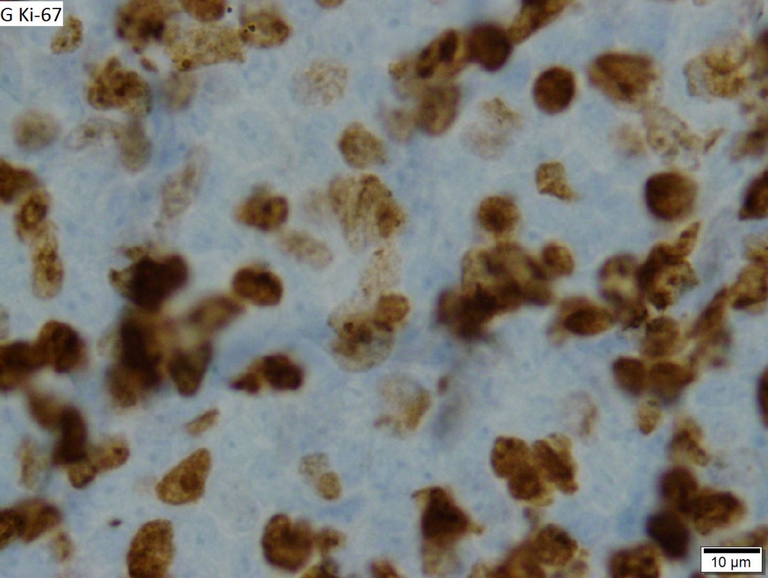
Of the 50 enrolled patients, four experienced disease progression after 17, 24, 34, and 35 months of treatment. The patients with disease progression included one with Ki-67 of less than 30%, and three with KI-67 of 30% or greater.
Grade 3-4 toxicities during induction including myelosuppression in 4%; fatigue, myalgia, and rashes in 8% each; and oral mucositis in 4%.
Dr. Wang said that future studies on minimal residual disease and clonal evolution are ongoing, and that data on more patients will be presented at the next annual meeting of the American Society of Hematology, scheduled for December 2019.
He also noted that the WINDOW-2 trial, in which ibrutinib and rituximab are followed by veneotclax and hyper-CVAD chemotherapy in patients with newly diagnosed MCL, is open and rapidly enrolling patients.
The study is supported by the National Cancer Institute. Dr. Wang reported financial relationships with Janssen, Pharmacyclics, and other companies.
SOURCE: Wang M et al. ICML-15, Abstract 12.
REPORTING FROM 15-ICML
Combo produces responses in triple-class refractory myeloma
Selinexor plus low-dose dexamethasone can produce responses in patients with triple-class refractory multiple myeloma, according to the phase 2 STORM trial.
The combination produced a response rate of 26% in patients who were refractory to at least one proteasome inhibitor, one immunomodulatory agent, and daratumumab.
The most common grade 3/4 adverse events in this trial were thrombocytopenia (59%), anemia (44%), hyponatremia (22%), and neutropenia (21%).
Ajai Chari, MD, of the Mount Sinai School of Medicine, New York, and colleagues reported these results in the New England Journal of Medicine.
The STORM trial included 123 patients with multiple myeloma who had previously received bortezomib, carfilzomib, lenalidomide, pomalidomide, daratumumab, and an alkylating agent. Their disease was refractory to at least one proteasome inhibitor, one immunomodulatory drug, and daratumumab.
The patients had received a median of 7 (range, 3-18) prior treatment regimens, and their median time since diagnosis was 6.6 years (range, 1.1-23.4 years). The median age at baseline was 65.2 years (range, 40-86 years), 58% of patients were men, and 53% had high-risk cytogenetics. In addition, 36% of patients had thrombocytopenia and 16% had neutropenia at baseline.
The patients received oral selinexor at 80 mg twice weekly plus dexamethasone at 20 mg twice weekly until disease progression, death, or discontinuation. Doses were modified in response to adverse events.
Results
In total, 96% of patients (118/123) discontinued treatment. The most common reasons for discontinuation were disease progression (n = 65) and adverse events (n = 38).
Of the 122 patients evaluable for efficacy, 26% achieved a partial response or better, and 39% had a minimal response or better. There were 24 partial responses, 16 minimal responses, 6 very good partial responses, and 2 stringent complete responses. Forty-eight patients had stable disease.
The median duration of response was 4.4 months, the median progression-free survival was 3.7 months, and the median overall survival was 8.6 months.
The median overall survival was 15.6 months in responders, 5.9 months in patients with stable disease, and 1.7 months in those who progressed.
All 123 patients were evaluable for safety, and 63% of them experienced serious adverse events. Pneumonia (11%) and sepsis (9%) were the most common serious events.
The most common treatment-emergent nonhematologic adverse events were fatigue (73%), nausea (72%), decreased appetite (56%), decreased weight (50%), diarrhea (46%), vomiting (38%), hyponatremia (37%), upper respiratory tract infection (23%), constipation (22%), and dyspnea (22%).
Treatment-emergent hematologic adverse events included thrombocytopenia (73%), anemia (67%), neutropenia (40%), leukopenia (33%), and lymphopenia (16%).
Eighty percent of patients had adverse events leading to dose modification or interruption. The most common of these were thrombocytopenia (43%), fatigue (16%), and neutropenia (11%).
“Because most patients involved in the study were older and frail, with limited end-organ reserve and at increased risk for adverse events, dose modifications were anticipated and were specified along with supportive care in the protocol,” the researchers wrote.
“The adverse events that were observed in the study were a function of dose, schedule, and baseline clinical characteristics (e.g., cytopenias). Thrombocytopenia … was reversible and was managed with dose interruptions and thrombopoietin-receptor agonists.”
There were 28 deaths on study, with 16 patients dying of disease progression and 12 dying from an adverse event. Two of the fatal adverse events were considered treatment related – sepsis in one patient and pneumonia with concurrent disease progression in another patient.
The researchers reported ties with Karyopharm Therapeutics, which sponsored the study, and many other companies.
SOURCE: Chari A et al. N Engl J Med 2019;381:727-38.
FROM NEW ENGLAND JOURNAL OF MEDICINE
Key clinical point:
Major finding: The response rate was 26%, which included 24 partial responses, 6 very good partial responses, and 2 stringent complete responses.
Study details: A phase 2 trial of 123 patients with multiple myeloma refractory to at least one proteasome inhibitor, one immunomodulatory drug, and daratumumab (triple-class refractory).
Disclosures: The researchers reported ties with Karyopharm Therapeutics, which sponsored the study, and many other companies.
Source: Chari A et al. N Engl J Med 2019;381:727-38.
Obinutuzumab-lenalidomide combo shows promise in relapsed/refractory FL
Obinutuzumab plus lenalidomide showed manageable safety and activity in patients with relapsed/refractory follicular lymphoma (FL), according to results from a phase 2 trial.
“The results of this phase 2 study show that induction therapy with obinutuzumab and lenalidomide followed by maintenance therapy with obinutuzumab is effective for many patients with relapsed or refractory follicular lymphoma,” wrote Franck Morschhauser, MD, PhD, of the University of Lille, France, and colleagues. The results were published in The Lancet Haematology.
The multicenter, single-arm study comprised 89 patients, 88 of whom were assessed for safety and 86 for efficacy. All eligible study patients received a minimum of one prior rituximab-based therapy before receiving obinutuzumab.
Study participants received intravenous obinutuzumab 1,000 mg for six 28-day cycles, in addition to oral lenalidomide 20 mg as induction therapy.
Maintenance therapy (year 1) consisted of oral lenalidomide 10 mg on days 2-22 of each cycle for a maximum of 12 28-day cycles plus obinutuzumab 1,000 mg on day 1 of alternate cycles (total of six infusions). Maintenance therapy (year 2) consisted of obinutuzumab 1,000 mg alone on day 1 for six 56-day cycles.
The primary outcome was the proportion of patients who achieved an overall response at the end of induction therapy. Secondary outcomes included various survival parameters and safety.
After analysis, the researchers found that the proportion of patients who achieved an overall response at induction end was 79% (95% confidence interval, 69-87). In addition, 38% of patients (95% CI, 28-50) had achieved a complete response at the end of induction therapy.
The progression-free survival, event-free survival, and overall survival rates were 65% (95% CI, 54-74), 62% (95% CI, 51-72), and 87% (95% CI, 78-93), respectively, at 2 years (no P values were reported).
“The results suggest that obinutuzumab plus lenalidomide is active as shown by 2-year outcomes (progression-free survival and overall survival) in the overall patient group,” the researchers wrote.
With respect to safety, basal cell carcinoma (6%), febrile neutropenia (5%), and infusion-related reactions (3%) were the most frequently reported serious toxicities. One patient died because of therapy-related febrile neutropenia.
The Lymphoma Academic Research Organisation, Celgene, and Roche funded the study. The authors reported financial affiliations with the study sponsors and several other companies.
SOURCE: Morschhauser F et al. Lancet Haematol. 2019 Jul 8. doi: 10.1016/S2352-3026(19)30089-4.
FROM THE LANCET HAEMATOLOGY
Brentuximab vedotin plus nivolumab shows positive outcomes in PMBL
Combination brentuximab vedotin and nivolumab showed manageable safety and high activity in patients with relapsed/refractory primary mediastinal B-cell lymphoma (PMBL), according to results from a phase 2 trial.
“We evaluated whether the combination of nivolumab and [brentuximab vedotin] was safe and synergistically effective in patients with [relapsed/refractory] PMBL,” Pier Luigi Zinzani, MD, PhD, of the University of Bologna (Italy), and colleagues wrote in the Journal of Clinical Oncology.
The CheckMate 436 study is a multicenter, open-label, phase 1-2 study that included patients with relapsed/refractory disease who had previously received autologous stem cell transplantation (ASCT) or had two or more previous chemotherapy regimens for those ineligible for ASCT.
The phase 2 component evaluated the safety and efficacy of the two-drug combo in an expansion cohort of 30 patients. Study participants received intravenous brentuximab vedotin at 1.8 mg/kg and nivolumab at 240 mg every 3 weeks until cancer progression or intolerable adverse effects.
The primary outcomes were the investigator-evaluated objective response rate and safety. Secondary outcomes included progression-free survival, complete remission rate, overall duration of response, among other measures.
After analysis, the researchers reported that 53% of patients had grade 3 or 4 treatment-related toxicities following a median of five treatment cycles. The most common treatment-related toxicities were neutropenia (30%) and peripheral neuropathy (27%).
Five patients died during the study follow-up, four because of disease progression and one as a result of sepsis that was not considered related to treatment.
At a median follow-up of 11.1 months, the objective response rate was 73% in study participants, including 11 patients (37%) who achieved a complete response and 11 patients (37%) who had a partial response. An additional three patients had stable disease.
The median progression-free survival, duration of response, and overall survival were not reached in this study.
“The combination of nivolumab and [brentuximab vedotin] may be synergistic and is highly active in patients with [relapsed/refractory] PMBL, serving as a potential bridge to other consolidative therapies of curative intent,” the researchers wrote.
The study was funded by Bristol-Myers Squibb and Seattle Genetics. The authors reported financial affiliations with the study sponsors and several other companies.
SOURCE: Zinzani PL et al. J Clin Oncol. 2019 Aug 9. doi: 10.1200/JCO.19.01492.
FROM THE JOURNAL OF CLINICAL ONCOLOGY
Key clinical point: Brentuximab vedotin plus nivolumab showed manageable safety and positive activity in patients with relapsed/refractory primary mediastinal B-cell lymphoma (PMBL).
Major finding: At 11.1 months, the objective response rate was 73% in study participants, including 37% of patients who achieved a complete response and 37% who had a partial response.
Study details: A phase 2 study of 30 patients with relapsed/refractory PMBL.
Disclosures: The study was funded by Bristol-Myers Squibb and Seattle Genetics. The authors reported financial affiliations with the study sponsors and several other companies.
Source: Zinzani PL et al. J Clin Oncol. 2019 Aug 9. doi: 10.1200/JCO.19.01492.
Ibrutinib/rituximab effective, safe as frontline treatment for older patients with MCL
LUGANO, SWITZERLAND – The chemotherapy-free combination of ibrutinib (Imbruvica) and rituximab is highly effective as frontline therapy for older, transplant-ineligible patients with nonblastoid mantle cell lymphoma, according to investigators.
In a phase 2 study of patients with a median age of 71 years, 38 of 41 patients (93%) had an objective response, and the regimen was both safe and easy to administer, reported Preetesh Jain, MD, PhD, of the University of Texas MD Anderson Cancer Center in Houston.
“The adverse event profile was generally favorable, with specific monitoring recommended for patients with cardiovascular comorbidities and a history of atrial fibrillation,” he said at the International Conference on Malignant Lymphoma.
The investigators enrolled 48 patients aged 65 years and older with previously untreated mantle cell lymphoma (MCL), of whom 41 were evaluable for the primary endpoints of overall response rate (ORR) and safety. The patients had good performance status and normal organ function, with the largest tumor size less than 10 cm. Patients with atrial fibrillation could participate, if the fibrillation was controlled. Patients with Ki-67 protein levels of 50% or greater and blastoid/pleomorphic histology were excluded.
Patients were treated with ibrutinib 560 mg orally daily for each 28-day cycle, with therapy continued until disease progression, or until therapy was stopped for any other reason. Patients also received intravenous rituximab 375 mg/m2 on days 1, 8, 15 and 22, plus or minus one day for cycle 1, on day 1 of cycles 3-8, and on day 1 of every other cycle for up to 2 years.
Of the 41 patients evaluable for response, 26 (64%) had a complete response (CR) and 12 (29%) had a partial response. Three additional patients had stable disease, for an objective response rate of 93%.
Of 34 patients with PET scans, all had negative scans. Of 37 patients evaluable for minimal residual disease (MRD) by flow cytometry, 21 (58%) were MRD negative.
Patients with low or intermediate Mantle Cell Lymphoma International Prognostic Index (MIPI) scores had a higher ORR (100% vs. 89% for patients with high MIPI scores), and patients with low Ki-67 levels had a higher response rate than that of patients with KI-67 of 30% or greater (80% vs. 87%).
Neither median 3-year progression-free survival nor median 3-year overall survival have been reached, with respective 3-year rates of 87% and 95%.
Four patients experienced disease progression at 4, 10, 13 and 33 months of treatment. Three of these patients had disease that had transformed to blastoid/pleomorphic variant, two had Ki-67 of 30% or greater, one had mutations in TP53, and one had FAT1 and SF3B1 mutations.
Two patients died after ibrutinib therapy, one who had discontinued therapy because of bleeding, and the other who died on treatment at 13 months from transformed disease. Both of these patients had high Ki-67 levels.
Grade 3 or 4 hematological adverse events were neutropenia in four patients, and thrombocytopenia in two patients. There were no cases of grade 3 or 4 anemia.
Grade 3 or 4 nonhematological adverse events were fatigue, myalgia, and atrial fibrillation in seven patients each, diarrhea in six patients, and petechiae/bleeding in three patients.
Patients will continue to be followed for late adverse events, secondary cancers, and relapses, and further studies on clonal evolution, mutation profiling, and MRD are ongoing and will be reported at a later date, Dr. Jain said.
The National Cancer Institute supported the study. Dr. Jain reported having no financial disclosures.
SOURCE: Jain P et al. 15-ICML. Abstract 011.
REPORTING FROM 15-ICML
