User login
Polatuzumab outperforms pinatuzumab in non-Hodgkin lymphoma
Favorable results from a phase 2 trial have prompted further development of polatuzumab vedotin in non-Hodgkin lymphoma.
In the ROMULUS trial, polatuzumab vedotin plus rituximab (R-pola) produced more durable responses than did pinatuzumab vedotin plus rituximab (R-pina) in patients with relapsed or refractory diffuse large B-cell lymphoma (DLBCL) or follicular lymphoma (FL).
Researchers also observed a more favorable benefit-risk profile with R-pola.
Franck Morschhauser, MD, of Centre Hospitalier Régional Universitaire de Lille, France, and his colleagues described these findings in the Lancet Haematology.
The ROMULUS trial included 81 DLBCL patients and 42 FL patients. They were randomized to receive R-pola or R-pina (rituximab at 375 mg/m2 plus either antibody-drug conjugate at 2.4 mg/kg) every 21 days until disease progression or unacceptable toxicity for up to 1 year.
Among DLBCL patients, the median age was 69 years in the R-pina arm and 68 years in the R-pola arm. Among FL patients, the median age was 59 years in the R-pina arm and 67 years in the R-pola arm.
Seventy-six percent of DLBCL patients randomized to R-pina were refractory to their last treatment, as were 80% of DLBCL patients assigned to R-pola, 52% of FL patients assigned to R-pina, and 35% of FL patients assigned to R-pola.
The median number of prior systemic therapies was three in the R-pina DLBCL arm, the R-pola DLBCL arm, and the R-pina FL arm. The median number of prior therapies was two in the R-pola FL arm.
Response and survival
Among the DLBCL patients, R-pina produced an objective response rate (ORR) of 60% and a complete response (CR) rate of 26%. R-pola produced an ORR of 54% and a CR rate of 21%. The median duration of response was 6.2 months in the R-pina arm and 13.4 months in the R-pola arm.
The median progression-free survival in the DLBCL cohort was 5.4 months for the R-pina arm and 5.6 months for the R-pola arm. The median overall survival was 16.5 months and 20.1 months, respectively.
In the FL cohort, R-pina produced an ORR of 62% and a CR rate of 5%. R-pola produced an ORR of 70% and a CR rate of 45%. The median duration of response was 6.5 months in the R-pina arm and 9.4 months in the R-pola arm.
The median progression-free survival in the FL cohort was 12.7 months for the R-pina arm and 15.3 months for the R-pola arm. The 2-year overall survival rate was 90.5% and 87.8%, respectively. The median overall survival was not reached in either arm.
“Patients treated with R-pola tended to have longer durations of response than those receiving R-pina (particularly those with relapsed or refractory diffuse large B-cell lymphoma), and the results for R-pola compared favorably with other novel antilymphoma agents,” Dr. Morschhauser and his colleagues wrote.
Safety
Among DLBCL patients, serious adverse events (AEs) occurred in 50.0% of those in the R-pina arm and 35.9% of those in the R-pola arm. Among FL patients, serious AEs occurred in 28.6% of those the R-pina arm and 35.0% of those in the R-pola arm.
Ten grade 5 AEs occurred in nine DLBCL patients who received R-pina (21.4%). These events included two cases of sepsis, influenza and pneumonia in the same patient, general physical health deterioration including one death attributed to disease progression, and one case each of Clostridium difficile sepsis, respiratory failure, urosepsis, and sudden death.
There was one grade 5 AE in a FL patient who received R-pola. The 84-year-old patient died of pulmonary congestion 64 days after the last of 12 cycles of treatment.
There were no fatal AEs in the other arms.
“These findings make pola a promising novel candidate for further clinical evaluation in combination regimens in treatment-refractory patients and also in a first-line setting in B-cell non-Hodgkin lymphoma,” Dr. Morschhauser and his colleagues wrote.
Polatuzumab vedotin was chosen by the study funder for further development in non-Hodgkin lymphoma, partly because of longer durations of response, compared with pinatuzumab vedotin.
Polatuzumab vedotin is currently under investigation in the phase 3 POLARIX study. The drug is being combined with rituximab, cyclophosphamide, doxorubicin, and prednisone and compared to rituximab-cyclophosphamide, doxorubicin, vincristine, and prednisone (R-CHOP) in patients with DLBCL.
The ROMULUS study was funded by F Hoffmann-La Roche. The study authors reported relationships with Roche and other companies.
SOURCE: Morschhauser F et al. Lancet Haematol. 2019 Mar 29. doi: 10.1016/S2352-3026(19)30026-2.
FROM LANCET HAEMATOLOGY
Early data support R-BAC for post-BTKi mantle cell lymphoma
GLASGOW – Patients with relapsed or refractory mantle cell lymphoma (MCL) who experience disease progression on a Bruton’s tyrosine kinase inhibitor (BTKi) may respond best to a combination of rituximab, bendamustine, and cytarabine (R-BAC), based on early results from an ongoing retrospective study.

Findings from the study, which were presented at the annual meeting of the British Society for Haematology, showed that R-BAC after BTKi failure had an overall response rate (ORR) of 90.5%.
This is a “remarkable response rate” according to the investigators, who cited previously reported response rates for other treatments ranging from 29% to 53%.
Treatment of relapsed/refractory MCL patients in the post-BTKi setting is an area of unmet clinical need, said senior author Simon Rule, MD, of the University of Plymouth, England. He noted that there is currently no consensus regarding best treatment strategy for this patient population.
Dr. Rule said that he and his colleagues have collected data on 30 patients so far, of which 22 were included in this early data release.
All patients received R-BAC between 2016 and 2018 at treatment centers in Italy and the United Kingdom. Treatment consisted of rituximab (375 mg/m2 or 500 mg) on day 1, bendamustine 70 mg/m2 on days 1 and 2, and cytarabine 500 mg/m2 on days 1 through 3, given in a 28-day cycle.
Patients received R-BAC immediately after BTKi failure. Data were drawn from hospital records.
Analysis showed that the median patient age was 65 years, with a range from 43 to 79 years. Most patients were men (81.8%), 55.0% were high risk based on the Mantle Cell Lymphoma International Prognostic Index, and 22.7% had blastoid morphology.
Patients had a median of two prior systemic therapies, with a range from one to six lines. First-line therapies included rituximab in combination with HDAC (high-dose cytarabine containing regimen), CHOP, CVP, or ibrutinib. Nine patients (42.9%) had allogeneic stem cell transplantation (ASCT) after induction treatment.
For BTKi therapy, most patients received ibrutinib (n = 18), while the remainder received acalabrutinib, tirabrutinib or M7583. Most patients discontinued BTKi therapy because of disease progression (90.9%); two patients stopped because of a lack of response (9.1%).
The median number of R-BAC cycles received was four. Two patients started with attenuated doses and seven patients reduced doses after the first cycle. More than 70% of patients completed R-BAC treatment.
The estimated median progression-free survival was 7.3 months and estimated median overall survival was 11.2 months.
Although the investigators reported a complete response rate of 57.1%, they noted that this figure “may be exaggerated” because of a lack of bone marrow biopsy; however, they suggested that the overall response rate (90.5%) “should be accurate.”
During the course of treatment, 31.8% of patients required inpatient admission, 22.7% developed neutropenic fever, and 77.8% required transfusion support. No treatment-related deaths occurred.
“This population, enriched for patients with high risk features, showed remarkable response rates to R-BAC,” the investigators wrote. “The treatment had acceptable toxicity, maintained efficacy at attenuated doses, and was used successfully as a bridge to ASCT in over 20% of patients.”
The investigators suggested that R-BAC should be considered a new standard of care in the United Kingdom for bendamustine-naive patients who are unable to be enrolled in clinical trials. “The high response rate makes it particularly appealing for patients considered candidates for consolidation ASCT,” they wrote.
In an interview, Dr. Rule added perspective to these findings.
“There’s been an obsession with venetoclax, that that’s the answer, but it really isn’t,” Dr. Rule said. “So people are looking for a new drug. I guess what I do differently to most people is I use CHOP frontline rather than bendamustine. To me, that’s the best way of sequencing the therapies, whereas if you use [bendamustine and rituximab] up front, which a lot of people do, particularly in the [United] States, your R-BAC might not be so effective.”
However, Dr. Rule said that first-line therapies appear to have minimal impact on R-BAC efficacy. “Even if you’ve had bendamustine, even if you’ve had high-dose cytarabine, even if you’ve had an allogeneic stem cell transplant, [R-BAC] still works,” he said.
Where patients have issues with tolerability, Dr. Rule noted that dose reductions are possible without sacrificing efficacy.
He offered an example of such a scenario. “My oldest patient was about 80 with blastoid disease, relapsing,” Dr. Rule said. “After ibrutinib, I gave him just a single dose of bendamustine at 70 mg, a single dose of cytarabine at 500 mg, just 1 day, and he had that six times, probably 3 weeks apart. He’s been in complete remission for over a year.”
With data on 30 patients collected, Dr. Rule said that he and his colleagues plan to present more extensive findings at the European Hematology Association Congress, held June 13-16 in Amsterdam.
The investigators reported having no conflicts of interest.
GLASGOW – Patients with relapsed or refractory mantle cell lymphoma (MCL) who experience disease progression on a Bruton’s tyrosine kinase inhibitor (BTKi) may respond best to a combination of rituximab, bendamustine, and cytarabine (R-BAC), based on early results from an ongoing retrospective study.
Findings from the study, which were presented at the annual meeting of the British Society for Haematology, showed that R-BAC after BTKi failure had an overall response rate (ORR) of 90.5%.
This is a “remarkable response rate” according to the investigators, who cited previously reported response rates for other treatments ranging from 29% to 53%.
Treatment of relapsed/refractory MCL patients in the post-BTKi setting is an area of unmet clinical need, said senior author Simon Rule, MD, of the University of Plymouth, England. He noted that there is currently no consensus regarding best treatment strategy for this patient population.
Dr. Rule said that he and his colleagues have collected data on 30 patients so far, of which 22 were included in this early data release.
All patients received R-BAC between 2016 and 2018 at treatment centers in Italy and the United Kingdom. Treatment consisted of rituximab (375 mg/m2 or 500 mg) on day 1, bendamustine 70 mg/m2 on days 1 and 2, and cytarabine 500 mg/m2 on days 1 through 3, given in a 28-day cycle.
Patients received R-BAC immediately after BTKi failure. Data were drawn from hospital records.
Analysis showed that the median patient age was 65 years, with a range from 43 to 79 years. Most patients were men (81.8%), 55.0% were high risk based on the Mantle Cell Lymphoma International Prognostic Index, and 22.7% had blastoid morphology.
Patients had a median of two prior systemic therapies, with a range from one to six lines. First-line therapies included rituximab in combination with HDAC (high-dose cytarabine containing regimen), CHOP, CVP, or ibrutinib. Nine patients (42.9%) had allogeneic stem cell transplantation (ASCT) after induction treatment.
For BTKi therapy, most patients received ibrutinib (n = 18), while the remainder received acalabrutinib, tirabrutinib or M7583. Most patients discontinued BTKi therapy because of disease progression (90.9%); two patients stopped because of a lack of response (9.1%).
The median number of R-BAC cycles received was four. Two patients started with attenuated doses and seven patients reduced doses after the first cycle. More than 70% of patients completed R-BAC treatment.
The estimated median progression-free survival was 7.3 months and estimated median overall survival was 11.2 months.
Although the investigators reported a complete response rate of 57.1%, they noted that this figure “may be exaggerated” because of a lack of bone marrow biopsy; however, they suggested that the overall response rate (90.5%) “should be accurate.”
During the course of treatment, 31.8% of patients required inpatient admission, 22.7% developed neutropenic fever, and 77.8% required transfusion support. No treatment-related deaths occurred.
“This population, enriched for patients with high risk features, showed remarkable response rates to R-BAC,” the investigators wrote. “The treatment had acceptable toxicity, maintained efficacy at attenuated doses, and was used successfully as a bridge to ASCT in over 20% of patients.”
The investigators suggested that R-BAC should be considered a new standard of care in the United Kingdom for bendamustine-naive patients who are unable to be enrolled in clinical trials. “The high response rate makes it particularly appealing for patients considered candidates for consolidation ASCT,” they wrote.
In an interview, Dr. Rule added perspective to these findings.
“There’s been an obsession with venetoclax, that that’s the answer, but it really isn’t,” Dr. Rule said. “So people are looking for a new drug. I guess what I do differently to most people is I use CHOP frontline rather than bendamustine. To me, that’s the best way of sequencing the therapies, whereas if you use [bendamustine and rituximab] up front, which a lot of people do, particularly in the [United] States, your R-BAC might not be so effective.”
However, Dr. Rule said that first-line therapies appear to have minimal impact on R-BAC efficacy. “Even if you’ve had bendamustine, even if you’ve had high-dose cytarabine, even if you’ve had an allogeneic stem cell transplant, [R-BAC] still works,” he said.
Where patients have issues with tolerability, Dr. Rule noted that dose reductions are possible without sacrificing efficacy.
He offered an example of such a scenario. “My oldest patient was about 80 with blastoid disease, relapsing,” Dr. Rule said. “After ibrutinib, I gave him just a single dose of bendamustine at 70 mg, a single dose of cytarabine at 500 mg, just 1 day, and he had that six times, probably 3 weeks apart. He’s been in complete remission for over a year.”
With data on 30 patients collected, Dr. Rule said that he and his colleagues plan to present more extensive findings at the European Hematology Association Congress, held June 13-16 in Amsterdam.
The investigators reported having no conflicts of interest.
REPORTING FROM BSH 2019
ASCO, CCO issue multiple myeloma treatment guidelines
New clinical practice guidelines, jointly released by two leading cancer organizations, provide nearly 50 specific recommendations for the management of newly diagnosed and relapsed multiple myeloma patients.
The guidelines from the American Society of Clinical Oncology (ASCO) and Cancer Care Ontario (CCO) were authored by a panel of 21 experts in medical oncology, surgery, radiation oncology, and advocacy who reviewed 124 relevant studies published between 2005 and 2018.
“The treatment of multiple myeloma has changed significantly in the last 5 years. Since 2015, four new drugs have been approved, thus providing more options and adding to the complexity of treatment options,” the expert panel wrote in the Journal of Clinical Oncology.
The recommendations are intended to put in context recent randomized trials and drug advances, according to the experts, led by cochairs Joseph Mikhael, MD, of City of Hope Cancer Center, Phoenix, and the International Myeloma Foundation, North Hollywood, Calif., and Tom Martin, MD, of the University of California, San Francisco.
Specifically, the recently approved agents include the proteasome inhibitor ixazomib, the histone deacetylase inhibitor panobinostat, and the monoclonal antibodies daratumumab and elotuzumab, directed at CD38 and SLAMF7, respectively.
There are 20 specific recommendations for newly diagnosed, transplant-eligible patients with multiple myeloma; 10 recommendations for newly diagnosed, transplant-ineligible patients; and 16 recommendations related to relapsed disease in the ASCO/CCO guidelines.
All transplant-eligible patients should be offered up-front autologous stem cell transplant (ASCT), according to the guidelines. By contrast, allogeneic transplant is not routinely recommended but “may be considered” in select high-risk patients, and tandem transplant “should not be routinely recommended,” the expert panelists said in their report.
Lenalidomide maintenance therapy should be routinely offered to standard-risk, transplant-eligible patients, according to the panel, whereas bortezomib maintenance could be considered in those who are intolerant of lenalidomide or can’t receive that immunomodulatory drug.
“Evidence is emerging for the use of ixazomib as maintenance therapy and may also be considered,” the panel members said, citing the TOURMALINE-MM3 study results presented at the 2018 annual meeting of the American Society of Hematology and recently published in the Lancet.
Although minimal residual disease (MRD)–negative status is linked to improved outcomes, there is insufficient evidence that MRD can be used today to modify maintenance therapy based on depth of response in transplant-eligible patients, according to the guidelines. Likewise, in transplant-ineligible patients, MRD shouldn’t be used to guide treatment goals in clinical practice, the authors said.
Triplet therapies such as bortezomib, lenalidomide, and dexamethasone (VRd) can be considered for transplant ineligible patients, as can the combination of daratumumab and bortezomib plus melphalan and prednisone that was approved by the Food and Drug Administration in May 2018.
For patients with biochemically relapsed myeloma and high-risk disease – defined as early relapse and presence of high-risk cytogenetics – treatment should begin immediately, whereas close observation may be appropriate for patients with asymptomatic, slowly progressive relapse.
Triplets containing two novel therapies should be administered on first relapse, and should continue until disease progression, the expert panel advised.
If it was not already done after primary induction, ASCT should be offered to relapsed, transplant-eligible myeloma patients.
The expert panel reported numerous financial relationships with industry, including Celgene, Sanofi, AbbVie, TeneoBio, Roche, June Therapeutics, and others.
SOURCE: Mikhael J et al. J Clin Oncol. 2019 Apr 1. doi: 10.1200/JCO.18.02096.
FROM THE JOURNAL OF CLINICAL ONCOLOGY
Real world responses mirror TOURMALINE-MM1 data
Glasgow – Patients with relapsed or refractory multiple myeloma (RRMM) who were treated with a combination of the oral protease inhibitor ixazomib with lenalidomide and dexamethasone (IRd) in routine clinical practice had similar responses to clinical trial patients, according to a global observational study.
Real-world progression-free survival (PFS) and overall response (OR) rates closely approximated data from the TOURMALINE‑MM1 trial, reported lead author Gordon Cook, MB ChB, PhD, clinical director of hematology at the University of Leeds (England).
Tolerability appeared slightly higher in routine clinical practice, and in agreement with previous real-world studies for RRMM, patients who received IRd in earlier lines of therapy had better outcomes than did those who received IRd in later lines of therapy. “The translation of clinical trial data into the real world is really important because we practice in the real world,” Dr. Cook said at the annual meeting of the British Society for Haematology. “We know that trials are really important for establishing efficacy and safety of drugs so they can get licensed and market access, but [clinical trials] often don’t tell us about the true effectiveness of the drugs and tolerability because the populations in trials are often different from [patients in] the real world.”
This situation leads to an evidence gap, which the present trial, dubbed INSIGHT MM aims to fill. INSIGHT is the largest global, prospective, observational trial for multiple myeloma conducted to date, with ongoing enrollment of about 4,200 patients from 15 countries with newly diagnosed or refractory/relapsed multiple myeloma. Dr. Cook estimated that recruitment would be complete by June of 2019.
“The aim of [INSIGHT MM] is to evaluate real-world treatment and outcomes [in multiple myeloma] over 5 years and beyond,” Dr. Cook said.
In combination with interim data from INSIGHT MM (n = 50), Dr. Cook reported patient outcomes from the Czech Registry of Monoclonal Gammopathies (n = 113), a similar database. Unlike INSIGHT MM, which includes patients treated with between one and three prior lines of therapy, the Czech registry does not cap the number of prior therapies. Overall, in the data presented by Dr. Cook, nine countries were represented; about 90% of which were European, although approximately 10% of patients were treated in the United States and about 1% were treated in Taiwan.
The median age of diagnosis was 67 years, with about 14% of patients over the age of 75 years. Median time from diagnosis to initiation of IRd was about 3.5 years (42.6 months), at which point 71% of patients had an Eastern Cooperative Oncology Group performance status of at least 1.
About two-thirds of the patients (65%) had IgG multiple myeloma, and 14% had extramedullary disease. The most common prior therapy was bortezomib (89%), followed by transplant (61%), thalidomide (42%), lenalidomide (21%), carfilzomib (11%), daratumumab (3%), and pomalidomide (2%).
Half of the patients received IRd as second-line therapy, while the other half received the treatment third-line (30%), or fourth-line or later (20%). Median duration of therapy was just over 1 year (14 months), with 62% of patients still receiving therapy at data cutoff.
Dr. Cook cautioned that with a median follow-up of 9.3 months, data are still immature. However, the results so far suggest strong similarities in tolerability and efficacy when comparing real-world and clinical trial administration of IRd.
Routine clinical use was associated with an overall response rate of 74%, compared with 78% in the TOURMALINE‑MM1 trial. Again, showing high similarity, median PFS rates were 20.9 months and 20.6 months for the present data set and the TOURMALINE‑MM1 trial, respectively.
Just 4% of patients permanently discontinued ixazomib in the real-world study, compared with 17% in the clinical trial, suggesting that IRd may be better tolerated in routine clinical practice than the trial data indicated.
“IRd is effective in this setting,” Dr. Cook said. “Bear in mind that patients in the real-world database were further down the line in terms of the treatment pathway, they had prior heavier exposure to bortezomib and lenalidomide, and their performance status was slightly less impressive than it was in [TOURMALINE‑MM1]; therefore, to see this level of response in the real world is very pleasing.”
When asked by an attendee if clinical trials should push for inclusion of patients more representative of real-world populations, Dr. Cook said no. “I think the way we conduct phase 3 clinical trials, in particular, has to be the way it is in order for us to ensure that we can actually get the absolute efficacy and the safety, and that has to be done by a refined population, I’m afraid,” he said.
However, Dr. Cook supported efforts to improve reliability of data for clinicians at the time of drug licensing.
“We should be running real-world exposure in parallel with phase 3 studies, which is harder to do but just requires a bit of imagination,” Dr. Cook said.
The study was funded by Takeda. The investigators reported financial relationships with Takeda and other companies.
SOURCE: Cook G et al. BSH 2019, Abstract OR-018.
Reporting from BSH 2019
Powerful breast-implant testimony constrained by limited evidence
What’s the role of anecdotal medical histories in the era of evidence-based medicine?
But the anecdotal histories fell short of producing a clear committee consensus on dramatic, immediate changes in FDA policy, such as joining a renewed ban on certain types of breast implants linked with a rare lymphoma, a step recently taken by 38 other countries, including 33 European countries acting in concert through the European Union.
The disconnect between gripping testimony and limited panel recommendations was most stark for a complication that’s been named Breast Implant Illness (BII) by patients on the Internet. Many breast implant recipients have reported life-changing symptoms that appeared after implant placement, most often fatigue, joint and muscle pain, brain fog, neurologic symptoms, immune dysfunction, skin manifestations, and autoimmune disease or symptoms. By my count, 22 people spoke about their harrowing experiences with BII symptoms out of the 77 who stepped to the panel’s public-comment mic during 4 hours of public testimony over 2-days of hearings, often saying that they had experienced dramatic improvements after their implants came out. The meeting of the General and Plastic Surgery Devices Panel of the Medical Devices Advisory Committee also heard presentations from two experts who ran some of the first reported studies on BII, or a BII-like syndrome called Autoimmune Syndrome Induced by Adjuvants (ASIA) described by Jan W.C. Tervaert, MD, professor of medicine and director of rheumatology at the University of Alberta in Edmonton. Dr. Tervaert and his associates published their findings about ASIA in the rheumatology literature last year (Clin Rheumatol. 2018 Feb;37[2]:483-93), and during his talk before the FDA panel, he said that silicone breast implants and the surgical mesh often used with them could be ASIA triggers.
Panel members seemed to mostly believe that the evidence they heard about BII did no more than hint at a possible association between breast implants and BII symptoms that required additional study. Many agreed on the need to include mention of the most common BII-linked patient complaints in informed consent material, but some were reluctant about even taking that step.

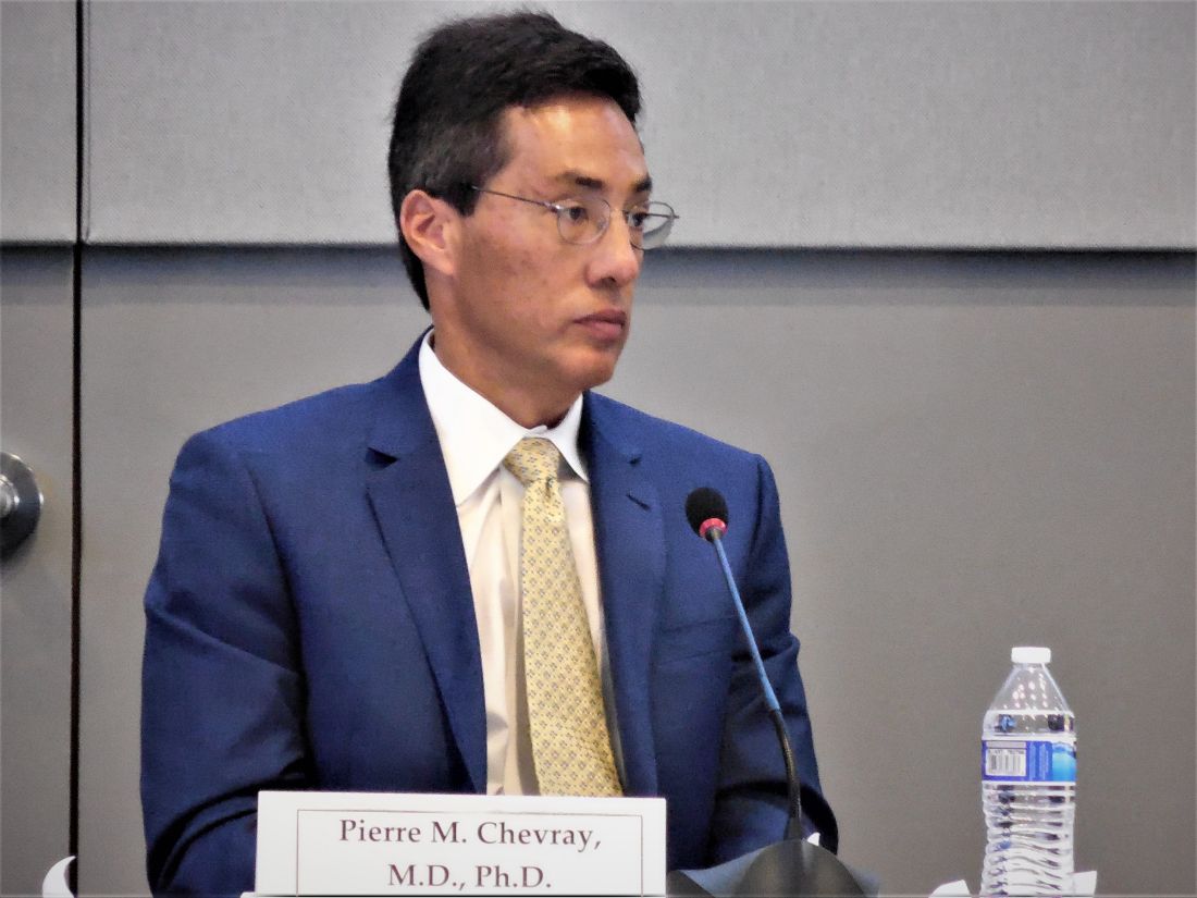
“I do not mention BII to patients. It’s not a disease; it’s a constellation of symptoms,” said panel member and plastic surgeon Pierre M. Chevray, MD, from Houston Methodist Hospital. The evidence for BII “is extremely anecdotal,” he said in an interview at the end of the 2-day session. Descriptions of BII “have been mainly published on social media. One reason why I don’t tell patients [about BII as part of informed consent] is because right now the evidence of a link is weak. We don’t yet even have a definition of this as an illness. A first step is to define it,” said Dr. Chevray, who has a very active implant practice. Other plastic surgeons were more accepting of BII as a real complication, although they agreed it needs much more study. During the testimony period, St. Louis plastic surgeon Patricia A. McGuire, MD, highlighted the challenge of teasing apart whether real symptoms are truly related to implants or are simply common ailments that accumulate during middle-age in many women. Dr. McGuire and some of her associates published an assessment of the challenges and possible solutions to studying BII that appeared shortly before the hearing (Plast Reconstr Surg. 2019 March;143[3S]:74S-81S),
Consensus recommendations from the panel to the FDA to address BII included having a single registry that would include all U.S. patients who receive breast implants (recently launched as the National Breast Implant Registry), inclusion of a control group, and collection of data at baseline and after regular follow-up intervals that includes a variety of measures relevant to autoimmune and rheumatologic disorders. Several panel members cited inadequate postmarketing safety surveillance by manufacturers in the years since breast implants returned to the U.S. market, and earlier in March, the FDA issued warning letters to two of the four companies that market U.S. breast implants over their inadequate long-term safety follow-up.
The panel’s decisions about the other major implant-associated health risk it considered, breast implant associated anaplastic large cell lymphoma (BIA-ALCL), faced a different sort of challenge. First described as linked to breast implants in 2011, today there is little doubt that BIA-ALCL is a consequence of breast implants, what several patients derisively called a “man-made cancer.” The key issue the committee grappled with was whether the calculated incidence of BIA-ALCL was at a frequency that warranted a ban on at least selected breast implant types. Mark W. Clemens, MD, a plastic surgeon at MD Anderson Cancer Center in Houston, told the panel that he calculated the Allergan Biocell group of implants, which have textured surfaces that allows for easier and more stable placement in patients, linked with an incidence of BIA-ALCL that was sevenfold to eightfold higher than that with smooth implants. That’s against a background of an overall incidence of about one case for every 20,000 U.S. implant recipients, Dr. Clemens said.
Many testifying patients, including several of the eight who described a personal history of BIA-ALCL, called for a ban on the sale of at least some breast implants because of their role in causing lymphoma. That sentiment was shared by Dr. Chevray, who endorsed a ban on “salt-loss” implants (the method that makes Biocell implants) during his closing comments to his fellow panel members. But earlier during panel discussions, others on the committee pushed back against implant bans, leaving the FDA’s eventual decision on this issue unclear. Evidence presented during the hearings suggests that implants cause ALCL by triggering a local “inflammatory milieu” and that different types of implants can have varying levels of potency for producing this milieu.

Perhaps the closest congruence between what patients called for and what the committee recommended was on informed consent. “No doubt, patients feel that informed consent failed them,” concluded panel member Karen E. Burke, MD, a New York dermatologist who was one of two panel discussants for the topic.
In addition to many suggestions on how to improve informed consent and public awareness lobbed at FDA staffers during the session by panel members, the final public comment of the 2 days came from Laurie A. Casas, MD, a Chicago plastic surgeon affiliated with the University of Chicago and a member of the board of directors of the American Society of Aesthetic Plastic Surgery (also know as the Aesthetic Society). During her testimony, Dr. Casas said “Over the past 2 days, we heard that patients need a structured educational checklist for informed consent. The Aesthetic Society hears you,” and promised that the website of the Society’s publication, the Aesthetic Surgery Journal, will soon feature a safety checklist for people receiving breast implants that will get updated as new information becomes available. She also highlighted the need for a comprehensive registry and long-term follow-up of implant recipients by the plastic surgeons who treated them.
In addition to better informed consent, patients who came to the hearing clearly also hoped to raise awareness in the general American public about the potential dangers from breast implants and the need to follow patients who receive implants. The 2 days of hearing accomplished that in part just by taking place. The New York Times and The Washington Post ran at least a couple of articles apiece on implant safety just before or during the hearings, while a more regional paper, the Philadelphia Inquirer, ran one article, as presumably did many other newspapers, broadcast outlets, and websites across America. Much of the coverage focused on compelling and moving personal stories from patients.
Women who have been having adverse effects from breast implants “have felt dismissed,” noted panel member Natalie C. Portis, PhD, a clinical psychologist from Oakland, Calif., and the patient representative on the advisory committee. “We need to listen to women that something real is happening.”
Dr. Tervaert, Dr. Chevray, Dr. McGuire, Dr. Clemens, Dr. Burke, Dr. Casas, and Dr. Portis had no relevant commercial disclosures.
What’s the role of anecdotal medical histories in the era of evidence-based medicine?
But the anecdotal histories fell short of producing a clear committee consensus on dramatic, immediate changes in FDA policy, such as joining a renewed ban on certain types of breast implants linked with a rare lymphoma, a step recently taken by 38 other countries, including 33 European countries acting in concert through the European Union.
The disconnect between gripping testimony and limited panel recommendations was most stark for a complication that’s been named Breast Implant Illness (BII) by patients on the Internet. Many breast implant recipients have reported life-changing symptoms that appeared after implant placement, most often fatigue, joint and muscle pain, brain fog, neurologic symptoms, immune dysfunction, skin manifestations, and autoimmune disease or symptoms. By my count, 22 people spoke about their harrowing experiences with BII symptoms out of the 77 who stepped to the panel’s public-comment mic during 4 hours of public testimony over 2-days of hearings, often saying that they had experienced dramatic improvements after their implants came out. The meeting of the General and Plastic Surgery Devices Panel of the Medical Devices Advisory Committee also heard presentations from two experts who ran some of the first reported studies on BII, or a BII-like syndrome called Autoimmune Syndrome Induced by Adjuvants (ASIA) described by Jan W.C. Tervaert, MD, professor of medicine and director of rheumatology at the University of Alberta in Edmonton. Dr. Tervaert and his associates published their findings about ASIA in the rheumatology literature last year (Clin Rheumatol. 2018 Feb;37[2]:483-93), and during his talk before the FDA panel, he said that silicone breast implants and the surgical mesh often used with them could be ASIA triggers.
Panel members seemed to mostly believe that the evidence they heard about BII did no more than hint at a possible association between breast implants and BII symptoms that required additional study. Many agreed on the need to include mention of the most common BII-linked patient complaints in informed consent material, but some were reluctant about even taking that step.
“I do not mention BII to patients. It’s not a disease; it’s a constellation of symptoms,” said panel member and plastic surgeon Pierre M. Chevray, MD, from Houston Methodist Hospital. The evidence for BII “is extremely anecdotal,” he said in an interview at the end of the 2-day session. Descriptions of BII “have been mainly published on social media. One reason why I don’t tell patients [about BII as part of informed consent] is because right now the evidence of a link is weak. We don’t yet even have a definition of this as an illness. A first step is to define it,” said Dr. Chevray, who has a very active implant practice. Other plastic surgeons were more accepting of BII as a real complication, although they agreed it needs much more study. During the testimony period, St. Louis plastic surgeon Patricia A. McGuire, MD, highlighted the challenge of teasing apart whether real symptoms are truly related to implants or are simply common ailments that accumulate during middle-age in many women. Dr. McGuire and some of her associates published an assessment of the challenges and possible solutions to studying BII that appeared shortly before the hearing (Plast Reconstr Surg. 2019 March;143[3S]:74S-81S),
Consensus recommendations from the panel to the FDA to address BII included having a single registry that would include all U.S. patients who receive breast implants (recently launched as the National Breast Implant Registry), inclusion of a control group, and collection of data at baseline and after regular follow-up intervals that includes a variety of measures relevant to autoimmune and rheumatologic disorders. Several panel members cited inadequate postmarketing safety surveillance by manufacturers in the years since breast implants returned to the U.S. market, and earlier in March, the FDA issued warning letters to two of the four companies that market U.S. breast implants over their inadequate long-term safety follow-up.
The panel’s decisions about the other major implant-associated health risk it considered, breast implant associated anaplastic large cell lymphoma (BIA-ALCL), faced a different sort of challenge. First described as linked to breast implants in 2011, today there is little doubt that BIA-ALCL is a consequence of breast implants, what several patients derisively called a “man-made cancer.” The key issue the committee grappled with was whether the calculated incidence of BIA-ALCL was at a frequency that warranted a ban on at least selected breast implant types. Mark W. Clemens, MD, a plastic surgeon at MD Anderson Cancer Center in Houston, told the panel that he calculated the Allergan Biocell group of implants, which have textured surfaces that allows for easier and more stable placement in patients, linked with an incidence of BIA-ALCL that was sevenfold to eightfold higher than that with smooth implants. That’s against a background of an overall incidence of about one case for every 20,000 U.S. implant recipients, Dr. Clemens said.
Many testifying patients, including several of the eight who described a personal history of BIA-ALCL, called for a ban on the sale of at least some breast implants because of their role in causing lymphoma. That sentiment was shared by Dr. Chevray, who endorsed a ban on “salt-loss” implants (the method that makes Biocell implants) during his closing comments to his fellow panel members. But earlier during panel discussions, others on the committee pushed back against implant bans, leaving the FDA’s eventual decision on this issue unclear. Evidence presented during the hearings suggests that implants cause ALCL by triggering a local “inflammatory milieu” and that different types of implants can have varying levels of potency for producing this milieu.
Perhaps the closest congruence between what patients called for and what the committee recommended was on informed consent. “No doubt, patients feel that informed consent failed them,” concluded panel member Karen E. Burke, MD, a New York dermatologist who was one of two panel discussants for the topic.
In addition to many suggestions on how to improve informed consent and public awareness lobbed at FDA staffers during the session by panel members, the final public comment of the 2 days came from Laurie A. Casas, MD, a Chicago plastic surgeon affiliated with the University of Chicago and a member of the board of directors of the American Society of Aesthetic Plastic Surgery (also know as the Aesthetic Society). During her testimony, Dr. Casas said “Over the past 2 days, we heard that patients need a structured educational checklist for informed consent. The Aesthetic Society hears you,” and promised that the website of the Society’s publication, the Aesthetic Surgery Journal, will soon feature a safety checklist for people receiving breast implants that will get updated as new information becomes available. She also highlighted the need for a comprehensive registry and long-term follow-up of implant recipients by the plastic surgeons who treated them.
In addition to better informed consent, patients who came to the hearing clearly also hoped to raise awareness in the general American public about the potential dangers from breast implants and the need to follow patients who receive implants. The 2 days of hearing accomplished that in part just by taking place. The New York Times and The Washington Post ran at least a couple of articles apiece on implant safety just before or during the hearings, while a more regional paper, the Philadelphia Inquirer, ran one article, as presumably did many other newspapers, broadcast outlets, and websites across America. Much of the coverage focused on compelling and moving personal stories from patients.
Women who have been having adverse effects from breast implants “have felt dismissed,” noted panel member Natalie C. Portis, PhD, a clinical psychologist from Oakland, Calif., and the patient representative on the advisory committee. “We need to listen to women that something real is happening.”
Dr. Tervaert, Dr. Chevray, Dr. McGuire, Dr. Clemens, Dr. Burke, Dr. Casas, and Dr. Portis had no relevant commercial disclosures.
UNITY-NHL: Interim findings show activity, tolerability of umbralisib for R/R MZL
ATLANTA – The phosphoinositide 3-kinase (PI3K) delta inhibitor umbralisib is active and well tolerated as single-agent therapy in patients with relapsed or refractory marginal zone lymphoma, according to findings from the ongoing phase 2 UNITY-NHL study.
The best overall response rate (ORR) among 42 study participants with at least 9 months of follow-up was 52% by both independent review committee (IRC) and investigator assessment, and the complete response rate was 19%, Nathan H. Fowler, MD, reported at the annual meeting of the American Association for Cancer Research.
The ORR by IRC assessment for those with extranodal, nodal, and splenic disease was 57%, 42%, and 43%, respectively, and for those with prior chemo-immunotherapy and those refractory to their last line of therapy, the ORR was 53% and 38%, respectively, said Dr. Fowler of MD Anderson Cancer Center, Houston.
The clinical benefit rate was 88%, and the median duration of response and progression-free survival (PFS) have not been reached; 12-month PFS is estimated at 66%, he added.
Patients in the marginal zone lymphoma cohort of the multicohort trial, which is evaluating umbralisib both as monotherapy and as part of various combinations in patients with previously treated non-Hodgkin’s lymphoma, received single agent umbralisib at a dose of 800 mg daily until disease progression or unacceptable toxicity.
“Most patients who have responded continue on drug,” he said in a video interview.
Dr. Fowler also further discussed the findings to date with respect to response, adverse events, and future directions in the wake of the breakthrough therapy designation recently granted by the Food and Drug Administration for umbralisib based on these findings.
“Despite the fact that a lot of things work in the front line, when patients relapse, especially when they become refractory to rituximab, there are limited options available,” he said, noting that currently used treatments are associated with significant adverse event and short remission times. “We still need better options.”
REPORTING FROM AACR 2019
Combo could replace standard conditioning regimen for myeloma
Busulfan plus melphalan could replace melphalan alone as the standard conditioning regimen for multiple myeloma patients undergoing autologous hematopoietic cell transplant, according to researchers.

In a phase 3 trial, patients who received conditioning with busulfan plus melphalan had significantly longer median progression-free survival compared with patients who received melphalan alone – 64.7 months versus 43.5 months (P = .022).
However, there was no overall survival advantage with busulfan plus melphalan, and adverse events were more common with this regimen, reported Qaiser Bashir, MD, of the University of Texas MD Anderson Cancer Center in Houston, and his colleagues. The report is in The Lancet Haematology.
To their knowledge, the researchers wrote, this was the first randomized, phase 3 trial showing a significant progression-free survival benefit for busulfan plus melphalan versus the standard of care of melphalan 200 mg/m2 pretransplantation conditioning. “These data suggest that busulfan plus melphalan conditioning can serve as a useful platform for further improvement of transplant outcomes in patients with myeloma.”
The current trial (NCT01413178) enrolled 205 multiple myeloma patients who were eligible for transplant. They were randomized to conditioning with melphalan alone or busulfan plus melphalan.
In all, 98 patients received melphalan alone, given at 200 mg/m2 on day –2. The 104 patients who received busulfan plus melphalan started with a test dose of busulfan at 32 mg/m2, which was followed by pharmacokinetically adjusted doses on days –7, –6, –5, and –4 to achieve a target daily area under the curve of 5,000 mmol/minute. These patients received melphalan at 70 mg/m2 per day on days –2 and –1.
The median age at transplant was 57.9 years (range, 31.7-70.9 years) in the busulfan group and 57.6 years (range, 34.3-70.6 years) in the melphalan-alone group.
The most common induction regimen used was bortezomib, lenalidomide, and dexamethasone, which was given to 60% of the busulfan group and 57% of the melphalan-alone group.
Most patients responded to induction – 96% of patients in the busulfan group and 94% of those in the melphalan-alone group.
There was no treatment-related mortality within 100 days of transplant.
At 90 days after transplant, the response rate was 98% in the busulfan group and 97% in the melphalan-alone group. The rate of complete remission/stringent complete remission was 27% and 34%, respectively.
Most patients received posttransplant maintenance. The most common maintenance regimen consisted of lenalidomide monotherapy, which was given to 57% of the busulfan group and 58% of the melphalan-alone group.
Patients continued maintenance until disease progression or unacceptable toxicity. The median duration of maintenance was 16.0 months in the busulfan group and 10.1 months in the melphalan-alone group.
The median follow-up was 22.6 months in the busulfan group and 20.2 months in the melphalan-alone group.
Progression-free survival was superior in the busulfan group. Median progression-free survival was 64.7 months in the busulfan group and 43.5 months in the melphalan-alone group (hazard ratio = 0.53; P = .022). The 3-year progression-free survival rate was 72% and 50%, respectively.
The median overall survival was not reached in either group. The 3-year overall survival rate was 91% in the busulfan group and 89% in the melphalan-alone group.
There were 10 deaths in the busulfan group, 7 due to progression and 3 due to infection. All 7 deaths in the melphalan-alone group were due to progression.
Grade 3-4 nonhematologic toxicity was more common in the busulfan group, occurring in 84% of that group and 33% of the melphalan-alone group (P less than .0001).
Grade 2-3 mucositis occurred in 74% of the busulfan group and 14% of the melphalan-alone group. There were no cases of grade 4 mucositis.
One patient in the busulfan group had grade 4 cardiac toxicity, an acute myocardial infarction, and ventricular fibrillation. However, the patient recovered and was in remission at last follow-up.
Two patients in the busulfan group developed second primary malignancies. One patient developed squamous cell skin cancer and rectal adenocarcinoma, and the other developed melanoma and basal cell skin carcinoma.
Three patients in the melphalan-alone group developed second primary malignancies. Two patients had squamous cell skin cancers and one had myelodysplastic syndrome.
Dr. Bashir and his colleagues noted that this study has limitations, including insufficient data to assess minimal residual disease and its impact on survival. It is a single-center study and induction and maintenance therapies were not prespecified.
“These results should be verified in a cooperative group or a multicenter, randomized study to assess the generalizability of our findings,” the researchers wrote.
Dr. Bashir and his colleagues reported having no competing financial interests. The trial was sponsored by MD Anderson Cancer Center in collaboration with Otsuka Pharmaceutical Development & Commercialization. The work was funded in part by the National Institutes of Health.
SOURCE: Bashir Q et al. Lancet Haematol. 2019 Mar 22. doi: 10.1016/S2352-3026(19)30023-7.
Busulfan plus melphalan could replace melphalan alone as the standard conditioning regimen for multiple myeloma patients undergoing autologous hematopoietic cell transplant, according to researchers.
In a phase 3 trial, patients who received conditioning with busulfan plus melphalan had significantly longer median progression-free survival compared with patients who received melphalan alone – 64.7 months versus 43.5 months (P = .022).
However, there was no overall survival advantage with busulfan plus melphalan, and adverse events were more common with this regimen, reported Qaiser Bashir, MD, of the University of Texas MD Anderson Cancer Center in Houston, and his colleagues. The report is in The Lancet Haematology.
To their knowledge, the researchers wrote, this was the first randomized, phase 3 trial showing a significant progression-free survival benefit for busulfan plus melphalan versus the standard of care of melphalan 200 mg/m2 pretransplantation conditioning. “These data suggest that busulfan plus melphalan conditioning can serve as a useful platform for further improvement of transplant outcomes in patients with myeloma.”
The current trial (NCT01413178) enrolled 205 multiple myeloma patients who were eligible for transplant. They were randomized to conditioning with melphalan alone or busulfan plus melphalan.
In all, 98 patients received melphalan alone, given at 200 mg/m2 on day –2. The 104 patients who received busulfan plus melphalan started with a test dose of busulfan at 32 mg/m2, which was followed by pharmacokinetically adjusted doses on days –7, –6, –5, and –4 to achieve a target daily area under the curve of 5,000 mmol/minute. These patients received melphalan at 70 mg/m2 per day on days –2 and –1.
The median age at transplant was 57.9 years (range, 31.7-70.9 years) in the busulfan group and 57.6 years (range, 34.3-70.6 years) in the melphalan-alone group.
The most common induction regimen used was bortezomib, lenalidomide, and dexamethasone, which was given to 60% of the busulfan group and 57% of the melphalan-alone group.
Most patients responded to induction – 96% of patients in the busulfan group and 94% of those in the melphalan-alone group.
There was no treatment-related mortality within 100 days of transplant.
At 90 days after transplant, the response rate was 98% in the busulfan group and 97% in the melphalan-alone group. The rate of complete remission/stringent complete remission was 27% and 34%, respectively.
Most patients received posttransplant maintenance. The most common maintenance regimen consisted of lenalidomide monotherapy, which was given to 57% of the busulfan group and 58% of the melphalan-alone group.
Patients continued maintenance until disease progression or unacceptable toxicity. The median duration of maintenance was 16.0 months in the busulfan group and 10.1 months in the melphalan-alone group.
The median follow-up was 22.6 months in the busulfan group and 20.2 months in the melphalan-alone group.
Progression-free survival was superior in the busulfan group. Median progression-free survival was 64.7 months in the busulfan group and 43.5 months in the melphalan-alone group (hazard ratio = 0.53; P = .022). The 3-year progression-free survival rate was 72% and 50%, respectively.
The median overall survival was not reached in either group. The 3-year overall survival rate was 91% in the busulfan group and 89% in the melphalan-alone group.
There were 10 deaths in the busulfan group, 7 due to progression and 3 due to infection. All 7 deaths in the melphalan-alone group were due to progression.
Grade 3-4 nonhematologic toxicity was more common in the busulfan group, occurring in 84% of that group and 33% of the melphalan-alone group (P less than .0001).
Grade 2-3 mucositis occurred in 74% of the busulfan group and 14% of the melphalan-alone group. There were no cases of grade 4 mucositis.
One patient in the busulfan group had grade 4 cardiac toxicity, an acute myocardial infarction, and ventricular fibrillation. However, the patient recovered and was in remission at last follow-up.
Two patients in the busulfan group developed second primary malignancies. One patient developed squamous cell skin cancer and rectal adenocarcinoma, and the other developed melanoma and basal cell skin carcinoma.
Three patients in the melphalan-alone group developed second primary malignancies. Two patients had squamous cell skin cancers and one had myelodysplastic syndrome.
Dr. Bashir and his colleagues noted that this study has limitations, including insufficient data to assess minimal residual disease and its impact on survival. It is a single-center study and induction and maintenance therapies were not prespecified.
“These results should be verified in a cooperative group or a multicenter, randomized study to assess the generalizability of our findings,” the researchers wrote.
Dr. Bashir and his colleagues reported having no competing financial interests. The trial was sponsored by MD Anderson Cancer Center in collaboration with Otsuka Pharmaceutical Development & Commercialization. The work was funded in part by the National Institutes of Health.
SOURCE: Bashir Q et al. Lancet Haematol. 2019 Mar 22. doi: 10.1016/S2352-3026(19)30023-7.
FROM LANCET HAEMATOLOGY
FDA panel calls for changes to breast implant rupture screening
A Food and Drug Administration advisory panel urged the agency to switch its recommended screening method for silent breast implant ruptures from MRI to ultrasound and to push the first screening examination back from the current 3 years post implant to 5 years.
Members of the FDA’s General and Plastic Surgery Advisory Panel also made suggestions to the FDA regarding how it might improve communication about the risks of breast implants to the public in general and to people considering implants in particular.

The panel also discussed the sort of safety and efficacy assessments the FDA should require for acellular dermal matrix (ADM), also known as mesh, to add the material’s label for use during breast reconstruction or implant augmentation. Surgeons have used mesh routinely as a surgical aid at other body sites, such as the abdomen. Although ADM is now also widely used during breast surgery, it has never undergone testing or labeling for use in that setting.
The FDA convened the advisory committee meeting largely to assess and discuss data and concerns about two recently appreciated complications of breast implant placement – breast implant–associated anaplastic large-cell lymphoma (BIA-ALCL) and a still poorly defined and described constellation of autoimmune and rheumatoid-like symptoms reported anecdotally by some breast implant recipients called Breast Implant Illness (BII). But agency officials asked the panel to also address these other issues related to the safety of breast implants and implant surgery.
The revised screening recommendations were primarily a response to a lack of compliance with current FDA recommendations to screen for breast implant rupture with MRI starting 3 years after placement and then every 2 years.
The problem is that a screening MRI costs about $1,500-$2,000 and is generally not covered by insurance when done for this purpose, although it is often covered when used to investigate a suspected rupture. The result is that less than 5% of implanted patients comply with the recommended screening schedule, noted committee chair Frank R. Lewis Jr., MD, executive director, emeritus, of the American Board of Surgery in Philadelphia.
“Effectively it’s a useless recommendation,” he said. “Ultrasound is far easier, quicker, and cheaper” and seems effective for screening.
The advisory panel recommended starting ultrasound screening 5 years after implantation, based on MRI screening data showing that virtually all ruptures don’t occur until after 5 years, and then following with ultrasound screening every 3 years after that. The panel recommended using MRI when the ultrasound result is equivocal or when the patient has symptoms suggesting rupture.
After hearing testimony during the sessions from several dozen women who told horror stories of the complications they experienced from breast implants, panel member Karen E. Burke, MD, PhD, spoke for many on the panel when she said “no doubt patients feel that the informed consent process failed them, that they were not aware of the risks.”
Dr. Burke suggested that patients must be informed so that they realize that breast implants are not static objects that will always sit unchanged in their body for the rest of their lives, that certain factors such as allergy or family history of tissue disease might predispose them to autoimmune-type reactions and that the diverse symptoms described for BII are possible sequelae.
A black box warning for the potential of developing anaplastic large-cell lymphoma should also go into the label, said Dr. Burke, a dermatologist who practices in New York City.
Dr. Lewis ridiculed the information booklets that implant manufacturers currently provide for patients as too long and dense. “They were not constructed to inform patients in the best way; they were constructed to provide legal protection.” He called for creating a two- or three-page list of potential adverse effects and points to consider.
Other panel members suggested public service advertisements similar to what is used to inform consumers about the risk from cigarettes. Dr. Burke recommended getting the word out about BII to other medical specialties that are more likely to see affected patients first, such as rheumatologists, immunologists, and dermatologists. She vowed to speak about these complications at an upcoming meeting of the American Academy of Dermatology. But other panel members noted that BII right now remains without any official medical definition nor clear causal link to breast implants.

The question of exactly what safety and efficacy data the FDA might require from manufacturers seeking a breast surgery indication for ADM was less clear.
Binita Ashar, MD, director of the FDA’s Division of Surgical Devices, highlighted the agency’s dilemma about considering data for a breast surgery indication. “The challenge for us is that we can’t expect a control arm because everyone today is using” mesh, she explained. “We’re looking for guidance on how to understand the risk-to-benefit profile” of ADM.
A plastic surgeon on the advisory panel, Pierre M. Chevray, MD, PhD, from Houston Methodist Hospital summarized the way ADM mesh reached its current niche in routine, U.S. breast surgery.
About 20 years ago, plastic surgeons began using mesh during implant surgery to improve eventual breast cosmesis. Surgeons began to wrap the implant in mesh and then attached the mesh to the pectoral muscle so that the implant could go on top of the muscle and not beneath it. It greatly diminished capsular contraction around the implant over time, reduced the risk for implant movement, and allowed for more natural positioning of the breast with the implant inside, he said.
Another factor in the growing use of mesh was heavy promotion by manufacturers to a generation of plastic surgeons, Dr. Chevray said. But use of ADM may also lead to a slightly increased rate of seromas and infections.
“The benefit from mesh is hard to prove and is questionable” because it largely depends on a subjective assessment by a surgeon or patient, Dr. Chevray said. “The cost [of ADM] is substantial, but no data have shown that outcomes are better” with its use. Despite that, “nearly every surgeon uses mesh” these days, he noted.
A Food and Drug Administration advisory panel urged the agency to switch its recommended screening method for silent breast implant ruptures from MRI to ultrasound and to push the first screening examination back from the current 3 years post implant to 5 years.
Members of the FDA’s General and Plastic Surgery Advisory Panel also made suggestions to the FDA regarding how it might improve communication about the risks of breast implants to the public in general and to people considering implants in particular.
The panel also discussed the sort of safety and efficacy assessments the FDA should require for acellular dermal matrix (ADM), also known as mesh, to add the material’s label for use during breast reconstruction or implant augmentation. Surgeons have used mesh routinely as a surgical aid at other body sites, such as the abdomen. Although ADM is now also widely used during breast surgery, it has never undergone testing or labeling for use in that setting.
The FDA convened the advisory committee meeting largely to assess and discuss data and concerns about two recently appreciated complications of breast implant placement – breast implant–associated anaplastic large-cell lymphoma (BIA-ALCL) and a still poorly defined and described constellation of autoimmune and rheumatoid-like symptoms reported anecdotally by some breast implant recipients called Breast Implant Illness (BII). But agency officials asked the panel to also address these other issues related to the safety of breast implants and implant surgery.
The revised screening recommendations were primarily a response to a lack of compliance with current FDA recommendations to screen for breast implant rupture with MRI starting 3 years after placement and then every 2 years.
The problem is that a screening MRI costs about $1,500-$2,000 and is generally not covered by insurance when done for this purpose, although it is often covered when used to investigate a suspected rupture. The result is that less than 5% of implanted patients comply with the recommended screening schedule, noted committee chair Frank R. Lewis Jr., MD, executive director, emeritus, of the American Board of Surgery in Philadelphia.
“Effectively it’s a useless recommendation,” he said. “Ultrasound is far easier, quicker, and cheaper” and seems effective for screening.
The advisory panel recommended starting ultrasound screening 5 years after implantation, based on MRI screening data showing that virtually all ruptures don’t occur until after 5 years, and then following with ultrasound screening every 3 years after that. The panel recommended using MRI when the ultrasound result is equivocal or when the patient has symptoms suggesting rupture.
After hearing testimony during the sessions from several dozen women who told horror stories of the complications they experienced from breast implants, panel member Karen E. Burke, MD, PhD, spoke for many on the panel when she said “no doubt patients feel that the informed consent process failed them, that they were not aware of the risks.”
Dr. Burke suggested that patients must be informed so that they realize that breast implants are not static objects that will always sit unchanged in their body for the rest of their lives, that certain factors such as allergy or family history of tissue disease might predispose them to autoimmune-type reactions and that the diverse symptoms described for BII are possible sequelae.
A black box warning for the potential of developing anaplastic large-cell lymphoma should also go into the label, said Dr. Burke, a dermatologist who practices in New York City.
Dr. Lewis ridiculed the information booklets that implant manufacturers currently provide for patients as too long and dense. “They were not constructed to inform patients in the best way; they were constructed to provide legal protection.” He called for creating a two- or three-page list of potential adverse effects and points to consider.
Other panel members suggested public service advertisements similar to what is used to inform consumers about the risk from cigarettes. Dr. Burke recommended getting the word out about BII to other medical specialties that are more likely to see affected patients first, such as rheumatologists, immunologists, and dermatologists. She vowed to speak about these complications at an upcoming meeting of the American Academy of Dermatology. But other panel members noted that BII right now remains without any official medical definition nor clear causal link to breast implants.
The question of exactly what safety and efficacy data the FDA might require from manufacturers seeking a breast surgery indication for ADM was less clear.
Binita Ashar, MD, director of the FDA’s Division of Surgical Devices, highlighted the agency’s dilemma about considering data for a breast surgery indication. “The challenge for us is that we can’t expect a control arm because everyone today is using” mesh, she explained. “We’re looking for guidance on how to understand the risk-to-benefit profile” of ADM.
A plastic surgeon on the advisory panel, Pierre M. Chevray, MD, PhD, from Houston Methodist Hospital summarized the way ADM mesh reached its current niche in routine, U.S. breast surgery.
About 20 years ago, plastic surgeons began using mesh during implant surgery to improve eventual breast cosmesis. Surgeons began to wrap the implant in mesh and then attached the mesh to the pectoral muscle so that the implant could go on top of the muscle and not beneath it. It greatly diminished capsular contraction around the implant over time, reduced the risk for implant movement, and allowed for more natural positioning of the breast with the implant inside, he said.
Another factor in the growing use of mesh was heavy promotion by manufacturers to a generation of plastic surgeons, Dr. Chevray said. But use of ADM may also lead to a slightly increased rate of seromas and infections.
“The benefit from mesh is hard to prove and is questionable” because it largely depends on a subjective assessment by a surgeon or patient, Dr. Chevray said. “The cost [of ADM] is substantial, but no data have shown that outcomes are better” with its use. Despite that, “nearly every surgeon uses mesh” these days, he noted.
AT AN FDA ADVISORY PANEL MEETING
FDA panel leans toward more robust breast implant surveillance
SILVER SPRING, MD. – A mandatory, comprehensive approach to collecting adverse event data from breast implant recipients was favored during a March 25 hearing by a Food and Drug Administration advisory panel that oversees surgical devices.
This additional data could offer more complete information during the informed consent process for breast implants and potentially validate a new, autoimmune-like syndrome – breast implant illness (BII).
On the first day of a scheduled 2-day hearing, the advisory panel held no votes and took no formal actions. After a day of expert presentations and comments from more than 40 members of the public – mostly personal stories from affected patients and from plastic surgeons who place breast implants, panel members discussed a handful of questions from the FDA about relevant data to collect to better define the risks posed to breast implant recipients from breast-implant associated anaplastic large cell lymphoma (BIA-ALCL) and BII.
The advisory panel meeting took place as reports recently appeared documenting the scope of BIA-ALCL (Plast Reconstr Surg. 2019 March;143[3S]:65S-73S) and how to diagnose and manage BIA-ALCL (Aesthetic Surg J. 2019 March;39[S1}:S3-S13), and the existence of BII (Plast Reconstr Surg. 2019 March;143[3S]:74S-81S).
During the day’s two public comment periods, the panel heard from several women who gave brief accounts of developing and dealing with BIA-ALCL or BII.
“We think it’s important that all breast implant patients be aware of the risk for BIA-ALCL,” said Binita Ashar, MD, director of the FDAs Division of Surgical Devices. The FDA “is asking the panel what further steps need to be taken to understand the BIA-ALCL risk,” said Dr. Ashar as she opened the meeting of the General and Plastic Surgery Devices Panel of the Medical Devices Advisory Committee.

While the agency, as well as the plastic surgery community, have acknowledged the existence of BIA-ALCL since 2011, only recently have good data emerged on the scope of the complication. During the hearing, Mark W. Clemens, MD, a plastic surgeon at MD Anderson Cancer Center in Houston, reported on his analysis of 457 unique cases of BIA-ALCL reported to the FDA since 2011. He found that the vast majority of cases had occurred in women who had received textured implants while a relatively small minority were linked with the placement of smooth implants.
Further scrutiny of the reported details of each case showed that none of the lymphomas were linked with a confirmed instance of “pure” smooth implant exposure. He also estimated the U.S. incidence of BIA-ALCL as roughly one case for every 20,000 implants. Complete, en bloc removal of the implant seems to be the most effective way to treat the cancer; most explanted patients have a good prognosis, he said.
Despite the apparent link between textured implants specifically and onset of BIA-ALCL, some panel members did not see a ban on textured implants as the answer.

Texturing the implant helps to stabilize the implant in position. Without texturing “we would need to use something else to stabilize the implant, or there would be a tsunami of reoperations,” said panel member Mary H. McGrath, MD, professor of surgery at the University of California, San Francisco. The main alternative to texturing for stabilizing implants is to wrap them in place using surgical mesh, but that approach may also cause problems.
“Instead of just taking textured implants off the market, we need to also look at their advantages. A critical issue is informed consent,” said panel member Marc E. Lippman, MD, a professor of medicine at Georgetown University, Washington. Banning smooth implants based on what’s known so far “would be an extraordinary over reaction,” he said during the first day’s session.
Current U.S. anecdotal experience suggests that a ban may not even be necessary because “plastic surgeons are more and more walking away from textured implants” because of the apparent link to BIA-ALCL, Dr. McGrath said.
BII has been a more recent and more controversial complication of breast implants. As recently as September 2018, Dr. Ashar said in a written statement that “the agency continues to believe that the weight of the currently available scientific evidence does not conclusively demonstrate an association between breast implants and connective tissue diseases,” the types of symptoms that characterize BII.
While the panel heard no new, conclusive evidence of a causal link between breast implants and the range of symptoms that some implant recipients report and is now collectively known as BII, several participants seemed convinced that the syndrome was real and needed better surveillance and study.
“It’s in the same family as chronic fatigue syndrome and fibromyalgia. It’s not a diagnosis, but a set of symptoms.” said Benjamin O. Anderson, MD, a surgical oncologist and professor of surgery at the University of Washington in Seattle and a panel member. “It’s a giant challenge. BII is a constellation of difficult symptoms. We need to think about how we ask patients, what are your symptoms?”

Frank R. Lewis Jr., MD, committee chair, said a more standardized measure of the most common BII symptoms is needed. “That may be exceedingly difficult, with as many as a hundred reported symptoms,” said Dr. Lewis, executive director, emeritus, of the American Board of Surgery in Philadelphia.
The hearing featured results from some of the most research projects aimed at fleshing out an understanding of BII.
Diana Zuckerman, PhD, president of the National Center for Health Research, reported data she and her associates collected in an online survey completed in late 2018 and early 2019 by 449 women who had approached the Center for help in getting health insurance coverage for medically-necessary explantation of their breast implants.
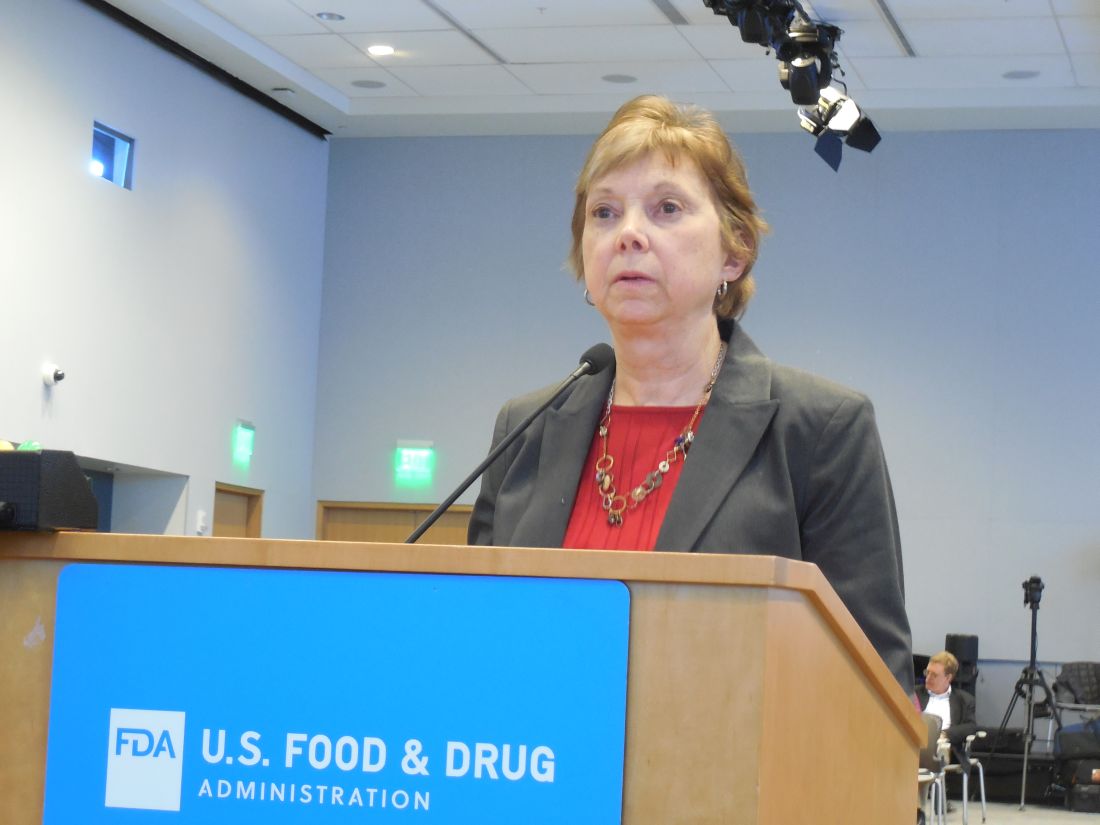
Their most common symptoms included joint, muscle or back pain, weakness or stiffness; fatigue; “brain fog;” and anxiety and depression. More than two-thirds of the respondents had a family history and 3% had a personal history of an autoimmune disease, and 61% said their symptoms improved after their implants were removed, Dr. Zuckerman reported during her presentation to the panel.
During the discussion, panel members seemed intent on expanding mandatory, routine surveillance to all breast implants placed in U.S. practice.
Andrea L. Pusic, MD, president of the Plastic Surgery Foundation, summarized the recent launch of the National Breast Implant Registry by the Foundation and its parent organization, the American Society of Plastic Surgeons. These organizations, and plastic surgeons in general, would be amenable to collecting the data the FDA deemed necessary to better track BIA-ALCL and BII, said Dr. Pusic, professor of surgery at Harvard Medical School and chief of plastic and reconstructive surgery at Brigham and Women’s Hospital in Boston.
“Plastic surgeons are willing to enter these data because we know they are important,” she told the FDA panel.
Dr. Ashar, Dr. Clemens, Dr. McGrath, Dr. Lippman, Dr. Anderson, Dr. Lewis, Dr. Zuckerman, and Dr. Pusic reported having no relevant commercial disclosures.
SILVER SPRING, MD. – A mandatory, comprehensive approach to collecting adverse event data from breast implant recipients was favored during a March 25 hearing by a Food and Drug Administration advisory panel that oversees surgical devices.
This additional data could offer more complete information during the informed consent process for breast implants and potentially validate a new, autoimmune-like syndrome – breast implant illness (BII).
On the first day of a scheduled 2-day hearing, the advisory panel held no votes and took no formal actions. After a day of expert presentations and comments from more than 40 members of the public – mostly personal stories from affected patients and from plastic surgeons who place breast implants, panel members discussed a handful of questions from the FDA about relevant data to collect to better define the risks posed to breast implant recipients from breast-implant associated anaplastic large cell lymphoma (BIA-ALCL) and BII.
The advisory panel meeting took place as reports recently appeared documenting the scope of BIA-ALCL (Plast Reconstr Surg. 2019 March;143[3S]:65S-73S) and how to diagnose and manage BIA-ALCL (Aesthetic Surg J. 2019 March;39[S1}:S3-S13), and the existence of BII (Plast Reconstr Surg. 2019 March;143[3S]:74S-81S).
During the day’s two public comment periods, the panel heard from several women who gave brief accounts of developing and dealing with BIA-ALCL or BII.
“We think it’s important that all breast implant patients be aware of the risk for BIA-ALCL,” said Binita Ashar, MD, director of the FDAs Division of Surgical Devices. The FDA “is asking the panel what further steps need to be taken to understand the BIA-ALCL risk,” said Dr. Ashar as she opened the meeting of the General and Plastic Surgery Devices Panel of the Medical Devices Advisory Committee.
While the agency, as well as the plastic surgery community, have acknowledged the existence of BIA-ALCL since 2011, only recently have good data emerged on the scope of the complication. During the hearing, Mark W. Clemens, MD, a plastic surgeon at MD Anderson Cancer Center in Houston, reported on his analysis of 457 unique cases of BIA-ALCL reported to the FDA since 2011. He found that the vast majority of cases had occurred in women who had received textured implants while a relatively small minority were linked with the placement of smooth implants.
Further scrutiny of the reported details of each case showed that none of the lymphomas were linked with a confirmed instance of “pure” smooth implant exposure. He also estimated the U.S. incidence of BIA-ALCL as roughly one case for every 20,000 implants. Complete, en bloc removal of the implant seems to be the most effective way to treat the cancer; most explanted patients have a good prognosis, he said.
Despite the apparent link between textured implants specifically and onset of BIA-ALCL, some panel members did not see a ban on textured implants as the answer.
Texturing the implant helps to stabilize the implant in position. Without texturing “we would need to use something else to stabilize the implant, or there would be a tsunami of reoperations,” said panel member Mary H. McGrath, MD, professor of surgery at the University of California, San Francisco. The main alternative to texturing for stabilizing implants is to wrap them in place using surgical mesh, but that approach may also cause problems.
“Instead of just taking textured implants off the market, we need to also look at their advantages. A critical issue is informed consent,” said panel member Marc E. Lippman, MD, a professor of medicine at Georgetown University, Washington. Banning smooth implants based on what’s known so far “would be an extraordinary over reaction,” he said during the first day’s session.
Current U.S. anecdotal experience suggests that a ban may not even be necessary because “plastic surgeons are more and more walking away from textured implants” because of the apparent link to BIA-ALCL, Dr. McGrath said.
BII has been a more recent and more controversial complication of breast implants. As recently as September 2018, Dr. Ashar said in a written statement that “the agency continues to believe that the weight of the currently available scientific evidence does not conclusively demonstrate an association between breast implants and connective tissue diseases,” the types of symptoms that characterize BII.
While the panel heard no new, conclusive evidence of a causal link between breast implants and the range of symptoms that some implant recipients report and is now collectively known as BII, several participants seemed convinced that the syndrome was real and needed better surveillance and study.
“It’s in the same family as chronic fatigue syndrome and fibromyalgia. It’s not a diagnosis, but a set of symptoms.” said Benjamin O. Anderson, MD, a surgical oncologist and professor of surgery at the University of Washington in Seattle and a panel member. “It’s a giant challenge. BII is a constellation of difficult symptoms. We need to think about how we ask patients, what are your symptoms?”
Frank R. Lewis Jr., MD, committee chair, said a more standardized measure of the most common BII symptoms is needed. “That may be exceedingly difficult, with as many as a hundred reported symptoms,” said Dr. Lewis, executive director, emeritus, of the American Board of Surgery in Philadelphia.
The hearing featured results from some of the most research projects aimed at fleshing out an understanding of BII.
Diana Zuckerman, PhD, president of the National Center for Health Research, reported data she and her associates collected in an online survey completed in late 2018 and early 2019 by 449 women who had approached the Center for help in getting health insurance coverage for medically-necessary explantation of their breast implants.
Their most common symptoms included joint, muscle or back pain, weakness or stiffness; fatigue; “brain fog;” and anxiety and depression. More than two-thirds of the respondents had a family history and 3% had a personal history of an autoimmune disease, and 61% said their symptoms improved after their implants were removed, Dr. Zuckerman reported during her presentation to the panel.
During the discussion, panel members seemed intent on expanding mandatory, routine surveillance to all breast implants placed in U.S. practice.
Andrea L. Pusic, MD, president of the Plastic Surgery Foundation, summarized the recent launch of the National Breast Implant Registry by the Foundation and its parent organization, the American Society of Plastic Surgeons. These organizations, and plastic surgeons in general, would be amenable to collecting the data the FDA deemed necessary to better track BIA-ALCL and BII, said Dr. Pusic, professor of surgery at Harvard Medical School and chief of plastic and reconstructive surgery at Brigham and Women’s Hospital in Boston.
“Plastic surgeons are willing to enter these data because we know they are important,” she told the FDA panel.
Dr. Ashar, Dr. Clemens, Dr. McGrath, Dr. Lippman, Dr. Anderson, Dr. Lewis, Dr. Zuckerman, and Dr. Pusic reported having no relevant commercial disclosures.
REPORTING FROM AN FDA ADVISORY COMMITTEE MEETING

Rituximab boosts survival in primary CNS lymphoma
For patients with primary central nervous system lymphoma (PCNSL), adding rituximab to combination high-dose methotrexate and temozolomide significantly boosted the 5-year overall survival rate, according to a retrospective study.
The triplet combination could be a safe and effective first-line option for patients with PCNSL, particularly the frail and elderly, who may have issues with toxicity when receiving current standard care, reported lead author Cui Chen, MD, of Sun Yat-Sen University Cancer Center in Guangzhou, China, and his colleagues.
“An increasing number of studies and meta‐analyses have investigated the effect of rituximab in PCNSL, indicating that rituximab can robustly enhance the response rate and possibly improve survival,” the investigators wrote in Cancer Medicine. “However, data regarding the addition of rituximab to [methotrexate and temozolomide] for PCNSL are limited, and no study has directly compared the efficacy of [rituximab/high-dose methotrexate/temozolomide] to that of [high-dose methotrexate/temozolomide].”
The study involved 62 patients with untreated PCNSL who were diagnosed between 2005 and 2015. Out of the 62 patients, 32 received rituximab/high-dose methotrexate/temozolomide (RMT) and 30 received high-dose methotrexate/temozolomide (MT). Patients received up to eight cycles of therapy, with discontinuation upon disease progression or toxicity.
The results showed that patients treated with RMT had significantly better outcomes than those who received MT, first marked by objective response rates, which were 93.7% for RMT and 69.0% for MT.
Survival rates also showed the advantage of rituximab. For the RMT group, 2-year and 5-year progression-free survival rates were 81.3% and 53.3%, respectively, compared with 46.5% and 29.1% for patients receiving MT.
Most importantly, rituximab boosted overall survival to a significant and notable extent, with higher rates at 2 years (82.3% vs. 65.7%) and 5 years (82.3% vs. 50.0%).
Efficacy did not diminish safety, as no significant differences in toxicity were found between treatment types. The most common grade 3-4 toxicities were hematologic; most commonly, this entailed neutropenia, which occurred in about one-quarter of patients.
“Given its outstanding efficacy and favorable toxicity, we consider RMT to be a feasible and safe therapeutic approach as a first‐line treatment for PCNSL. Moreover, RMT is an ideal regimen for elderly patients and frail populations who may not tolerate [whole‐brain radiation therapy] or [autologous stem‐cell transplantation],” the researchers wrote.
The study was funded by the Natural Science Foundation of Guangdong Province. The researchers reported having no conflicts of interest.
SOURCE: Chen C et al. Cancer Med. 2019 Mar 1. doi: 10.1002/cam4.1906.
FROM CANCER MEDICINE