User login
Three steps identify causes of most stillbirths
SAN FRANCISCO – The cause of stillbirth can be identified in two-thirds of cases by checking the placental histology, conducting an autopsy, and karyotype testing.
That’s a "major, major take-home point" that’s "very different than what I was taught" in medical training, Dr. Yair J. Blumenfeld said at a meeting on antepartum and intrapartum management sponsored by the University of California, San Francisco.
That finding from an important 2011 study and other new data in the past 5 years suggests that perhaps clinicians should take a staggered approach when ordering tests to search for the etiology of a stillbirth. "Maybe I shouldn’t do a $2,000 workup for thrombophilia and anticardiolipin antibodies if the autopsy showed me that there’s an underlying structural abnormality, or if there’s an abnormal karyotype," he suggested.

In general, a growing proportion of stillbirths is being attributed to maternal, fetal, or placental causes, shrinking the proportion relegated to "idiopathic" or unexplained stillbirth. The idea that most stillbirths are idiopathic is "somewhat old thinking" at this point, said Dr. Blumenfeld of Stanford (Calif.) University.
The Eunice Kennedy Shriver National Institute of Child Health and Human Development created the Stillbirth Collaborative Research Network, which developed a new system of determining the causes of stillbirth and tested it in a multicenter, population-based case-control study in five U.S. states during 2006-2008. Potential etiologies for each stillbirth were graded as a possible or probable cause of death based on a complete evaluation including autopsy, placental pathology, medical records, maternal interview, karyotype, and other laboratory tests.
Investigators found a probable cause in 61% of the 512 stillbirths from 500 women and a possible or probable cause in 76%. More than one possible or probable cause was found in 31% of stillbirths, showing some overlap in the causes of stillbirth. The leading causes of antepartum stillbirths were obstetric complications in 29% and placental pathology in 23%, although some of the causes of stillbirth varied significantly by race (JAMA 2011;306:2459-68).
Obstetric complications were less likely to be the cause of stillbirths in white women (in 22%) or Hispanic women (25%), compared with black women (44%) or women of other races (41%). Infection as a cause of stillbirth also was less likely in whites (7%) or Hispanics (8%), compared with blacks (25%) or other races (22%). Hispanics and whites, however, had higher rates of umbilical cord complications as a cause of stillbirth (13% for each), compared with blacks (4%) or other races (5%).
Among the clinically indicated tests for stillbirths, the placental histology identified a cause of stillbirth 52% of the time. An autopsy found a cause in 31% of cases, and karyotype testing identified a cause 9% of the time. Eight other screening tests found a cause for stillbirth in 0.4%-4.8% of cases, depending on the test. These included screens for antibodies, toxicology, or blood glucose; tests for syphilis, parvovirus, lupus anticoagulant, or anticardiolipin antibody; or detection of fetal blood in fetal-maternal hemorrhage.
"I’m not saying we shouldn’t do these things, but I think in today’s health care climate, especially with health care economics, you should start to think about maybe a staggered approach" in order to control costs, Dr. Blumenfeld said.
Controversy surrounds several topics in the search for stillbirth etiologies: whether chromosomal microarrays are better than karyotype testing; whether or not to order screening for thrombophilias and antiphospholipid antibodies; and what to do if the parents reject an autopsy.
In a study of 532 stillbirths, chromosomal microarray testing yielded results more often than did karyotyping – in 87% of cases vs. 71% – and detected aneuploidy or copy number variants more often, in 8.8% of cases vs. 6.5% (New Engl. J. Med. 2012;367:2185-93). Whether that difference is worth a price tag of approximately $2,000 for microarray testing remains to be seen, but "we’re going to see a lot more studies" of stillbirths using this and other new technologies, Dr. Blumenfeld said.
Practice bulletins from the American College of Obstetricians and Gynecologists are "all over the map" when it comes to deciding whether or not to test for thrombophilias and antiphospholipid antibodies when there’s a stillbirth," he said. "It’s still very, very controversial." It’s probably wise to use the results of autopsy, karyotyping, and placental histology to help decide whether to pursue these other tests, and to talk with patients about their family history of thrombophilia, what the placenta looked like, and other factors that could guide decision-making, he added.
How health care providers counseled parents affected parents’ decision to accept or decline an autopsy of their stillborn infant in 22% of cases, according to one study of 460 parents, 354 obstetricians, 21 perinatal pathologists, and 2,256 midwives (BJOG 2012;119:987-97). Altogether, 62% of parents agreed to an autopsy.
Parents who decline an autopsy still are likely to consent to a "fetal virtuopsy" – a physical exam and MRI or CT imaging of the stillborn infant, a separate study of 96 mothers suggests. Although 62% consented to autopsy, 99% consented to a virtuopsy. In a few cases, the MRI detected abnormalities that were missed on autopsy (Ultrasound Obstet. Gynecol. 2012;39:659-65).
"Clearly, this is not standard, but I think we’re going to see a lot more studies taking this kind of approach to women who are not accepting of an autopsy," Dr. Blumenfeld said. "Go back to your home institutions, find your favorite pediatrician, geneticist, or dysmorphologist, and ask them, ‘Are you willing to come and look at this stillbirth once it is born, and try to get some information just by looking at the infant?’ I guarantee that you will be able to find somebody like that in your institution. It’s something that we do at Stanford."
National statistics from 2006 suggest that 6 of every 1,000 live births will be stillbirths, a rate similar to the prevalence of congenital heart disease, he said. In 2006, there were 25,972 fetal deaths after 20 weeks’ gestation in the United States (Natl. Vital Stat. Rep. 2012:60;1-23). Long-term trends show that the rate of stillbirths has declined after 28 weeks’ gestation but not between 20 and 27 weeks’ gestation.
Dr. Blumenfeld reported having no financial disclosures.
On Twitter @sherryboschert
SAN FRANCISCO – The cause of stillbirth can be identified in two-thirds of cases by checking the placental histology, conducting an autopsy, and karyotype testing.
That’s a "major, major take-home point" that’s "very different than what I was taught" in medical training, Dr. Yair J. Blumenfeld said at a meeting on antepartum and intrapartum management sponsored by the University of California, San Francisco.
That finding from an important 2011 study and other new data in the past 5 years suggests that perhaps clinicians should take a staggered approach when ordering tests to search for the etiology of a stillbirth. "Maybe I shouldn’t do a $2,000 workup for thrombophilia and anticardiolipin antibodies if the autopsy showed me that there’s an underlying structural abnormality, or if there’s an abnormal karyotype," he suggested.
In general, a growing proportion of stillbirths is being attributed to maternal, fetal, or placental causes, shrinking the proportion relegated to "idiopathic" or unexplained stillbirth. The idea that most stillbirths are idiopathic is "somewhat old thinking" at this point, said Dr. Blumenfeld of Stanford (Calif.) University.
The Eunice Kennedy Shriver National Institute of Child Health and Human Development created the Stillbirth Collaborative Research Network, which developed a new system of determining the causes of stillbirth and tested it in a multicenter, population-based case-control study in five U.S. states during 2006-2008. Potential etiologies for each stillbirth were graded as a possible or probable cause of death based on a complete evaluation including autopsy, placental pathology, medical records, maternal interview, karyotype, and other laboratory tests.
Investigators found a probable cause in 61% of the 512 stillbirths from 500 women and a possible or probable cause in 76%. More than one possible or probable cause was found in 31% of stillbirths, showing some overlap in the causes of stillbirth. The leading causes of antepartum stillbirths were obstetric complications in 29% and placental pathology in 23%, although some of the causes of stillbirth varied significantly by race (JAMA 2011;306:2459-68).
Obstetric complications were less likely to be the cause of stillbirths in white women (in 22%) or Hispanic women (25%), compared with black women (44%) or women of other races (41%). Infection as a cause of stillbirth also was less likely in whites (7%) or Hispanics (8%), compared with blacks (25%) or other races (22%). Hispanics and whites, however, had higher rates of umbilical cord complications as a cause of stillbirth (13% for each), compared with blacks (4%) or other races (5%).
Among the clinically indicated tests for stillbirths, the placental histology identified a cause of stillbirth 52% of the time. An autopsy found a cause in 31% of cases, and karyotype testing identified a cause 9% of the time. Eight other screening tests found a cause for stillbirth in 0.4%-4.8% of cases, depending on the test. These included screens for antibodies, toxicology, or blood glucose; tests for syphilis, parvovirus, lupus anticoagulant, or anticardiolipin antibody; or detection of fetal blood in fetal-maternal hemorrhage.
"I’m not saying we shouldn’t do these things, but I think in today’s health care climate, especially with health care economics, you should start to think about maybe a staggered approach" in order to control costs, Dr. Blumenfeld said.
Controversy surrounds several topics in the search for stillbirth etiologies: whether chromosomal microarrays are better than karyotype testing; whether or not to order screening for thrombophilias and antiphospholipid antibodies; and what to do if the parents reject an autopsy.
In a study of 532 stillbirths, chromosomal microarray testing yielded results more often than did karyotyping – in 87% of cases vs. 71% – and detected aneuploidy or copy number variants more often, in 8.8% of cases vs. 6.5% (New Engl. J. Med. 2012;367:2185-93). Whether that difference is worth a price tag of approximately $2,000 for microarray testing remains to be seen, but "we’re going to see a lot more studies" of stillbirths using this and other new technologies, Dr. Blumenfeld said.
Practice bulletins from the American College of Obstetricians and Gynecologists are "all over the map" when it comes to deciding whether or not to test for thrombophilias and antiphospholipid antibodies when there’s a stillbirth," he said. "It’s still very, very controversial." It’s probably wise to use the results of autopsy, karyotyping, and placental histology to help decide whether to pursue these other tests, and to talk with patients about their family history of thrombophilia, what the placenta looked like, and other factors that could guide decision-making, he added.
How health care providers counseled parents affected parents’ decision to accept or decline an autopsy of their stillborn infant in 22% of cases, according to one study of 460 parents, 354 obstetricians, 21 perinatal pathologists, and 2,256 midwives (BJOG 2012;119:987-97). Altogether, 62% of parents agreed to an autopsy.
Parents who decline an autopsy still are likely to consent to a "fetal virtuopsy" – a physical exam and MRI or CT imaging of the stillborn infant, a separate study of 96 mothers suggests. Although 62% consented to autopsy, 99% consented to a virtuopsy. In a few cases, the MRI detected abnormalities that were missed on autopsy (Ultrasound Obstet. Gynecol. 2012;39:659-65).
"Clearly, this is not standard, but I think we’re going to see a lot more studies taking this kind of approach to women who are not accepting of an autopsy," Dr. Blumenfeld said. "Go back to your home institutions, find your favorite pediatrician, geneticist, or dysmorphologist, and ask them, ‘Are you willing to come and look at this stillbirth once it is born, and try to get some information just by looking at the infant?’ I guarantee that you will be able to find somebody like that in your institution. It’s something that we do at Stanford."
National statistics from 2006 suggest that 6 of every 1,000 live births will be stillbirths, a rate similar to the prevalence of congenital heart disease, he said. In 2006, there were 25,972 fetal deaths after 20 weeks’ gestation in the United States (Natl. Vital Stat. Rep. 2012:60;1-23). Long-term trends show that the rate of stillbirths has declined after 28 weeks’ gestation but not between 20 and 27 weeks’ gestation.
Dr. Blumenfeld reported having no financial disclosures.
On Twitter @sherryboschert
EXPERT ANALYSIS FROM A MEETING ON ANTEPARTUM AND INTRAPARTUM MANAGEMENT

Initial prenatal care visit is myth-busting time
SAN FRANCISCO – Dr. Meg Autry typically spends a first prenatal care visit talking about what her pregnant patient may have heard – the myths and the facts – regarding the dos and don’ts during pregnancy.
She’s not alone. Patients hear plenty of prenatal myths perpetuated not only by their peers but by some health care providers, she said at a meeting on antepartum and intrapartum management sponsored by the University of California, San Francisco.
An informal electronic poll of the physicians, nurses, and nurse-midwives at the meeting showed that 63% discuss diet at a new patient visit, 17% talk about environmental exposures, 12% discuss exercise, and 8% discuss dietary supplements. These topics were generated more by the health care providers than by the patients, they said.
Deli meats were the leading dietary topic, discussed by 34%, followed by cooked fish, sushi, vegetarian diets, and cheese, in that order. Most of the questions about exercise come from women who exercise a lot, 73% of respondents said, than from women who don’t exercise enough (27%). Iron led the list of supplements discussed (by 36%), followed by omega-3 fatty acids, and (in a third-place tie) calcium and vitamin D.
Dr. Autry, professor of ob.gyn. and reproductive sciences at the university, offered the following myth busters and evidence-based advice for prenatal care. "My goal is to give you a basis for what you talk about with patients," she said.
• Fish: A food that’s high in quality protein, low in saturated fat, and full of beneficial omega-3 fatty acids comes with a downside: Methylmercury that can impair fetal and newborn motor and cognitive skills. Two prospective studies of dietary fish’s effects in pregnancy produced conflicting results. One study that found adverse effects from daily eating of fish was conducted in the Danish Faeroe Islands, where much of what they ate was whale. The other study, in the Pacific Ocean’s Seychelles Islands, found infant neurodevelopmental benefits when mothers ate fish in 12 meals/week, including types of fish that were more similar to those eaten in the United States, Dr. Autry said.
The Food and Drug Administration and Environmental Protection Agency in 2004 advised consumers to eat up to 12 ounces (two average meals) per week of a variety of fish that are low in mercury and to check local advisories about the safety of locally caught fish.
Encourage women to eat fish, she said, "but don’t eat fish that live for a long time and that eat lots of other fish," such as shark, swordfish, king mackerel, tilefish, whale, or albacore tuna.
• Sushi: Very rare infectious diseases from raw fish are virtually a nonproblem in the United States, because most sushi is flash frozen, which kills most pathogens. "I don’t think you need to tell patients they need to stop eating sushi," she said. "Talk to them more about mercury" and talk about the benefits of eating moderate amounts of fish.
• Supplements: There’s no evidence that taking a supplement to get omega-3 fatty acids is beneficial, especially compared with eating fish. "We’re just supposed to eat it, we’re not supposed to have a pill to fix everything," she said.
A standard prenatal vitamin supplies the iron and other vitamins and minerals needed, and it’s good to advise patients to eat foods that promote iron absorption and are high in vitamin C, such as strawberries or broccoli. Warn women that their prenatal vitamin should be their maximum dose of vitamin A, a known teratogen. There’s no good evidence that taking extra calcium or vitamin D supplements generally is helpful in pregnancy, but the American College of Obstetricians and Gynecologists recommends considering vitamin D supplementation in women at risk of deficiency.
• Cheese: A third of U.S. cases of listeriosis occurs in pregnant women and is associated with miscarriage and stillbirth. Nationally, the bacteria Listeria reaches people most commonly through hot dogs. In California, it’s the queso fresco. The FDA and/or the U.S. Department of Agriculture advise pregnant women not to eat hot dogs or luncheon meats unless they’re reheating to steaming, and to avoid soft cheeses; refrigerated pâtés or meat spreads; smoked seafood; raw or unpasteurized milk; and raw or undercooked meats.
• Caffeine: Approximately 85% of U.S. women report eating or drinking caffeine-containing food or drinks, Dr. Autry said. Concerns that caffeine consumption might be associated with low birth weight, congenital anomalies, delay in conception, or miscarriage were poorly designed and confounded by an association between caffeine intake and cigarette smoking. More recent studies predominantly have been negative, and a randomized, controlled trial found no association between moderate caffeine intake and gestational age or birth weight (BMJ 2007;334:409).
• Alcohol: Bad news for the 10% of pregnant women who report ingesting alcohol and especially for the 2% who binge drink during pregnancy: There’s no safe level of alcohol intake during pregnancy. Federal data suggest that 1 in 6,000 U.S. newborns have fetal alcohol syndrome or fetal alcohol spectrum disorder.
• Nicotine: The tricky problem with nicotine is not just that it’s "associated with everything bad," Dr. Autry said, but that people know it’s bad, so an estimated 25%-50% of pregnant women don’t disclose that they smoke. Smoking in pregnancy is associated with miscarriage, abruption, ectopic pregnancy, preterm delivery, and more. Among mothers who quit smoking during pregnancy, 90% relapse after delivery. "It’s really important to continue the smoking cessation discussion during pregnancy," she said. "It’s important to say, ‘If you go back to smoking, don’t do it in the house, because it’s bad for the kid.’"
• Hot tubs: Soaking during the first trimester, or any time in pregnancy in water heated to 100 F or higher, is potentially teratogenic, two studies suggest. Maternal hyperthermia from hot tubs has been associated with first trimester fetal loss and with a nearly doubling in risk for neural tube defects.
• Exercise: The general recommendation to get 30 minutes or more of moderate exercise on most days applies to pregnant women unless they have some medical or obstetric complication. Exercise is believed to help prevent gestational diabetes, reduce the risk for preeclampsia and premature labor, and decrease the risk for postpartum depression. ACOG recommends avoiding scuba diving, contact sports, and supine activities or motionless standings. Yoga is fine, but avoid so-called "hot yoga," Dr. Autry said.
For high-performance athletes, exertion at high altitudes appears to be safe. There is no pregnancy-specific maximum heart rate.
"Just don’t do anything in pregnancy that you wouldn’t do before," Dr. Autry said. If you’ve never run a marathon, pregnancy is not the time to start.
• Hair dye: There are no data to support the idea of teratogenic effects from the chemicals in hair dyes. But it’s probably still a good idea to look at the labels and choose products with ingredients that are less long acting and organic, if possible, she said.
For resources on environmental exposures during pregnancy, see the university’s Program on Reproductive Health and the Environment.
Dr. Autry reported having no financial disclosures.
On Twitter @sherryboschert
EXPERT ANALYSIS AT A MEETING ON ANTEPARTUM AND INTRAPARTUM MANAGEMENT

No benefit seen with nifedipine maintenance tocolysis
SAN FRANCISCO – A large randomized, double-blind placebo-controlled study in the Netherlands supports earlier studies that found no benefit from routine maintenance tocolysis using nifedipine in women with arrested preterm labor.
What to do when preterm labor stops is "a million dollar question that many of us have faced," Dr. Deirdre J. Lyell said at a meeting on antepartum and intrapartum management sponsored by the University of California, San Francisco. Surveys suggest that more than a quarter of maternal-fetal medicine specialists routinely use maintenance tocolysis, most often with nifedipine, despite its questionable utility.

The Dutch study of 406 women "puts the question to bed," said Dr. Lyell, a maternal-fetal medicine ob.gyn. at Stanford (Calif.) University. She was not involved in the study.
The multicenter trial in these women was underpowered to show a statistically significant difference in its primary outcome – a composite of perinatal adverse outcomes – because of a lower-than-expected rate of adverse events in the control group, so it couldn’t exclude a possible benefit from nifedipine. However, "its use for maintenance tocolysis does not appear beneficial at this time," the investigators wrote (JAMA 2013;309:41-7).
Women in the study had arrested preterm labor at 24-34 weeks’ gestation. They were randomized to maintenance tocolysis with 20 mg nifedipine or placebo every 4-6 hours. Delivery occurred at 35 weeks in the nifedipine group and at 35 weeks and 2 days in the control group. The number of days of pregnancy after starting treatment was 34 and 33, respectively, and the two groups had similar rates of recurrent preterm labor. Twelve percent in the nifedipine group and 14% in the control group developed one of the problems in the primary composite outcome: perinatal death, chronic lung disease, neonatal sepsis, intraventricular hemorrhage greater than grade 2, periventricular leukomalacia greater than grade 1, or necrotizing enterocolitis.
"There’s no evidence these medications were able to delay delivery," Dr. Lyell said.
Three previous randomized trials also found no benefit from maintenance tocolysis with nifedipine. One nonblinded study of 74 U.S. women found no difference in outcomes (Am. J. Obstet. Gynecol. 1999;181:822-7). Neither did a nonblinded Iranian study that randomized 73 women but did not report how the randomization was conducted (J. Perinat. Med. 2004;32:220-4). Dr. Lyell and her associates then conducted a double-blind placebo-controlled trial in 68 women, which again was negative (Obstet. Gynecol. 2008;112:1221-6).
These studies were small, "but we still haven’t seen anything robust to say this is something we should do," Dr. Lyell said.
Nifedipine can produce maternal side effects. In a trial comparing intravenous magnesium sulfate with oral nifedipine for acute tocolysis, nifedipine caused headache in 24% of women who received the drug, vomiting in 5%, hypotension in 5%, shortness of breath in 5%, and lethargy in 5% (Obstet. Gynecol. 2007;110:61-7).
Beta-mimetics such as terbutaline have a much longer list of maternal, fetal, and neonatal side effects, and two randomized controlled trials have shown that maintenance tocolysis using a terbutaline pump was not more effective than giving saline to prevent recurrent preterm labor, Dr. Lyell said.
A 2008 survey of 827 members of the Society of Maternal-Fetal Medicine found that 29% routinely use maintenance tocolysis and 31% will use it if the patient desires. Among members who employ maintenance tocolysis, nifedipine was the first choice of 79% (Obstet. Gynecol. 2008;112:42-7).
In an informal electronic survey of the physicians, nurses, and midwives at the meeting, 6% said they routinely use maintenance tocolysis, 67% occasionally use it, and 26% never use it. In a repeat survey after Dr. Lyell’s talk, none favored routine maintenance tocolysis, 53% said they would use it occasionally, and 47% said they would no longer use it.
The Netherlands Organization for Health Research and Development funded the study. Dr. Lyell reported having no relevant financial disclosures.
On Twitter @sherryboschert
SAN FRANCISCO – A large randomized, double-blind placebo-controlled study in the Netherlands supports earlier studies that found no benefit from routine maintenance tocolysis using nifedipine in women with arrested preterm labor.
What to do when preterm labor stops is "a million dollar question that many of us have faced," Dr. Deirdre J. Lyell said at a meeting on antepartum and intrapartum management sponsored by the University of California, San Francisco. Surveys suggest that more than a quarter of maternal-fetal medicine specialists routinely use maintenance tocolysis, most often with nifedipine, despite its questionable utility.
The Dutch study of 406 women "puts the question to bed," said Dr. Lyell, a maternal-fetal medicine ob.gyn. at Stanford (Calif.) University. She was not involved in the study.
The multicenter trial in these women was underpowered to show a statistically significant difference in its primary outcome – a composite of perinatal adverse outcomes – because of a lower-than-expected rate of adverse events in the control group, so it couldn’t exclude a possible benefit from nifedipine. However, "its use for maintenance tocolysis does not appear beneficial at this time," the investigators wrote (JAMA 2013;309:41-7).
Women in the study had arrested preterm labor at 24-34 weeks’ gestation. They were randomized to maintenance tocolysis with 20 mg nifedipine or placebo every 4-6 hours. Delivery occurred at 35 weeks in the nifedipine group and at 35 weeks and 2 days in the control group. The number of days of pregnancy after starting treatment was 34 and 33, respectively, and the two groups had similar rates of recurrent preterm labor. Twelve percent in the nifedipine group and 14% in the control group developed one of the problems in the primary composite outcome: perinatal death, chronic lung disease, neonatal sepsis, intraventricular hemorrhage greater than grade 2, periventricular leukomalacia greater than grade 1, or necrotizing enterocolitis.
"There’s no evidence these medications were able to delay delivery," Dr. Lyell said.
Three previous randomized trials also found no benefit from maintenance tocolysis with nifedipine. One nonblinded study of 74 U.S. women found no difference in outcomes (Am. J. Obstet. Gynecol. 1999;181:822-7). Neither did a nonblinded Iranian study that randomized 73 women but did not report how the randomization was conducted (J. Perinat. Med. 2004;32:220-4). Dr. Lyell and her associates then conducted a double-blind placebo-controlled trial in 68 women, which again was negative (Obstet. Gynecol. 2008;112:1221-6).
These studies were small, "but we still haven’t seen anything robust to say this is something we should do," Dr. Lyell said.
Nifedipine can produce maternal side effects. In a trial comparing intravenous magnesium sulfate with oral nifedipine for acute tocolysis, nifedipine caused headache in 24% of women who received the drug, vomiting in 5%, hypotension in 5%, shortness of breath in 5%, and lethargy in 5% (Obstet. Gynecol. 2007;110:61-7).
Beta-mimetics such as terbutaline have a much longer list of maternal, fetal, and neonatal side effects, and two randomized controlled trials have shown that maintenance tocolysis using a terbutaline pump was not more effective than giving saline to prevent recurrent preterm labor, Dr. Lyell said.
A 2008 survey of 827 members of the Society of Maternal-Fetal Medicine found that 29% routinely use maintenance tocolysis and 31% will use it if the patient desires. Among members who employ maintenance tocolysis, nifedipine was the first choice of 79% (Obstet. Gynecol. 2008;112:42-7).
In an informal electronic survey of the physicians, nurses, and midwives at the meeting, 6% said they routinely use maintenance tocolysis, 67% occasionally use it, and 26% never use it. In a repeat survey after Dr. Lyell’s talk, none favored routine maintenance tocolysis, 53% said they would use it occasionally, and 47% said they would no longer use it.
The Netherlands Organization for Health Research and Development funded the study. Dr. Lyell reported having no relevant financial disclosures.
On Twitter @sherryboschert
EXPERT ANALYSIS AT A MEETING ON ANTEPARTUM AND INTRAPARTUM MANAGEMENT

Ten steps reduce dangerous chemicals in pregnancy
SAN FRANCISCO – Pregnant women, more than anyone, face a challenge that Rachel Carson described in her 1962 book "Silent Spring": "What we have to face is not an occasional dose of poison which has accidentally got into some article of food, but a persistent and continuous poisoning of the whole human environment."
Modern science suggests that this is truer than ever, and that the danger especially applies to fetuses, Dr. Marya G. Zlatnik said.

One study reported that 43 of 163 (26%) unwanted chemicals could be detected in 99% of pregnant women in the United States, even though some chemicals had been banned for years (Environ. Health Perspect. 2011;119:878-85).
What’s a woman – and her physician – to do? Physicians can recommend 10 relatively simple steps that their pregnant patients can take today to protect themselves and their babies, she said at a meeting on antepartum and intrapartum management sponsored by the University of California, San Francisco.
• Buy organic. It’s not cheap, but buying organic foods such as fruit and produce make a big difference in reducing exposure to pesticides, said Dr. Zlatnik, a high-risk ob.gyn. and perinatologist at the university. One study of 23 schoolchildren substituted organic food for their conventional diets and found a dramatic drop in organophosphorus pesticide levels in their urine while eating organically, but not before or after (Environ. Health Perspect. 2006;114:260-3).
• Don’t microwave plastic. Taking that plastic tub of leftovers directly from the refrigerator to the microwave oven may be convenient, but it can contaminate your food with bisphenol A (BPA) or other endocrine disrupting chemicals that may be in the plastic container. Why does that matter? A growing number of studies suggest that fetal exposure to BPA increases the risks for multiple health problems.
One study of 244 mothers and their children showed that each 10-fold increase in maternal serum BPA levels was associated with more anxious and depressed behavior in the children by age 3 years (Pediatrics 2011;128:873-882). Another study in mice found that BPA may increase the risk for metabolic disorders and diabetes and that the fetal development stage was the critical window of susceptibility to BPA exposure (PLoS One 2013;8:e64143). Swedish investigators showed in a separate study that BPA increases angiogenesis in human endometrial cells in vitro, suggesting that the chemical could disturb normal endometrial functioning related to pregnancy and fertility.
• Avoid plastic contact with food/drinks. Plastic storage containers for food aren’t the only problem. Even without heating in a microwave, plastic can leach BPA or other endocrine-disrupting chemicals into your food or drinks. And plastic is ubiquitous – it’s in the linings of most canned foods, in polycarbonate water bottles, and in the plastic pouches or soft-box linings of wet foods. Try to avoid any food item with substantial plastic content.
• Don’t accept the receipt. Carbonless receipts – the kind you get at the ATM or in any store where you pay by credit card – commonly contain BPA, phthalates, or other endocrine disruptors. In response to concerns about BPA, some stores have started to use BPA-free receipts, "but many times when the BPA is removed, another phthalate gets added, so don’t even take the receipts," Dr. Zlatnik advised. Workers who have to handle carbonless receipts should wear gloves and/or wash their hands frequently, she added.
• Limit pesticide use around the home. A good principle in general, this is especially important during pregnancy.
• Be thoughtful about personal care products. Cosmetics and other personal care products can contain BPA or other endocrine disruptors. Skip the lipstick during pregnancy if you can’t verify that it’s free of harmful chemicals, for example.
• Mop daily. Dust mopping or damp mopping to remove dust that may contain lead or potentially harmful chemicals. "Along those lines, don’t wear shoes in the house," because they track these substances into the home, Dr. Zlatnik said.
• Replace foam furniture. Ubiquitous laws require furniture or mattresses containing foam to be treated with flame-retardant chemicals to reduce fire risk, but now it’s becoming clear that polybrominated diphenyl ethers (PBDEs) in the those chemicals can pose health risks.
Maternal prenatal PBDE concentrations were associated with impaired attention in children at age 5 years, poorer fine-motor coordination at ages 5 and 7 years, and lower scores on some sections of IQ tests at age 7 years in a study of 323 mothers and children (Environ. Health Perspect. 2013;121:257-62). A separate study of 210 cord blood samples found lower scores on tests of mental and physical development (including IQ) at 12-72 months of age in children who had higher concentrations of PBDEs at birth (Environ. Health Perspect. 2010;118:712-9). Serum levels of PBDE in the second trimester of pregnancy were associated with thyroid-stimulating hormone levels in a study of 25 pregnant Californians (Environ. Sci. Technol. 2011;45:7896-905).
• Quit smoking. Avoid second-hand smoke. Remind patients that toxins in cigarette smoke increase the risk for miscarriage, infertility, and preterm birth, among other problems.
• Avoid lead. This can be simple and inexpensive, such as dust mopping daily and avoiding lead-containing lipsticks, or complicated and expensive, such as staying away from jobs or hobbies with exposure to lead, or lead paint–abatement remedies. Women with a history of lead exposure can take calcium supplements to minimize the release of lead stored in bone.
"This is not a comprehensive list, but these are some relatively simple things to do" to avoid troublesome environmental chemicals during pregnancy, Dr. Zlatnik said.
She reported having no financial disclosures.
On Twitter @sherryboschert
SAN FRANCISCO – Pregnant women, more than anyone, face a challenge that Rachel Carson described in her 1962 book "Silent Spring": "What we have to face is not an occasional dose of poison which has accidentally got into some article of food, but a persistent and continuous poisoning of the whole human environment."
Modern science suggests that this is truer than ever, and that the danger especially applies to fetuses, Dr. Marya G. Zlatnik said.
One study reported that 43 of 163 (26%) unwanted chemicals could be detected in 99% of pregnant women in the United States, even though some chemicals had been banned for years (Environ. Health Perspect. 2011;119:878-85).
What’s a woman – and her physician – to do? Physicians can recommend 10 relatively simple steps that their pregnant patients can take today to protect themselves and their babies, she said at a meeting on antepartum and intrapartum management sponsored by the University of California, San Francisco.
• Buy organic. It’s not cheap, but buying organic foods such as fruit and produce make a big difference in reducing exposure to pesticides, said Dr. Zlatnik, a high-risk ob.gyn. and perinatologist at the university. One study of 23 schoolchildren substituted organic food for their conventional diets and found a dramatic drop in organophosphorus pesticide levels in their urine while eating organically, but not before or after (Environ. Health Perspect. 2006;114:260-3).
• Don’t microwave plastic. Taking that plastic tub of leftovers directly from the refrigerator to the microwave oven may be convenient, but it can contaminate your food with bisphenol A (BPA) or other endocrine disrupting chemicals that may be in the plastic container. Why does that matter? A growing number of studies suggest that fetal exposure to BPA increases the risks for multiple health problems.
One study of 244 mothers and their children showed that each 10-fold increase in maternal serum BPA levels was associated with more anxious and depressed behavior in the children by age 3 years (Pediatrics 2011;128:873-882). Another study in mice found that BPA may increase the risk for metabolic disorders and diabetes and that the fetal development stage was the critical window of susceptibility to BPA exposure (PLoS One 2013;8:e64143). Swedish investigators showed in a separate study that BPA increases angiogenesis in human endometrial cells in vitro, suggesting that the chemical could disturb normal endometrial functioning related to pregnancy and fertility.
• Avoid plastic contact with food/drinks. Plastic storage containers for food aren’t the only problem. Even without heating in a microwave, plastic can leach BPA or other endocrine-disrupting chemicals into your food or drinks. And plastic is ubiquitous – it’s in the linings of most canned foods, in polycarbonate water bottles, and in the plastic pouches or soft-box linings of wet foods. Try to avoid any food item with substantial plastic content.
• Don’t accept the receipt. Carbonless receipts – the kind you get at the ATM or in any store where you pay by credit card – commonly contain BPA, phthalates, or other endocrine disruptors. In response to concerns about BPA, some stores have started to use BPA-free receipts, "but many times when the BPA is removed, another phthalate gets added, so don’t even take the receipts," Dr. Zlatnik advised. Workers who have to handle carbonless receipts should wear gloves and/or wash their hands frequently, she added.
• Limit pesticide use around the home. A good principle in general, this is especially important during pregnancy.
• Be thoughtful about personal care products. Cosmetics and other personal care products can contain BPA or other endocrine disruptors. Skip the lipstick during pregnancy if you can’t verify that it’s free of harmful chemicals, for example.
• Mop daily. Dust mopping or damp mopping to remove dust that may contain lead or potentially harmful chemicals. "Along those lines, don’t wear shoes in the house," because they track these substances into the home, Dr. Zlatnik said.
• Replace foam furniture. Ubiquitous laws require furniture or mattresses containing foam to be treated with flame-retardant chemicals to reduce fire risk, but now it’s becoming clear that polybrominated diphenyl ethers (PBDEs) in the those chemicals can pose health risks.
Maternal prenatal PBDE concentrations were associated with impaired attention in children at age 5 years, poorer fine-motor coordination at ages 5 and 7 years, and lower scores on some sections of IQ tests at age 7 years in a study of 323 mothers and children (Environ. Health Perspect. 2013;121:257-62). A separate study of 210 cord blood samples found lower scores on tests of mental and physical development (including IQ) at 12-72 months of age in children who had higher concentrations of PBDEs at birth (Environ. Health Perspect. 2010;118:712-9). Serum levels of PBDE in the second trimester of pregnancy were associated with thyroid-stimulating hormone levels in a study of 25 pregnant Californians (Environ. Sci. Technol. 2011;45:7896-905).
• Quit smoking. Avoid second-hand smoke. Remind patients that toxins in cigarette smoke increase the risk for miscarriage, infertility, and preterm birth, among other problems.
• Avoid lead. This can be simple and inexpensive, such as dust mopping daily and avoiding lead-containing lipsticks, or complicated and expensive, such as staying away from jobs or hobbies with exposure to lead, or lead paint–abatement remedies. Women with a history of lead exposure can take calcium supplements to minimize the release of lead stored in bone.
"This is not a comprehensive list, but these are some relatively simple things to do" to avoid troublesome environmental chemicals during pregnancy, Dr. Zlatnik said.
She reported having no financial disclosures.
On Twitter @sherryboschert
EXPERT ANALYSIS FROM A MEETING ON ANTEPARTUM AND INTRAPARTUM MANAGEMENT

Ask pregnant patients about alternative products, practices
SAN FRANCISCO – Only 29% of pregnant women who were taking complementary and alternative medication products, such as dietary supplements, had discussed them with their doctor, judging from the findings of one recent study of 500 patients.
"This is really important. We should talk to our patients about their use of integrative medicine," Margaret A. Chesney, Ph.D. said.
The safety of most complementary and alternative medication (CAM) products on the market is assumed, not proved. The products are not standardized, their labeling may not be accurate, and some products are contaminated, particularly dietary supplement products from China, she said at a meeting on antepartum and intrapartum management sponsored by the University of California, San Francisco.

The study found that 28% of 250 obstetric patients in a faculty-led practice and 9% of 250 obstetric patients in a resident-led practice were using CAM products. Of the women using the products, 29% said they had spoken with their health care provider about their use (J. Reprod. Med. 2012;57:390-6). Older women and whites were more likely to use CAM products, which may be related to socioeconomic status, said Dr. Chesney, director of the university’s Osher Center for Integrative Medicine.
Published studies of the utility of CAM during pregnancy are few and preliminary, but their findings suggest that ginger may help nausea in pregnancy, yoga might help reduce pain and discomfort, and mindfulness-based stress reduction techniques could reduce pregnancy-related anxiety and depression. Acupuncture, on the other hand, did not seem to help with infertility, and there’s no evidence to support claims of it helping labor pain, she said.
Ginger was effective as vitamin B6 in reducing first-trimester nausea, retching, and vomiting in a randomized, controlled trial involving 291 patients who were less than 16-weeks pregnant. The women took either 350 mg ginger or 25 mg vitamin B6 three times per day for 3 weeks. Nausea and vomiting were assessed at 7, 14, and 21 days (Obstet. Gynecol. 103:639-45). The investigators made their own ginger pills by loading capsules with crystallized ginger, Dr. Chesney noted.
The findings need to be replicated, "but there are not a lot of bad side effects from ginger, so it’s something to consider" for patients experiencing nausea in pregnancy, she said.
A review of three poorly designed randomized, controlled trials of yoga in 298 pregnancy women suggests that yoga might significantly reduce pain, discomfort, and perceived stress and improve scores for physical domain in quality of life measures during pregnancy, but the studies’ designs did not meet guidelines from the Consolidated Standard of Reporting Trials.
Despite claims that acupuncture can help improve fertility, there was no difference in pregnancy rates in 160 women who were undergoing in vitro fertilization who were randomized to treatment with acupuncture or a sham procedure, a randomized, double-blind study found (Fertil. Steril. 2011;95:583-7). No good evidence is available to support separate claims that acupuncture can reduce labor pain, Dr. Chesney added.
A pilot study of 27 pregnant women who participated in nine weekly classes and a retreat to learn mindfulness-based stress reduction techniques found that their scores for mindfulness and positive affect significantly increased, with decreases in scores for pregnancy anxiety, depression, and negative affect at the end of the trial compared with the start (J. Child Fam. Stud. 2010;19:190-202).
That preliminary evidence was good enough that the National Institutes of Health are funding a larger study of mindfulness-based stress reduction in pregnancy, she said.
Dr. Chesney reported having no financial disclosures.
On Twitter @sherryboschert
SAN FRANCISCO – Only 29% of pregnant women who were taking complementary and alternative medication products, such as dietary supplements, had discussed them with their doctor, judging from the findings of one recent study of 500 patients.
"This is really important. We should talk to our patients about their use of integrative medicine," Margaret A. Chesney, Ph.D. said.
The safety of most complementary and alternative medication (CAM) products on the market is assumed, not proved. The products are not standardized, their labeling may not be accurate, and some products are contaminated, particularly dietary supplement products from China, she said at a meeting on antepartum and intrapartum management sponsored by the University of California, San Francisco.
The study found that 28% of 250 obstetric patients in a faculty-led practice and 9% of 250 obstetric patients in a resident-led practice were using CAM products. Of the women using the products, 29% said they had spoken with their health care provider about their use (J. Reprod. Med. 2012;57:390-6). Older women and whites were more likely to use CAM products, which may be related to socioeconomic status, said Dr. Chesney, director of the university’s Osher Center for Integrative Medicine.
Published studies of the utility of CAM during pregnancy are few and preliminary, but their findings suggest that ginger may help nausea in pregnancy, yoga might help reduce pain and discomfort, and mindfulness-based stress reduction techniques could reduce pregnancy-related anxiety and depression. Acupuncture, on the other hand, did not seem to help with infertility, and there’s no evidence to support claims of it helping labor pain, she said.
Ginger was effective as vitamin B6 in reducing first-trimester nausea, retching, and vomiting in a randomized, controlled trial involving 291 patients who were less than 16-weeks pregnant. The women took either 350 mg ginger or 25 mg vitamin B6 three times per day for 3 weeks. Nausea and vomiting were assessed at 7, 14, and 21 days (Obstet. Gynecol. 103:639-45). The investigators made their own ginger pills by loading capsules with crystallized ginger, Dr. Chesney noted.
The findings need to be replicated, "but there are not a lot of bad side effects from ginger, so it’s something to consider" for patients experiencing nausea in pregnancy, she said.
A review of three poorly designed randomized, controlled trials of yoga in 298 pregnancy women suggests that yoga might significantly reduce pain, discomfort, and perceived stress and improve scores for physical domain in quality of life measures during pregnancy, but the studies’ designs did not meet guidelines from the Consolidated Standard of Reporting Trials.
Despite claims that acupuncture can help improve fertility, there was no difference in pregnancy rates in 160 women who were undergoing in vitro fertilization who were randomized to treatment with acupuncture or a sham procedure, a randomized, double-blind study found (Fertil. Steril. 2011;95:583-7). No good evidence is available to support separate claims that acupuncture can reduce labor pain, Dr. Chesney added.
A pilot study of 27 pregnant women who participated in nine weekly classes and a retreat to learn mindfulness-based stress reduction techniques found that their scores for mindfulness and positive affect significantly increased, with decreases in scores for pregnancy anxiety, depression, and negative affect at the end of the trial compared with the start (J. Child Fam. Stud. 2010;19:190-202).
That preliminary evidence was good enough that the National Institutes of Health are funding a larger study of mindfulness-based stress reduction in pregnancy, she said.
Dr. Chesney reported having no financial disclosures.
On Twitter @sherryboschert
EXPERT ANALYSIS FROM A MEETING ON ANTEPARTUM AND INTRAPARTUM MANAGEMENT

Maternal safety blueprint outlined
SAN FRANCISCO – Every birthing facility in the United States should have specific practices and equipment to optimize maternal safety in pregnancy, according to a recent consensus meeting of national medical organizations.
These include so-called "safety bundles," or safety initiatives, of protocols and equipment for preventing and managing obstetric hemorrhage, venous thromboembolism, and severe hypertension, as well as for supporting patients, families, and staff. Specific early-warning criteria should trigger a maternal evaluation, and facilities should regularly review severe maternal morbidity from a systems perspective.
Motivated by climbing maternal mortality rates in the United States in recent decades, the recommendations build on preliminary success from California efforts to improve maternal safety, said Dr. Elliott K. Main, who cochaired the "National Maternal Health Initiative: Strategies to Improve Maternal Health and Safety" consensus meeting in New Orleans in May 2013.

Maternal mortality rates declined in California since 1970, but started increasing again around 2000, so that by 2005, the rate of 17 maternal deaths/100,000 live births was similar to rates in the early 1970s. Nationally, U.S. maternal mortality rates increased from 1980 to 2008, in contrast with decreases in many other developed countries, he said at a meeting on antepartum and intrapartum management sponsored by the University of California, San Francisco.
The U.S. rates of pregnancy-related deaths from hemorrhage or hypertensive disorders of pregnancy decreased from the periods of 1987-1990 to 1998-2005, but increased for deaths associated with cardiomyopathy or other cardiovascular conditions, one study showed (Obstet. Gynecol. 2010;116:1302-9). Other data suggest that hemorrhage and preeclampsia cause the lion’s share of morbidity.
The Joint Commission in 2010 issued Sentinel Alert #44 to suggest ways that birthing centers might better improve maternal morbidity and mortality, and Dr. Main directed ongoing work by the California Maternal Quality Care Collaborative (CMQCC) to identify and address common causes of pregnancy-related deaths that have a good chance of being altered to improve outcomes.Free "toolkits" of best practices with guidelines, protocols, sample policies, and more are available on the CMQCC website .
Representatives of more than 30 organizations participated in the May consensus meeting, including the American College of Obstetricians and Gynecologists (ACOG), the American Academy of Family Physicians, the Society for Maternal-Fetal Medicine, the American Hospital Association, the Centers for Disease Control and Prevention, and the Joint Commission.
Dr. Main, director of maternal-fetal medicine at California Pacific Medical Center, San Francisco, described some of the "safety bundles" that emerged from the consensus meeting and will be recommended nationally. "We’re not expecting you to implement all of these at once, but these are what you want to be working toward," he said.
• Hemorrhage: Be ready for obstetric hemorrhage by having a hemorrhage cart of equipment with instructions for newer procedures such as intrauterine balloons and compression sutures. Partner with your local blood bank to make sure that blood products at the right ratios are available rapidly and reliably, he said. Hold hemorrhage-response drills regularly with post-drill debriefs or "huddles." Ensure rapid availability of medications, establish easy availability of special care resources, and educate the unit staff about hemorrhage protocols.
To better recognize obstetric hemorrhage, assess a patient’s risk on admission and late in labor. Use the Early Warning Tool (see below) for vital signs and symptoms, and get semiquantitative assessments of cumulative blood loss, with the emphasis on cumulative, Dr. Main said.
Response to obstetric hemorrhage should rely on a protocol standardized for your unit with checklists. Universal use of active management in the third stage of labor is important for hemorrhage prevention. To promote learning, establish a culture of post-hemorrhage debrief/huddles, and review all serious cases for systems issues.
In an informal poll of the physicians and nurses in the audience, 58% said they have a comprehensive, standardized protocol for obstetric hemorrhage in their hospital, 23% have one that could be improved, 11% don’t have one, and 8% had no clue.
• Hypertension: Debate continues about the definition of severe preeclampsia, and ACOG should be issuing a presidential task force statement later this year on the topic, Dr. Main said.
Meanwhile, key elements of a "safety bundle" for hypertension in pregnancy include having unit-standard protocols and policies for the treatment of severe hypertension and eclampsia, safe use of magnesium therapy, and managing magnesium overdose, he said. The birthing unit also should have an agreed-upon definition of severe preeclampsia, early warning tools employing vital signs and symptoms, and regular review of all hypertension cases with severe morbidity to look for systems issues.
The CMQCC is expected soon to publish a California Preeclampsia QI Toolkit for quality improvement, which will be tested in 26 hospitals, he added.
A poll of the audience found that 31% have a comprehensive, standardized protocol for severe pregnancy hypertension in their hospital, 22% have one that could be improved, 35% don’t have one, and 11% had not a clue.(The percentages added up to 99% rather than 100% when the survey results were displayed at the meeting.)
• Thromboembolism: The keys to preventing venous thromboembolism are to have protocols for use of a sequential compression device, pharmacologic prophylaxis for higher-risk mothers, and antenatal prophylaxis, Dr. Main said.
A draft safety bundle for prevention of venous thromboembolism during cesarean section calls for applying a sequential compression device prior to delivery. Add chemoprophylaxis to all who already are receiving prophylaxis or full anticoagulation, patients with a history of thromboembolism who are not already on chemoprophylaxis, mothers with a family history of venous thromboembolism and any thrombophilia, mothers who are morbidly obese, or any patients with a score of two or more for other, more minor risk factors.
Obesity is "the big risk factor in California," he said. Two-thirds of pregnant Californians who die from thomboembolism have a body mass index greater then 40 kg/m2.
An unexpectedly high proportion of the audience – 29% said that their hospital has a standardized protocol for using Lovenox (enoxaparin) for obstetric patients at higher risk for venous thromboembolism. Few local hospitals will have such protocols, Dr. Main noted, and if they have them, they’re usually very complicated. Another 18% at the meeting said they have such a protocol but it could be improved; 38% said they don’t have a protocol, and 15% had no clue.
• Warnings: Draft criteria for an Early Warning Tool that should trigger an evaluation of maternal safety include specific vital signs and important symptoms. Troubling vital signs include a systolic blood pressure below 90 mm Hg or above 160 mm Hg; diastolic blood pressure higher than 100 mm Hg; sustained heart rate below 50 or above 120 beats per minute; respiratory rate slower than 10 or faster than 30 breaths/minute; oxygen saturation less than 95% room air (at sea level); or oliguria less than 30 mL/hour for 2 hours.
Among symptoms, maternal agitation, confusion or unresponsiveness often is a sign of low oxygen saturation, Dr. Main said. A patient with hypertension who reports an unremitting headache is a red flag. Shortness of breath in a patient with preeclampsia or hypertension should raise big concerns about the development of pulmonary edema and cardiovascular problems.
As a core safety principal, however, the bedside clinician should always feel comfortable escalating concern at any point, because these criteria can’t address all scenarios, he added. These warning criteria are being rolled out in New York State hospitals for a trial, he said.
• Cardiovascular: There are not enough data yet to identify opportunities for safety improvements related to cardiovascular or cardiomyopathy risks in pregnancy, so instead of a safety bundle Dr. Main presented three clinical pearls from California’s work on maternal safety.
Morbid obesity plus hypertension equals high risk for cardiomyopathy, especially if the patient is African American and older than 35 years, he said. There are not many pregnant women with known underlying cardiovascular disease, but this group should be followed closely by a multidisciplinary team, perhaps in a tertiary care center. The third pearl was new to Dr. Main: the onset of wheezing in the third trimester, which is not likely to be asthma but cardiac in origin and deserves a patient referral for evaluation.
Dr. Main reported having no financial disclosures.
On Twitter @sherryboschert
SAN FRANCISCO – Every birthing facility in the United States should have specific practices and equipment to optimize maternal safety in pregnancy, according to a recent consensus meeting of national medical organizations.
These include so-called "safety bundles," or safety initiatives, of protocols and equipment for preventing and managing obstetric hemorrhage, venous thromboembolism, and severe hypertension, as well as for supporting patients, families, and staff. Specific early-warning criteria should trigger a maternal evaluation, and facilities should regularly review severe maternal morbidity from a systems perspective.
Motivated by climbing maternal mortality rates in the United States in recent decades, the recommendations build on preliminary success from California efforts to improve maternal safety, said Dr. Elliott K. Main, who cochaired the "National Maternal Health Initiative: Strategies to Improve Maternal Health and Safety" consensus meeting in New Orleans in May 2013.
Maternal mortality rates declined in California since 1970, but started increasing again around 2000, so that by 2005, the rate of 17 maternal deaths/100,000 live births was similar to rates in the early 1970s. Nationally, U.S. maternal mortality rates increased from 1980 to 2008, in contrast with decreases in many other developed countries, he said at a meeting on antepartum and intrapartum management sponsored by the University of California, San Francisco.
The U.S. rates of pregnancy-related deaths from hemorrhage or hypertensive disorders of pregnancy decreased from the periods of 1987-1990 to 1998-2005, but increased for deaths associated with cardiomyopathy or other cardiovascular conditions, one study showed (Obstet. Gynecol. 2010;116:1302-9). Other data suggest that hemorrhage and preeclampsia cause the lion’s share of morbidity.
The Joint Commission in 2010 issued Sentinel Alert #44 to suggest ways that birthing centers might better improve maternal morbidity and mortality, and Dr. Main directed ongoing work by the California Maternal Quality Care Collaborative (CMQCC) to identify and address common causes of pregnancy-related deaths that have a good chance of being altered to improve outcomes.Free "toolkits" of best practices with guidelines, protocols, sample policies, and more are available on the CMQCC website .
Representatives of more than 30 organizations participated in the May consensus meeting, including the American College of Obstetricians and Gynecologists (ACOG), the American Academy of Family Physicians, the Society for Maternal-Fetal Medicine, the American Hospital Association, the Centers for Disease Control and Prevention, and the Joint Commission.
Dr. Main, director of maternal-fetal medicine at California Pacific Medical Center, San Francisco, described some of the "safety bundles" that emerged from the consensus meeting and will be recommended nationally. "We’re not expecting you to implement all of these at once, but these are what you want to be working toward," he said.
• Hemorrhage: Be ready for obstetric hemorrhage by having a hemorrhage cart of equipment with instructions for newer procedures such as intrauterine balloons and compression sutures. Partner with your local blood bank to make sure that blood products at the right ratios are available rapidly and reliably, he said. Hold hemorrhage-response drills regularly with post-drill debriefs or "huddles." Ensure rapid availability of medications, establish easy availability of special care resources, and educate the unit staff about hemorrhage protocols.
To better recognize obstetric hemorrhage, assess a patient’s risk on admission and late in labor. Use the Early Warning Tool (see below) for vital signs and symptoms, and get semiquantitative assessments of cumulative blood loss, with the emphasis on cumulative, Dr. Main said.
Response to obstetric hemorrhage should rely on a protocol standardized for your unit with checklists. Universal use of active management in the third stage of labor is important for hemorrhage prevention. To promote learning, establish a culture of post-hemorrhage debrief/huddles, and review all serious cases for systems issues.
In an informal poll of the physicians and nurses in the audience, 58% said they have a comprehensive, standardized protocol for obstetric hemorrhage in their hospital, 23% have one that could be improved, 11% don’t have one, and 8% had no clue.
• Hypertension: Debate continues about the definition of severe preeclampsia, and ACOG should be issuing a presidential task force statement later this year on the topic, Dr. Main said.
Meanwhile, key elements of a "safety bundle" for hypertension in pregnancy include having unit-standard protocols and policies for the treatment of severe hypertension and eclampsia, safe use of magnesium therapy, and managing magnesium overdose, he said. The birthing unit also should have an agreed-upon definition of severe preeclampsia, early warning tools employing vital signs and symptoms, and regular review of all hypertension cases with severe morbidity to look for systems issues.
The CMQCC is expected soon to publish a California Preeclampsia QI Toolkit for quality improvement, which will be tested in 26 hospitals, he added.
A poll of the audience found that 31% have a comprehensive, standardized protocol for severe pregnancy hypertension in their hospital, 22% have one that could be improved, 35% don’t have one, and 11% had not a clue.(The percentages added up to 99% rather than 100% when the survey results were displayed at the meeting.)
• Thromboembolism: The keys to preventing venous thromboembolism are to have protocols for use of a sequential compression device, pharmacologic prophylaxis for higher-risk mothers, and antenatal prophylaxis, Dr. Main said.
A draft safety bundle for prevention of venous thromboembolism during cesarean section calls for applying a sequential compression device prior to delivery. Add chemoprophylaxis to all who already are receiving prophylaxis or full anticoagulation, patients with a history of thromboembolism who are not already on chemoprophylaxis, mothers with a family history of venous thromboembolism and any thrombophilia, mothers who are morbidly obese, or any patients with a score of two or more for other, more minor risk factors.
Obesity is "the big risk factor in California," he said. Two-thirds of pregnant Californians who die from thomboembolism have a body mass index greater then 40 kg/m2.
An unexpectedly high proportion of the audience – 29% said that their hospital has a standardized protocol for using Lovenox (enoxaparin) for obstetric patients at higher risk for venous thromboembolism. Few local hospitals will have such protocols, Dr. Main noted, and if they have them, they’re usually very complicated. Another 18% at the meeting said they have such a protocol but it could be improved; 38% said they don’t have a protocol, and 15% had no clue.
• Warnings: Draft criteria for an Early Warning Tool that should trigger an evaluation of maternal safety include specific vital signs and important symptoms. Troubling vital signs include a systolic blood pressure below 90 mm Hg or above 160 mm Hg; diastolic blood pressure higher than 100 mm Hg; sustained heart rate below 50 or above 120 beats per minute; respiratory rate slower than 10 or faster than 30 breaths/minute; oxygen saturation less than 95% room air (at sea level); or oliguria less than 30 mL/hour for 2 hours.
Among symptoms, maternal agitation, confusion or unresponsiveness often is a sign of low oxygen saturation, Dr. Main said. A patient with hypertension who reports an unremitting headache is a red flag. Shortness of breath in a patient with preeclampsia or hypertension should raise big concerns about the development of pulmonary edema and cardiovascular problems.
As a core safety principal, however, the bedside clinician should always feel comfortable escalating concern at any point, because these criteria can’t address all scenarios, he added. These warning criteria are being rolled out in New York State hospitals for a trial, he said.
• Cardiovascular: There are not enough data yet to identify opportunities for safety improvements related to cardiovascular or cardiomyopathy risks in pregnancy, so instead of a safety bundle Dr. Main presented three clinical pearls from California’s work on maternal safety.
Morbid obesity plus hypertension equals high risk for cardiomyopathy, especially if the patient is African American and older than 35 years, he said. There are not many pregnant women with known underlying cardiovascular disease, but this group should be followed closely by a multidisciplinary team, perhaps in a tertiary care center. The third pearl was new to Dr. Main: the onset of wheezing in the third trimester, which is not likely to be asthma but cardiac in origin and deserves a patient referral for evaluation.
Dr. Main reported having no financial disclosures.
On Twitter @sherryboschert
EXPERT ANALYSIS AT A MEETING ON ANTEPARTUM AND INTRAPARTUM MANAGEMENT

Children of diabetic mothers at greater risk of overweight as young adults
CHICAGO – At age 17, the children of mothers with diabetes during pregnancy are more likely to be overweight, according to an Israeli longitudinal study.
This was equally the case regardless of whether the mother had gestational or pregestational diabetes, Dr. Zvi Laron reported at the annual scientific sessions of the American Diabetes Association.
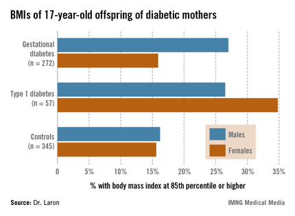
The Israeli data expand upon the earlier findings of EPOCH (Exploring Perinatal Outcomes among Children), a retrospective cohort study conducted by investigators at the University of Colorado, Denver. The Colorado investigators followed 95 children exposed to diabetes in utero – 87 whose mothers had gestational diabetes and 8 with type 1 diabetes – and 409 controls out to age 13 years, and determined that the children of diabetic mothers were more obese. Of note, the investigators found no significant differences between the children of diabetic mothers and controls in growth trajectories during infancy and early childhood, but a higher body mass index growth velocity among the offspring of diabetic mothers at ages 10-13 years (J. Pediatr. 2011;158:941-6).
The Israeli study tracked a much larger group of children of diabetic mothers later into adolescence, taking advantage of the fact that most Israeli youth have to report for compulsory military service at age 17.
Dr. Laron of Tel Aviv University and his coworkers identified 447 singleton term neonates of mothers with gestational diabetes and 97 whose mothers had type 1 diabetes born during 1987-1993 at the Helen Schneider Hospital for Women in Petach Tikva, Israel. The control group was composed of an equal number of singleton term babies born to nondiabetic mothers at the hospital during the same years.
Consistent with other reports, the average birth weight and length of the babies of diabetic mothers were significantly greater than in controls. For example, 24.3% of the baby boys and 18.4% of the girls of mothers with gestational diabetes were at or above the 90th percentile for birth weight for the Israeli population, compared with 6.8% of male and 6.0% of female controls.
Sixty-two percent of the original study population presented for military service at age 17; Arabs, as well as ultra-Orthodox Jewish men and women, are exempt from military service. The offspring exposed in utero to maternal diabetes didn’t differ from controls in terms of height, but the males with in utero exposure were significantly more likely to have a BMI at or above the 85th percentile nationally, and the females showed a similar albeit nonsignificant trend (see chart). Unlike in the Colorado study, however, the Israeli investigators didn’t have access to serial measurements that would have enabled them to determine the age at which the accelerated BMI growth trajectory occurred.
It is well established that adolescent overweight is associated with increased risk of subsequent obesity in adulthood. If the Colorado investigators are correct in their conclusion that exposure to diabetes in utero results in accelerated BMI growth occurring in late childhood, it would suggest that early childhood provides a window of opportunity for obesity prevention in these children. But a better understanding of the underlying mechanisms involved in the association between in utero exposure to diabetes and accelerated BMI growth trajectory occurring 10-13 years later may be needed before targeted preventive interventions can be developed and tested.
Dr. Laron reported having no conflicts of interest.
CHICAGO – At age 17, the children of mothers with diabetes during pregnancy are more likely to be overweight, according to an Israeli longitudinal study.
This was equally the case regardless of whether the mother had gestational or pregestational diabetes, Dr. Zvi Laron reported at the annual scientific sessions of the American Diabetes Association.
The Israeli data expand upon the earlier findings of EPOCH (Exploring Perinatal Outcomes among Children), a retrospective cohort study conducted by investigators at the University of Colorado, Denver. The Colorado investigators followed 95 children exposed to diabetes in utero – 87 whose mothers had gestational diabetes and 8 with type 1 diabetes – and 409 controls out to age 13 years, and determined that the children of diabetic mothers were more obese. Of note, the investigators found no significant differences between the children of diabetic mothers and controls in growth trajectories during infancy and early childhood, but a higher body mass index growth velocity among the offspring of diabetic mothers at ages 10-13 years (J. Pediatr. 2011;158:941-6).
The Israeli study tracked a much larger group of children of diabetic mothers later into adolescence, taking advantage of the fact that most Israeli youth have to report for compulsory military service at age 17.
Dr. Laron of Tel Aviv University and his coworkers identified 447 singleton term neonates of mothers with gestational diabetes and 97 whose mothers had type 1 diabetes born during 1987-1993 at the Helen Schneider Hospital for Women in Petach Tikva, Israel. The control group was composed of an equal number of singleton term babies born to nondiabetic mothers at the hospital during the same years.
Consistent with other reports, the average birth weight and length of the babies of diabetic mothers were significantly greater than in controls. For example, 24.3% of the baby boys and 18.4% of the girls of mothers with gestational diabetes were at or above the 90th percentile for birth weight for the Israeli population, compared with 6.8% of male and 6.0% of female controls.
Sixty-two percent of the original study population presented for military service at age 17; Arabs, as well as ultra-Orthodox Jewish men and women, are exempt from military service. The offspring exposed in utero to maternal diabetes didn’t differ from controls in terms of height, but the males with in utero exposure were significantly more likely to have a BMI at or above the 85th percentile nationally, and the females showed a similar albeit nonsignificant trend (see chart). Unlike in the Colorado study, however, the Israeli investigators didn’t have access to serial measurements that would have enabled them to determine the age at which the accelerated BMI growth trajectory occurred.
It is well established that adolescent overweight is associated with increased risk of subsequent obesity in adulthood. If the Colorado investigators are correct in their conclusion that exposure to diabetes in utero results in accelerated BMI growth occurring in late childhood, it would suggest that early childhood provides a window of opportunity for obesity prevention in these children. But a better understanding of the underlying mechanisms involved in the association between in utero exposure to diabetes and accelerated BMI growth trajectory occurring 10-13 years later may be needed before targeted preventive interventions can be developed and tested.
Dr. Laron reported having no conflicts of interest.
AT THE ADA ANNUAL SCIENTIFIC SESSIONS
Major finding: Of 17-year-old males born to diabetic mothers, 27% had a BMI at or above the 85th percentile nationally, compared with 16% of controls.
Data source: A retrospective study of 329 Israeli 17-year-olds born to diabetic mothers and 345 controls.
Disclosures: Dr. Laron reported having no conflicts of interest.

Home digital pregnancy test indicates week of conception
LONDON – A home digital pregnancy test is able to correctly estimate the likely week of conception, according to the results of a 10-month, prospective observational study of 1,505 nonpregnant U.S. women.
"There was a high [98%] level of agreement between the digital pregnancy test and standardized ultrasound," Sarah Johnson, Ph.D., said in an interview at the annual meeting of the European Society of Human Reproduction and Embryology.
The results of the digital pregnancy test were compared with ultrasound measurements performed at approximately 12 weeks after the woman had her last menstrual period (LMP).
"We received clearance from the Food and Drug Administration last December, and all being well it should be on the shelves in September," said Dr. Johnson, who is a scientific and medical affairs manager for SPD Development of Bedford, England. The digital pregnancy test is already available in the United Kingdom* based on data from a previous study (Curr. Med. Res. Opin. 2011;27:393-401).
Dr. Johnson noted that this is the first home pregnancy test to provide an accurate and early assessment of pregnancy at the time when a woman first suspects that she might be pregnant. It is also the first test to provide a digital readout, which is potentially simpler for women to understand than seeing a line in a window to indicate whether they are pregnant or not.
Similar to other home pregnancy tests, the digital pregnancy test measures levels of human chorionic gonadotrophin (hCG) excreted in the urine. Measuring hCG is a tried-and-tested method of confirming pregnancy as the hormone plays a critical role in early pregnancy, helping with the implantation of the embryo and preventing further oocyte development.
Unlike existing tests, however, the digital pregnancy test estimates the time since conception based on thresholds of hCG, which have been shown to rise rapidly after ovulation has occurred, and measurement of this hormone in the urine reflects the duration of pregnancy (Curr. Med. Res. Opin. 2009;25:741-8).
In using the digital home pregnancy test, the woman can either hold the test strip in the urine stream for 5 seconds or dip it into a urine sample collected in a clean container for 20 seconds. Results from the test are then given in a few minutes as a digital readout saying if the woman is "pregnant" or "not pregnant," with additional numbers displayed if conception was likely to be 1-2, 2-3, or 3+ weeks ago.
During the study, women were required to provide urine samples every day throughout their menstrual cycles, a random sample of which were then tested in the laboratory using the digital home pregnancy test.
A total of 250 women became pregnant during the study, with sufficient data on 153 women available for analysis. All of these women had ultrasound performed at approximately 11-13 weeks of their pregnancy to determine the gestational age according to the crown rump length measurement. More than 3,600 digital pregnancy tests were preformed in three batches, with the technicians blinded to the samples.
One of the interesting observations of the study is the inaccuracy of the LMP, Dr. Johnson said. All the women in the study planned on becoming pregnant and were carefully monitoring their menstrual cycles. Although they thought they knew exactly when their LMP was, the urine assessments suggested they were wrong by about 5 days.
"When a woman thinks she is pregnant, all she has is her last menstrual period, and that in so many cases is wrong, so this new test provides something that can provide greater accuracy," Dr. Johnson suggested.
SPD Development also has a digital ovulation test that is soon to be released in both the United States and the United Kingdom.
"Normal ovulation tests just measure luteinizing hormone [LH], so they tell you the best 2 days [to conceive]," Dr. Johnson said. The digital ovulation test still measures the LH surge, which indicates ovulation has occurred, but it also measures levels of estrone-3-glucuronide (E3G), which is a major urinary metabolite of estradiol. Levels of E3G rise a couple of days before the LH surge.
"By measuring [E3G], you are actually telling a woman about those additional fertile days during which she can also get pregnant before the 2 peak days," Dr. Johnson explained. Data presented in a poster at the annual meeting of ESHRE showed that the new digital ovulation test identified 4 or more fertile days in 80% of menstrual cycles.
The digital ovulation test will come as a kit with replaceable test strips. A woman urinates on the test strip, puts it into the reader, and the device then remembers the result and helps the woman identify her most fertile period.
The study was funded by SPD Development. Dr. Johnson is an employee of the company, which manufactures the Clearblue Digital Pregnancy Test With Conception Indicator and the Clearblue Digital Ovulation Test With Dual Hormone Indicator.
*Correction, 7/18/2013: An earlier version of this story misstated the availability of the test.
AT ESHRE 2013
Major finding: A home digital pregnancy test correctly indicated the week of conception in 98% of cases confirmed by standard gestational dating ultrasound.
Data source: A prospective, 10-month observational study of 1,505 U.S.-based, nonpregnant women wanting to conceive; 153 became pregnant, had used the digital pregnancy test with conception indicator, and had ultrasound scans to determine gestational age.
Disclosures: The study was funded by SPD Development. Dr. Johnson is an employee of the company, which manufactures the Clearblue Digital Pregnancy Test With Conception Indicator and the Clearblue Digital Ovulation Test With Dual Hormone Indicator.
UPDATE ON INFECTIOUS DISEASE
The past year has seen the publication of four studies with immediate relevance for clinicians:
- a retrospective, population-based cohort study that explores whether women who have chorioamnionitis in one pregnancy are at risk for the same type of infection in a subsequent pregnancy
- another retrospective cohort study that assesses the clinical utility of testing for gonorrhea and chlamydia before inserting an intrauterine device (IUD)
- an elegant primate experiment that highlights the value of azithromycin in subjects with chorioamnionitis
- a multicenter, randomized, nonblinded trial in seriously ill patients to determine whether daily bathing with chlorhexidine-impregnated washcloths can reduce the acquisition of multidrug-resistant organisms and the incidence of hospital-acquired bloodstream infection.
CHORIOAMNIONITIS IN ONE PREGNANCY IS LIKELY TO RECUR IN THE NEXT GENERATION
Cohen-Cline HN, Kahn TR, Hutter CM. A population-based study of the risk of repeat clinical chorioamnionitis in Washington State, 1989–2008. Am J Obstet Gynecol. 2012;207(6):473.e1–e7.
This retrospective, population-based cohort study (Level II evidence) is one of the few to examine the risk of recurrence for chorioamnionitis, and the findings are intriguing. Women who were infected during their first delivery were 3.43 times more likely to become infected in their second delivery than women who did not have chorioamnionitis in their first pregnancy (95% confidence interval [CI], 2.67–4.42; P <.001). This association persisted even after adjustment for potential confounders, such as age, ethnicity, presence of premature rupture of membranes (PROM), and internal fetal monitoring.
Chorioamnionitis is a common affliction
This infection complicates approximately 5% of term deliveries and a significantly higher percentage of preterm deliveries. The principal causative organisms are group B streptococci (GBS), Escherichia coli and other aerobic Gram-negative bacilli, both Gram-positive and Gram-negative anaerobes, and genital mycoplasmas.
The main risk factors for chorioamnionitis are:
- prematurity
- prolonged labor
- prolonged rupture of membranes
- multiple internal examinations
- internal fetal monitoring
- low socioeconomic status
- preexisting genital tract infection (eg, bacterial vaginosis, GBS colonization).
Infants delivered to infected mothers are at increased risk for sepsis, pneumonia, and meningitis. Severely infected infants, particularly those who are premature, are also at increased risk for cerebral palsy.
Details of the study
This investigation focused on women in Washington State who had a first pregnancy from 1989 through 2008 and then had at least one additional birth during the study period.
Participants included 6,219 women who had chorioamnionitis in their first pregnancy and 25,294 women who did not. Using logistic regression, Cohen-Cline and colleagues estimated the odds ratio for chorioamnionitis in the second delivery, taking into account the following potential confounders:
- maternal age
- ethnicity
- presence of PROM
- use of internal monitoring
- smoking.
As I stated above, women who had chorioamnionitis in their first pregnancy were 3.43 times as likely to have it again in their second pregnancy.
What this EVIDENCE means for practice
When a patient has a history of chorioamnionitis, we should do everything possible to reduce her risk for recurrent infection. For example, we should screen her for lower genital tract infections that predispose to chorioamnionitis:
-gonorrhea
-chlamydia
-bacterial vaginosis
-GBS.
If the patient has any of the first three infections, treat her immediately with the appropriate antibiotics. If she is colonized with GBS, administer one of the intrapartum antibiotic regimens recommended by the Centers for Disease Control and Prevention (CDC).
If the patient has a history of preterm PROM or spontaneous preterm delivery, initiate prophylaxis with progesterone and assess her cervical length periodically to determine whether cerclage is indicated.
During labor, make every effort to minimize the duration of ruptured membranes, the length of invasive monitoring, and the number of internal vaginal examinations.
At the earliest sign of intra-amniotic infection, treat the patient with broad-spectrum antibiotics, usually ampicillin plus gentamicin.
In low-risk populations, universal screening for sexually transmitted infections is probably unnecessary before IUD insertion
Sufrin CB, Postlethwaite D, Armstrong MA, et al. Neisseria gonorrhea and Chlamydia trachomatis screening at intrauterine device insertion and pelvic inflammatory disease. Obstet Gynecol. 2012;120(6):1314–1321.
This retrospective cohort study (Level II evidence) focused on women who had an IUD inserted in a managed-care practice at Kaiser Permanente of Northern California during a 5-year period. Sufrin and colleagues compared the incidence of pelvic inflammatory disease (PID) within 90 days after insertion among women who were, and were not, screened for gonorrhea and chlamydia.
Among 57,728 IUD insertions, 47% involved women who were unscreened within 1 year of the procedure. Among women who were screened, 19% were tested on the day of IUD insertion.
The overall risk of PID in the study cohort was very low—0.54% (95% CI, 0.48–0.60). Investigators were unable to identify any significant difference in the risk of PID between women who had no screening versus those who were screened. Among women who were screened, same-day screening was equivalent to prescreening.
Investigators concluded that the most reasonable protocol is to screen on the basis of risk factors on the same day as IUD insertion. If the patient has obvious evidence of endocervicitis (ie, mucopurulent discharge), IUD insertion should be delayed. Otherwise, if the patient has risk factors for infection, screening should be followed by IUD insertion.
If the screen is positive, the patient should be treated in accordance with the latest CDC recommendations, and the IUD can be left in place.
Sufrin and colleagues concluded that adherence to this protocol would be associated with a very low, and clinically acceptable, risk of PID.
STI screening need not be an obstacle to IUD use
The IUD is an excellent method of contraception, and it is suitable for most patients. It is particularly useful for women who have difficulty remembering to take a pill each day or to use a barrier method of contraception at each episode of coitus.
Obstacles to more widespread use of the IUD include:
- high initial cost
- misconceptions on the part of the patient about the mechanism of action and adverse effects of the device
- cumbersome protocols that require multiple physician visits for counseling and sexually transmitted infection (STI) testing before the device is inserted.
What this EVIDENCE means for practice
This study provides reassurance that, at least in a relatively affluent managed-care population, universal testing for STIs is probably not necessary. When testing is indicated, it can be performed on the same day that the IUD is inserted, minimizing the number of office visits.
What is less clear is whether the same protocol can be applied to a population with a significantly higher prevalence of STIs. In such a population, universal screening for gonorrhea and chlamydia may be more prudent. However, screening still can be performed on the same day as IUD insertion.
In a primate model of intra-amniotic infection with Ureaplasma, maternal azithromycin prolonged gestation
Grigsby PL, Novy MJ, Sadowsky DW, et al. Maternal azithromycin therapy for Ureaplasma intraamniotic infection delays preterm delivery and reduces fetal lung injury in a primate model. Am J Obstet Gynecol. 2012;207(6):475.e1–e14.
Grigsby and colleagues assessed the efficacy of azithromycin—with and without anti-inflammatory agents—in delaying preterm birth and minimizing fetal lung injury in a primate model. They found that azithromycin significantly prolonged gestation.
Details of the study
The study involved 16 chronically instrumented rhesus monkeys who received intra-amniotic inoculation with Ureaplasma parvum (107 colony-forming units/mL) and were then observed. When contractions began, as they invariably did, six monkeys received no treatment, five received intravenous (IV) azithromycin (12.5 mg/kg every 12 hours) for 10 days, and five received azithromycin plus dexamethasone and indomethacin.
Key outcome measures were the intra-amniotic concentration of proinflammatory mediators, the frequency of positive amniotic fluid cultures for U parvum, and the extent of histologic fetal lung injury.
In treated animals, the mean (SD) inoculation-to-delivery interval was 20.9 (1.4) days, compared with 13.7 (2.5) days in untreated monkeys (P <.05).
In addition, there was a twofold to threefold increase in the percentage of undelivered animals at 18 to 20 days after inoculation in the treatment group, compared with the no-treatment group. Treatment also significantly decreased the Ureaplasma colony count in the amniotic fluid, effectively eliminating the organism within 4 days.
In both treatment groups, the amniotic fluid concentration of proinflammatory mediators decreased significantly, compared with the untreated group. Treatment also significantly reduced the magnitude of deleterious histologic changes in the fetal lungs.
Somewhat surprisingly, dexamethasone and indomethacin did not enhance the treatment effect of azithromycin. Moreover, despite prolongation of pregnancy, all animals in the treatment group still delivered prematurely.
Why treatment should target genital mycoplasmas
Chorioamnionitis is an importance cause of preterm labor and preterm delivery. The principal pathogens are part of the normal vaginal flora: aerobic Gram-negative bacilli, aerobic Gram-positive cocci, anaerobes, and genital mycoplasmas.
Most treatment regimens for chorioamnionitis (eg, ampicillin plus gentamicin) do not specifically target the genital mycoplasmas. However, the most commonly recommended prophylactic antibiotic regimens for patients with preterm PROM include agents with specific action against mycoplasmas, namely erythromycin and azithromycin.
In this clinical setting, antibiotic prophylaxis prolongs the latency period and decreases the frequency of both maternal and fetal/neonatal infection.
This elegant basic science investigation sheds new light on the importance of the genital mycoplasmas in the pathogenesis of preterm labor and helps to explain why drugs like erythromycin and azithromycin may be so valuable in prolonging the latent period and reducing the frequency of infection and injury in the baby.
What this EVIDENCE means for practice
Because IV azithromycin rapidly achieved inhibitory concentrations in amniotic fluid and maintained these concentrations over 10 days of treatment, it significantly reduced the concentration of Ureaplasma in the amniotic fluid as well as the risk of histologic injury to the fetal lung.
Accordingly, I recommend that azithromycin remain a key component of the prophylactic regimen for patients with preterm PROM. It also may be advisable to add azithromycin to the usual combination of ampicillin plus gentamicin for empiric treatment of chorioamnionitis.
Daily bathing with chlorhexidine cloths can protect hospitalized patients from serious infection
Climo MW, Yokoe DS, Warren DK, et al. Effect of daily chlorhexidine bathing on hospital-acquired infection. N Engl J Med. 2013;368(6):533–542.
This multicenter, randomized, nonblinded trial of 7,727 seriously ill patients sought to determine whether daily bathing with chlorhexidine-impregnated washcloths can decrease the acquisition of multidrug-resistant organisms and the incidence of hospital-acquired bloodstream infection.
Each day, patients in eight ICUs and one bone-marrow transplant unit bathed themselves, or were bathed by nursing staff, with 2% chlorhexidine-impregnated cloths or non–antimicrobial washcloths. All body surfaces except the face were cleansed. After 6 months, each unit changed to the other method of bathing.
Investigators focused on two outcomes:
- the prevalence of colonization of the nares with methicillin-resistant Staphylococcus aureus (MRSA) or colonization of the perirectal area with vancomycin-resistant enterococci (VRE)
- the frequency of hospital-acquired bloodstream infection (bacterial or fungal) detected more than 48 hours after admission to the unit.
The overall rate of MRSA or VRE acquisition was reduced by 23% when patients were bathed with chlorhexidine (5.10 versus 6.60 cases per 100 patient-days; P = .03). The overall rate of hospital-acquired bloodstream infection was reduced by 28% during the intervention period (4.78 vs 6.60 cases per 1,000 patient-days; P = .006).
In particular, the rate of central-catheter–associated bloodstream infection was 53% lower during the intervention (1.55 vs 3.30 cases per 1,000 catheter-days; P = .004).
The intervention had the greatest impact on infections caused by Gram-positive and fungal organisms.
The protective effect of chlorhexidine bathing was greatest among patients who had the longest length of stay in the unit.
Chlorhexidine did not cause an increased frequency of skin reactions. Moreover, use of the antiseptic washes did not cause the emergence of MRSA or VRA isolates with high-level resistance.
This study is of great interest in light of a recent report that demonstrated that preoperative preparation of the skin with chlorhexidine was more effective than preparation with povidone-iodine in reducing the risk of surgical-site infections after major operative procedures.1 Not only is chlorhexidine highly active against the usual bacteria that colonize the skin of hospitalized patients, it also has residual antibacterial activity that further decreases the colonization of the patient’s skin by microbes.
What this EVIDENCE means for practice
This study has two clear implications for ObGyns. First, chlorhexidine washes should be used by all patients who are scheduled for surgery, particularly those undergoing procedures that carry a relatively high risk of postoperative wound infection, such as total abdominal hysterectomy, radical hysterectomy, and cesarean delivery. In morbidly obese patients, particular attention should be directed to the skin beneath the abdominal panniculus.
Second, when we have seriously ill obstetric or gynecologic patients, especially those with long-term indwelling catheters who require prolonged hospitalization, we should order daily bathing (excluding the face) with chlorhexidine.
Reference
1. Darouiche RO, Wall MJ Jr, Itani KM, et al. Chlorhexidine-alcohol versus povidone-iodine for surgical-site antisepsis. N Engl J Med. 2010;362(1):18–26.
The past year has seen the publication of four studies with immediate relevance for clinicians:
- a retrospective, population-based cohort study that explores whether women who have chorioamnionitis in one pregnancy are at risk for the same type of infection in a subsequent pregnancy
- another retrospective cohort study that assesses the clinical utility of testing for gonorrhea and chlamydia before inserting an intrauterine device (IUD)
- an elegant primate experiment that highlights the value of azithromycin in subjects with chorioamnionitis
- a multicenter, randomized, nonblinded trial in seriously ill patients to determine whether daily bathing with chlorhexidine-impregnated washcloths can reduce the acquisition of multidrug-resistant organisms and the incidence of hospital-acquired bloodstream infection.
CHORIOAMNIONITIS IN ONE PREGNANCY IS LIKELY TO RECUR IN THE NEXT GENERATION
Cohen-Cline HN, Kahn TR, Hutter CM. A population-based study of the risk of repeat clinical chorioamnionitis in Washington State, 1989–2008. Am J Obstet Gynecol. 2012;207(6):473.e1–e7.
This retrospective, population-based cohort study (Level II evidence) is one of the few to examine the risk of recurrence for chorioamnionitis, and the findings are intriguing. Women who were infected during their first delivery were 3.43 times more likely to become infected in their second delivery than women who did not have chorioamnionitis in their first pregnancy (95% confidence interval [CI], 2.67–4.42; P <.001). This association persisted even after adjustment for potential confounders, such as age, ethnicity, presence of premature rupture of membranes (PROM), and internal fetal monitoring.
Chorioamnionitis is a common affliction
This infection complicates approximately 5% of term deliveries and a significantly higher percentage of preterm deliveries. The principal causative organisms are group B streptococci (GBS), Escherichia coli and other aerobic Gram-negative bacilli, both Gram-positive and Gram-negative anaerobes, and genital mycoplasmas.
The main risk factors for chorioamnionitis are:
- prematurity
- prolonged labor
- prolonged rupture of membranes
- multiple internal examinations
- internal fetal monitoring
- low socioeconomic status
- preexisting genital tract infection (eg, bacterial vaginosis, GBS colonization).
Infants delivered to infected mothers are at increased risk for sepsis, pneumonia, and meningitis. Severely infected infants, particularly those who are premature, are also at increased risk for cerebral palsy.
Details of the study
This investigation focused on women in Washington State who had a first pregnancy from 1989 through 2008 and then had at least one additional birth during the study period.
Participants included 6,219 women who had chorioamnionitis in their first pregnancy and 25,294 women who did not. Using logistic regression, Cohen-Cline and colleagues estimated the odds ratio for chorioamnionitis in the second delivery, taking into account the following potential confounders:
- maternal age
- ethnicity
- presence of PROM
- use of internal monitoring
- smoking.
As I stated above, women who had chorioamnionitis in their first pregnancy were 3.43 times as likely to have it again in their second pregnancy.
What this EVIDENCE means for practice
When a patient has a history of chorioamnionitis, we should do everything possible to reduce her risk for recurrent infection. For example, we should screen her for lower genital tract infections that predispose to chorioamnionitis:
-gonorrhea
-chlamydia
-bacterial vaginosis
-GBS.
If the patient has any of the first three infections, treat her immediately with the appropriate antibiotics. If she is colonized with GBS, administer one of the intrapartum antibiotic regimens recommended by the Centers for Disease Control and Prevention (CDC).
If the patient has a history of preterm PROM or spontaneous preterm delivery, initiate prophylaxis with progesterone and assess her cervical length periodically to determine whether cerclage is indicated.
During labor, make every effort to minimize the duration of ruptured membranes, the length of invasive monitoring, and the number of internal vaginal examinations.
At the earliest sign of intra-amniotic infection, treat the patient with broad-spectrum antibiotics, usually ampicillin plus gentamicin.
In low-risk populations, universal screening for sexually transmitted infections is probably unnecessary before IUD insertion
Sufrin CB, Postlethwaite D, Armstrong MA, et al. Neisseria gonorrhea and Chlamydia trachomatis screening at intrauterine device insertion and pelvic inflammatory disease. Obstet Gynecol. 2012;120(6):1314–1321.
This retrospective cohort study (Level II evidence) focused on women who had an IUD inserted in a managed-care practice at Kaiser Permanente of Northern California during a 5-year period. Sufrin and colleagues compared the incidence of pelvic inflammatory disease (PID) within 90 days after insertion among women who were, and were not, screened for gonorrhea and chlamydia.
Among 57,728 IUD insertions, 47% involved women who were unscreened within 1 year of the procedure. Among women who were screened, 19% were tested on the day of IUD insertion.
The overall risk of PID in the study cohort was very low—0.54% (95% CI, 0.48–0.60). Investigators were unable to identify any significant difference in the risk of PID between women who had no screening versus those who were screened. Among women who were screened, same-day screening was equivalent to prescreening.
Investigators concluded that the most reasonable protocol is to screen on the basis of risk factors on the same day as IUD insertion. If the patient has obvious evidence of endocervicitis (ie, mucopurulent discharge), IUD insertion should be delayed. Otherwise, if the patient has risk factors for infection, screening should be followed by IUD insertion.
If the screen is positive, the patient should be treated in accordance with the latest CDC recommendations, and the IUD can be left in place.
Sufrin and colleagues concluded that adherence to this protocol would be associated with a very low, and clinically acceptable, risk of PID.
STI screening need not be an obstacle to IUD use
The IUD is an excellent method of contraception, and it is suitable for most patients. It is particularly useful for women who have difficulty remembering to take a pill each day or to use a barrier method of contraception at each episode of coitus.
Obstacles to more widespread use of the IUD include:
- high initial cost
- misconceptions on the part of the patient about the mechanism of action and adverse effects of the device
- cumbersome protocols that require multiple physician visits for counseling and sexually transmitted infection (STI) testing before the device is inserted.
What this EVIDENCE means for practice
This study provides reassurance that, at least in a relatively affluent managed-care population, universal testing for STIs is probably not necessary. When testing is indicated, it can be performed on the same day that the IUD is inserted, minimizing the number of office visits.
What is less clear is whether the same protocol can be applied to a population with a significantly higher prevalence of STIs. In such a population, universal screening for gonorrhea and chlamydia may be more prudent. However, screening still can be performed on the same day as IUD insertion.
In a primate model of intra-amniotic infection with Ureaplasma, maternal azithromycin prolonged gestation
Grigsby PL, Novy MJ, Sadowsky DW, et al. Maternal azithromycin therapy for Ureaplasma intraamniotic infection delays preterm delivery and reduces fetal lung injury in a primate model. Am J Obstet Gynecol. 2012;207(6):475.e1–e14.
Grigsby and colleagues assessed the efficacy of azithromycin—with and without anti-inflammatory agents—in delaying preterm birth and minimizing fetal lung injury in a primate model. They found that azithromycin significantly prolonged gestation.
Details of the study
The study involved 16 chronically instrumented rhesus monkeys who received intra-amniotic inoculation with Ureaplasma parvum (107 colony-forming units/mL) and were then observed. When contractions began, as they invariably did, six monkeys received no treatment, five received intravenous (IV) azithromycin (12.5 mg/kg every 12 hours) for 10 days, and five received azithromycin plus dexamethasone and indomethacin.
Key outcome measures were the intra-amniotic concentration of proinflammatory mediators, the frequency of positive amniotic fluid cultures for U parvum, and the extent of histologic fetal lung injury.
In treated animals, the mean (SD) inoculation-to-delivery interval was 20.9 (1.4) days, compared with 13.7 (2.5) days in untreated monkeys (P <.05).
In addition, there was a twofold to threefold increase in the percentage of undelivered animals at 18 to 20 days after inoculation in the treatment group, compared with the no-treatment group. Treatment also significantly decreased the Ureaplasma colony count in the amniotic fluid, effectively eliminating the organism within 4 days.
In both treatment groups, the amniotic fluid concentration of proinflammatory mediators decreased significantly, compared with the untreated group. Treatment also significantly reduced the magnitude of deleterious histologic changes in the fetal lungs.
Somewhat surprisingly, dexamethasone and indomethacin did not enhance the treatment effect of azithromycin. Moreover, despite prolongation of pregnancy, all animals in the treatment group still delivered prematurely.
Why treatment should target genital mycoplasmas
Chorioamnionitis is an importance cause of preterm labor and preterm delivery. The principal pathogens are part of the normal vaginal flora: aerobic Gram-negative bacilli, aerobic Gram-positive cocci, anaerobes, and genital mycoplasmas.
Most treatment regimens for chorioamnionitis (eg, ampicillin plus gentamicin) do not specifically target the genital mycoplasmas. However, the most commonly recommended prophylactic antibiotic regimens for patients with preterm PROM include agents with specific action against mycoplasmas, namely erythromycin and azithromycin.
In this clinical setting, antibiotic prophylaxis prolongs the latency period and decreases the frequency of both maternal and fetal/neonatal infection.
This elegant basic science investigation sheds new light on the importance of the genital mycoplasmas in the pathogenesis of preterm labor and helps to explain why drugs like erythromycin and azithromycin may be so valuable in prolonging the latent period and reducing the frequency of infection and injury in the baby.
What this EVIDENCE means for practice
Because IV azithromycin rapidly achieved inhibitory concentrations in amniotic fluid and maintained these concentrations over 10 days of treatment, it significantly reduced the concentration of Ureaplasma in the amniotic fluid as well as the risk of histologic injury to the fetal lung.
Accordingly, I recommend that azithromycin remain a key component of the prophylactic regimen for patients with preterm PROM. It also may be advisable to add azithromycin to the usual combination of ampicillin plus gentamicin for empiric treatment of chorioamnionitis.
Daily bathing with chlorhexidine cloths can protect hospitalized patients from serious infection
Climo MW, Yokoe DS, Warren DK, et al. Effect of daily chlorhexidine bathing on hospital-acquired infection. N Engl J Med. 2013;368(6):533–542.
This multicenter, randomized, nonblinded trial of 7,727 seriously ill patients sought to determine whether daily bathing with chlorhexidine-impregnated washcloths can decrease the acquisition of multidrug-resistant organisms and the incidence of hospital-acquired bloodstream infection.
Each day, patients in eight ICUs and one bone-marrow transplant unit bathed themselves, or were bathed by nursing staff, with 2% chlorhexidine-impregnated cloths or non–antimicrobial washcloths. All body surfaces except the face were cleansed. After 6 months, each unit changed to the other method of bathing.
Investigators focused on two outcomes:
- the prevalence of colonization of the nares with methicillin-resistant Staphylococcus aureus (MRSA) or colonization of the perirectal area with vancomycin-resistant enterococci (VRE)
- the frequency of hospital-acquired bloodstream infection (bacterial or fungal) detected more than 48 hours after admission to the unit.
The overall rate of MRSA or VRE acquisition was reduced by 23% when patients were bathed with chlorhexidine (5.10 versus 6.60 cases per 100 patient-days; P = .03). The overall rate of hospital-acquired bloodstream infection was reduced by 28% during the intervention period (4.78 vs 6.60 cases per 1,000 patient-days; P = .006).
In particular, the rate of central-catheter–associated bloodstream infection was 53% lower during the intervention (1.55 vs 3.30 cases per 1,000 catheter-days; P = .004).
The intervention had the greatest impact on infections caused by Gram-positive and fungal organisms.
The protective effect of chlorhexidine bathing was greatest among patients who had the longest length of stay in the unit.
Chlorhexidine did not cause an increased frequency of skin reactions. Moreover, use of the antiseptic washes did not cause the emergence of MRSA or VRA isolates with high-level resistance.
This study is of great interest in light of a recent report that demonstrated that preoperative preparation of the skin with chlorhexidine was more effective than preparation with povidone-iodine in reducing the risk of surgical-site infections after major operative procedures.1 Not only is chlorhexidine highly active against the usual bacteria that colonize the skin of hospitalized patients, it also has residual antibacterial activity that further decreases the colonization of the patient’s skin by microbes.
What this EVIDENCE means for practice
This study has two clear implications for ObGyns. First, chlorhexidine washes should be used by all patients who are scheduled for surgery, particularly those undergoing procedures that carry a relatively high risk of postoperative wound infection, such as total abdominal hysterectomy, radical hysterectomy, and cesarean delivery. In morbidly obese patients, particular attention should be directed to the skin beneath the abdominal panniculus.
Second, when we have seriously ill obstetric or gynecologic patients, especially those with long-term indwelling catheters who require prolonged hospitalization, we should order daily bathing (excluding the face) with chlorhexidine.
Reference
1. Darouiche RO, Wall MJ Jr, Itani KM, et al. Chlorhexidine-alcohol versus povidone-iodine for surgical-site antisepsis. N Engl J Med. 2010;362(1):18–26.
Reference
1. Darouiche RO, Wall MJ Jr, Itani KM, et al. Chlorhexidine-alcohol versus povidone-iodine for surgical-site antisepsis. N Engl J Med. 2010;362(1):18–26.
How to manage emergencies associated with tocolysis for preterm labor
CASE 1: Preterm labor with cervical changes
Ms. M, a 42-year-old woman pregnant with her second child, begins having contractions at 30 weeks’ gestation. Examination reveals that her cervix is dilated 2 cm and effaced 50%. She is given subcutaneous terbutaline to suppress her contractions. Thirty minutes later, she complains of shortness of breath and chest pain. An electrocardiogram reveals depression of the ST segment, and a chest radiograph shows mild pulmonary edema.
How should her symptoms be managed?
Preterm labor precedes delivery in about 50% of preterm births. Approximately 33% of women who have preterm labor will experience spontaneous resolution, and more than 50% of women who have preterm labor will deliver at term. Although the use of tocolytic therapy has proved to be effective at temporarily suppressing uterine activity, it has not been shown to delay delivery for more than a few hours or days.1
The American College of Obstetricians and Gynecologists (ACOG) recommends the use of tocolytics only when a delay in labor for approximately 48 hours would improve outcome. Therefore, tocolytic therapy should be reserved for the following circumstances:
- to stop the progress of labor long enough to administer antenatal corticosteroid therapy
- to prolong pregnancy when there is an underlying self-limiting condition that can cause labor, such as pyelonephritis
- to provide time for safe transport to a facility with a higher level of neonatal care.2
Tocolytics are generally not indicated before the fetus is viable, although we lack data from randomized, controlled trials to support a specific recommendation. The approach is clearer when the fetus is near the upper limits of viability. Most studies suggest that 34 weeks’ gestation is the threshold at which the perinatal morbidity and mortality associated with delivery are too low to justify the cost and potential complications of tocolysis.3
Women who experience preterm labor without cervical changes generally should not be treated with tocolytics.2 Contraindications to tocolytic therapy include:
- lethal fetal anomaly
- nonreassuring fetal status
- maternal disease
- maternal hemorrhage with hemodynamic instability.
Beta-adrenergic agonists carry many risks
These agents have been studied in several randomized, controlled trials. Although ritodrine was approved as tocolytic therapy by the US Food and Drug Administration (FDA), it has since been removed from the US market. Terbutaline is still available but lacks FDA approval as a tocolytic.
Maternal side effects associated with beta-adrenergic agonists are thought to arise from stimulation of the beta-1 and beta-2 adrenergic receptors. Stimulation of the former increases maternal heart rate and stroke volume, whereas stimulation of the beta-2 adrenergic receptors causes the relaxation of smooth muscle, including the muscles of the myometrium, blood vessels, and bronchial tree. The resulting symptoms may include maternal tachycardia, cardiac arrhythmias, palpitations, and metabolic aberrations (including hyperglycemia, hypokalemia, and hypotension). Common symptoms associated with the administration of a beta-adrenergic agonist include tremor, shortness of breath, and chest discomfort.4 Although pulmonary edema and myocardial ischemia are uncommon, they can occur even when there is no history of underlying maternal disease.
Terbutaline has been linked to maternal deaths
Sixteen maternal deaths were reported following initial marketing of terbutaline in 1976 until 2009. Three of the 16 cases involved outpatient use of terbutaline administered by a subcutaneous pump, and nine cases involved use of oral terbutaline alone or in addition to subcutaneous or IV terbutaline. In addition, 12 cases of serious maternal cardiovascular events were reported in association with terbutaline. These events included cardiac arrhythmias, myocardial infarction, pulmonary edema, hypertension, and tachycardia.
Because of these events, the FDA issued a black box warning for terbutaline that prohibits its use in the treatment of preterm labor for longer than 48 to 72 hours in the inpatient or outpatient setting because of the potential for serious maternal heart problems and death.5 Oral terbutaline should be avoided entirely in the prevention and treatment of preterm labor. However, the use of terbutaline for the management of acute tachysystole with an abnormal fetal heart-rate (FHR) pattern remains a reasonable course of treatment.6
Fetal tachycardia is the most common side effect of beta-adrenergic receptor agonists. For this reason, use of these drugs is not recommended when changes in FHR may be the first sign of fetal compromise, such as in patients with hemorrhage or infection. Neonatal hypoglycemia may also occur if maternal hyperglycemia is not controlled.7
Case 1 Resolved
Terbutaline is discontinued, and the patient’s pulmonary edema is treated with a single dose of furosemide. Electrolyte abnormalities resolve with discontinuation of medication. The patient stabilizes. Once her cardiorespiratory status improves, her contractions lessen and the cervix remains unchanged. She requires no further tocolysis and is discharged home. She presents again at 38 weeks in spontaneous labor.
CASE 2: Preterm labor treated with indomethacin
Ms. J, age 23, is 26 weeks’ pregnant with her first child. When she experienced preterm labor at 24.5 weeks’ gestation, she was given indomethacin. Now, ultrasonographic imaging reveals decreased amniotic fluid volume.
How should she be managed?
Indomethacin is a cyclooxygenase (COX) inhibitor. These drugs reduce prostaglandin production through the general inhibition of cyclooxygenase or by a specific receptor.8 Indomethacin is the most commonly used tocolytic in this class. It is a nonspecific COX inhibitor, as opposed to a COX-2 inhibitor. The latter has been associated with serious adverse outcomes in the nonobstetric population. COX-2 inhibitors now carry a black box warning or are no longer available.
Maternal contraindications for COX inhibitors include asthma, bleeding disorders, and significant renal dysfunction.
Although maternal side effects with COX inhibitors are usually mild, fetal side effects may be serious enough to cause perinatal morbidity or death.9
How indomethacin can lead to oligohydramnios
Maternal administration of indomethacin or ibuprofen can reduce fetal urine output and decrease the volume of amniotic fluid. In most cases, oligohydramnios occurs when indomethacin or ibuprofen has been given for more than 72 hours. For this reason, long-term use of a COX inhibitor should be accompanied by frequent monitoring of amniotic fluid volume by ultrasonography.
The most serious fetal complication associated with prolonged indomethacin administration (longer than 72 hours) is premature constriction of the ductus arteriosus. Ductal constriction appears to be contingent on gestational age. It has been described as early as 24 weeks’ gestation but is most common after 31 or 32 weeks. Therefore, indomethacin is not recommended for use after 32 weeks’ gestation.10
CASE 2 Resolved
The indomethacin is discontinued as soon as the decreased amniotic fluid is noted. The fluid volume returns to normal over the next 3 to 5 days. Because of the early gestational age, nifedipine is given to suppress contractions, and the patient has no further complications.
CASE 3: Preterm labor and magnesium intoxication
Ms. K experiences contractions and rapid cervical change at 32 weeks’ gestation. She is given magnesium for the preterm labor and fetal neuroprophylaxis, with nifedipine, a calcium-channel blocker, added as second-line tocolysis. Approximately 8 hours later, she reports difficulty breathing and moving.
How should her obstetrician proceed?
Calcium-channel blockers such as nifedipine are used for acute and maintenance tocolysis. This class of drugs is often selected for its relative ease of use and safety, as it has few maternal and fetal side effects. However, concomitant use of a calcium-channel blocker and magnesium sulfate can sometimes lead to neuromuscular blockade and significant respiratory depression, even necessitating mechanical ventilation.9 Treatment of these effects includes IV administration of 10% calcium gluconate (5–10 mEq), which usually reverses respiratory depression and heart block caused by magnesium intoxication. In extreme cases, peritoneal dialysis or hemodialysis may be required.
CASE 3 Resolved
The patient is given 10% calcium gluconate in the dosage described above, and she stabilizes. However, her contractions continue and she delivers at 32 weeks’ gestation. The infant does well in the NICU.
CASE 4: Preterm labor in a woman with kidney dysfunction
Ms. F, age 40, presents at 30 weeks’ gestation with regular contractions and cervical dilation of more than 3 cm. She also reports a history of kidney disease.
What steps are recommended prior to the initiation of magnesium therapy?
Magnesium sulfate has been used for more than 40 years to treat preterm labor and is still considered a first-line therapy in many centers. Although maternal side effects usually are mild, an adverse event may occur if the patient is not monitored carefully. An absence of deep-tendon reflexes should alert the clinician that magnesium levels need to be measured. Reflexes usually are lost at a serum level of 10 mEq/L or higher. When the magnesium level exceeds 13 mEq/L, cardiac arrest is a risk. IV calcium should be administered immediately in such patients.
Magnesium should be used with caution in patients with myocardial compromise. Because magnesium is eliminated by the kidneys, women with impaired renal function may experience magnesium toxicity at normal doses. If a patient has a creatinine level above 1 mg/dL, consider alternative treatment for her preterm labor. If magnesium is given, the normal loading dose (4–6 g) is appropriate, but the maintenance dose should be reduced.11
Fetal effects of magnesium sulfate
Recent studies indicate that predelivery magnesium may offer fetal neuroprotection. The minimum duration of administration for such neuroprotection is unknown but is less than 24 hours.8
Although magnesium can alter FHR patterns slightly, these changes are not clinically significant. Magnesium can also cause mild neonatal suppression at the time of delivery, but its effects quickly resolve with appropriate neonatal resuscitation. Long-term (>5 days) therapy is not recommended.
In May 2013, the FDA issued a warning about the risk of neonatal complications with long-term maternal magnesium administration. These complications include osteopenia, low calcium, and bone fracture. The pregnancy category for magnesium sulfate will be changed from “A” to “D” because of these teratogenic effects.12
CASE 4 Resolved
Because magnesium is mainly cleared by renal excretion, the clinician administers the medication with caution in this patient with reduced renal function. The clinician administers the same 4- to 6-g bolus that would be given a patient with normal kidney function, but the maintenance dose is reduced to 1 g. Magnesium levels are obtained every 12 hours or when clinically indicated.
Bottom line: Be ready to act
The short-term use of tocolytic therapy usually is not associated with maternal or fetal complications. After initial administration, maintenance tocolytic therapy probably does not prolong gestation.
Given the potential for harm without additional fetal benefit associated with extended therapy, I recommend that clinicians follow current clinical guidelines from ACOG for use of tocolytic agents. In the process, be vigilant for complications and be ready to act appropriately. Keep maternal and fetal conditions in mind when selecting a tocolytic agent.
9. US Food and Drug Administration. Terbutaline: Label Change—Warnings Against Use for Treatment of Preterm Labor. Published February 17, 2011. http://www.fda.gov/Safety/MedWatch/SafetyInformation/SafetyalertsforHumanMedicalProducts/ucm243843.htm. Accessed June 17, 2013.
12. US Food and Drug Administration. Magnesium Sulfate: Drug Safety Communication—Recommendation against Prolonged Use in Preterm Labor. Published May 30, 2013. http://www.fda.gov/Safety/MedWatch/SafetyInformation/SafetyAlertsforHumanMedicalProducts/ucm354603.htm. Accessed June 17, 2013.
CASE 1: Preterm labor with cervical changes
Ms. M, a 42-year-old woman pregnant with her second child, begins having contractions at 30 weeks’ gestation. Examination reveals that her cervix is dilated 2 cm and effaced 50%. She is given subcutaneous terbutaline to suppress her contractions. Thirty minutes later, she complains of shortness of breath and chest pain. An electrocardiogram reveals depression of the ST segment, and a chest radiograph shows mild pulmonary edema.
How should her symptoms be managed?
Preterm labor precedes delivery in about 50% of preterm births. Approximately 33% of women who have preterm labor will experience spontaneous resolution, and more than 50% of women who have preterm labor will deliver at term. Although the use of tocolytic therapy has proved to be effective at temporarily suppressing uterine activity, it has not been shown to delay delivery for more than a few hours or days.1
The American College of Obstetricians and Gynecologists (ACOG) recommends the use of tocolytics only when a delay in labor for approximately 48 hours would improve outcome. Therefore, tocolytic therapy should be reserved for the following circumstances:
- to stop the progress of labor long enough to administer antenatal corticosteroid therapy
- to prolong pregnancy when there is an underlying self-limiting condition that can cause labor, such as pyelonephritis
- to provide time for safe transport to a facility with a higher level of neonatal care.2
Tocolytics are generally not indicated before the fetus is viable, although we lack data from randomized, controlled trials to support a specific recommendation. The approach is clearer when the fetus is near the upper limits of viability. Most studies suggest that 34 weeks’ gestation is the threshold at which the perinatal morbidity and mortality associated with delivery are too low to justify the cost and potential complications of tocolysis.3
Women who experience preterm labor without cervical changes generally should not be treated with tocolytics.2 Contraindications to tocolytic therapy include:
- lethal fetal anomaly
- nonreassuring fetal status
- maternal disease
- maternal hemorrhage with hemodynamic instability.
Beta-adrenergic agonists carry many risks
These agents have been studied in several randomized, controlled trials. Although ritodrine was approved as tocolytic therapy by the US Food and Drug Administration (FDA), it has since been removed from the US market. Terbutaline is still available but lacks FDA approval as a tocolytic.
Maternal side effects associated with beta-adrenergic agonists are thought to arise from stimulation of the beta-1 and beta-2 adrenergic receptors. Stimulation of the former increases maternal heart rate and stroke volume, whereas stimulation of the beta-2 adrenergic receptors causes the relaxation of smooth muscle, including the muscles of the myometrium, blood vessels, and bronchial tree. The resulting symptoms may include maternal tachycardia, cardiac arrhythmias, palpitations, and metabolic aberrations (including hyperglycemia, hypokalemia, and hypotension). Common symptoms associated with the administration of a beta-adrenergic agonist include tremor, shortness of breath, and chest discomfort.4 Although pulmonary edema and myocardial ischemia are uncommon, they can occur even when there is no history of underlying maternal disease.
Terbutaline has been linked to maternal deaths
Sixteen maternal deaths were reported following initial marketing of terbutaline in 1976 until 2009. Three of the 16 cases involved outpatient use of terbutaline administered by a subcutaneous pump, and nine cases involved use of oral terbutaline alone or in addition to subcutaneous or IV terbutaline. In addition, 12 cases of serious maternal cardiovascular events were reported in association with terbutaline. These events included cardiac arrhythmias, myocardial infarction, pulmonary edema, hypertension, and tachycardia.
Because of these events, the FDA issued a black box warning for terbutaline that prohibits its use in the treatment of preterm labor for longer than 48 to 72 hours in the inpatient or outpatient setting because of the potential for serious maternal heart problems and death.5 Oral terbutaline should be avoided entirely in the prevention and treatment of preterm labor. However, the use of terbutaline for the management of acute tachysystole with an abnormal fetal heart-rate (FHR) pattern remains a reasonable course of treatment.6
Fetal tachycardia is the most common side effect of beta-adrenergic receptor agonists. For this reason, use of these drugs is not recommended when changes in FHR may be the first sign of fetal compromise, such as in patients with hemorrhage or infection. Neonatal hypoglycemia may also occur if maternal hyperglycemia is not controlled.7
Case 1 Resolved
Terbutaline is discontinued, and the patient’s pulmonary edema is treated with a single dose of furosemide. Electrolyte abnormalities resolve with discontinuation of medication. The patient stabilizes. Once her cardiorespiratory status improves, her contractions lessen and the cervix remains unchanged. She requires no further tocolysis and is discharged home. She presents again at 38 weeks in spontaneous labor.
CASE 2: Preterm labor treated with indomethacin
Ms. J, age 23, is 26 weeks’ pregnant with her first child. When she experienced preterm labor at 24.5 weeks’ gestation, she was given indomethacin. Now, ultrasonographic imaging reveals decreased amniotic fluid volume.
How should she be managed?
Indomethacin is a cyclooxygenase (COX) inhibitor. These drugs reduce prostaglandin production through the general inhibition of cyclooxygenase or by a specific receptor.8 Indomethacin is the most commonly used tocolytic in this class. It is a nonspecific COX inhibitor, as opposed to a COX-2 inhibitor. The latter has been associated with serious adverse outcomes in the nonobstetric population. COX-2 inhibitors now carry a black box warning or are no longer available.
Maternal contraindications for COX inhibitors include asthma, bleeding disorders, and significant renal dysfunction.
Although maternal side effects with COX inhibitors are usually mild, fetal side effects may be serious enough to cause perinatal morbidity or death.9
How indomethacin can lead to oligohydramnios
Maternal administration of indomethacin or ibuprofen can reduce fetal urine output and decrease the volume of amniotic fluid. In most cases, oligohydramnios occurs when indomethacin or ibuprofen has been given for more than 72 hours. For this reason, long-term use of a COX inhibitor should be accompanied by frequent monitoring of amniotic fluid volume by ultrasonography.
The most serious fetal complication associated with prolonged indomethacin administration (longer than 72 hours) is premature constriction of the ductus arteriosus. Ductal constriction appears to be contingent on gestational age. It has been described as early as 24 weeks’ gestation but is most common after 31 or 32 weeks. Therefore, indomethacin is not recommended for use after 32 weeks’ gestation.10
CASE 2 Resolved
The indomethacin is discontinued as soon as the decreased amniotic fluid is noted. The fluid volume returns to normal over the next 3 to 5 days. Because of the early gestational age, nifedipine is given to suppress contractions, and the patient has no further complications.
CASE 3: Preterm labor and magnesium intoxication
Ms. K experiences contractions and rapid cervical change at 32 weeks’ gestation. She is given magnesium for the preterm labor and fetal neuroprophylaxis, with nifedipine, a calcium-channel blocker, added as second-line tocolysis. Approximately 8 hours later, she reports difficulty breathing and moving.
How should her obstetrician proceed?
Calcium-channel blockers such as nifedipine are used for acute and maintenance tocolysis. This class of drugs is often selected for its relative ease of use and safety, as it has few maternal and fetal side effects. However, concomitant use of a calcium-channel blocker and magnesium sulfate can sometimes lead to neuromuscular blockade and significant respiratory depression, even necessitating mechanical ventilation.9 Treatment of these effects includes IV administration of 10% calcium gluconate (5–10 mEq), which usually reverses respiratory depression and heart block caused by magnesium intoxication. In extreme cases, peritoneal dialysis or hemodialysis may be required.
CASE 3 Resolved
The patient is given 10% calcium gluconate in the dosage described above, and she stabilizes. However, her contractions continue and she delivers at 32 weeks’ gestation. The infant does well in the NICU.
CASE 4: Preterm labor in a woman with kidney dysfunction
Ms. F, age 40, presents at 30 weeks’ gestation with regular contractions and cervical dilation of more than 3 cm. She also reports a history of kidney disease.
What steps are recommended prior to the initiation of magnesium therapy?
Magnesium sulfate has been used for more than 40 years to treat preterm labor and is still considered a first-line therapy in many centers. Although maternal side effects usually are mild, an adverse event may occur if the patient is not monitored carefully. An absence of deep-tendon reflexes should alert the clinician that magnesium levels need to be measured. Reflexes usually are lost at a serum level of 10 mEq/L or higher. When the magnesium level exceeds 13 mEq/L, cardiac arrest is a risk. IV calcium should be administered immediately in such patients.
Magnesium should be used with caution in patients with myocardial compromise. Because magnesium is eliminated by the kidneys, women with impaired renal function may experience magnesium toxicity at normal doses. If a patient has a creatinine level above 1 mg/dL, consider alternative treatment for her preterm labor. If magnesium is given, the normal loading dose (4–6 g) is appropriate, but the maintenance dose should be reduced.11
Fetal effects of magnesium sulfate
Recent studies indicate that predelivery magnesium may offer fetal neuroprotection. The minimum duration of administration for such neuroprotection is unknown but is less than 24 hours.8
Although magnesium can alter FHR patterns slightly, these changes are not clinically significant. Magnesium can also cause mild neonatal suppression at the time of delivery, but its effects quickly resolve with appropriate neonatal resuscitation. Long-term (>5 days) therapy is not recommended.
In May 2013, the FDA issued a warning about the risk of neonatal complications with long-term maternal magnesium administration. These complications include osteopenia, low calcium, and bone fracture. The pregnancy category for magnesium sulfate will be changed from “A” to “D” because of these teratogenic effects.12
CASE 4 Resolved
Because magnesium is mainly cleared by renal excretion, the clinician administers the medication with caution in this patient with reduced renal function. The clinician administers the same 4- to 6-g bolus that would be given a patient with normal kidney function, but the maintenance dose is reduced to 1 g. Magnesium levels are obtained every 12 hours or when clinically indicated.
Bottom line: Be ready to act
The short-term use of tocolytic therapy usually is not associated with maternal or fetal complications. After initial administration, maintenance tocolytic therapy probably does not prolong gestation.
Given the potential for harm without additional fetal benefit associated with extended therapy, I recommend that clinicians follow current clinical guidelines from ACOG for use of tocolytic agents. In the process, be vigilant for complications and be ready to act appropriately. Keep maternal and fetal conditions in mind when selecting a tocolytic agent.
9. US Food and Drug Administration. Terbutaline: Label Change—Warnings Against Use for Treatment of Preterm Labor. Published February 17, 2011. http://www.fda.gov/Safety/MedWatch/SafetyInformation/SafetyalertsforHumanMedicalProducts/ucm243843.htm. Accessed June 17, 2013.
12. US Food and Drug Administration. Magnesium Sulfate: Drug Safety Communication—Recommendation against Prolonged Use in Preterm Labor. Published May 30, 2013. http://www.fda.gov/Safety/MedWatch/SafetyInformation/SafetyAlertsforHumanMedicalProducts/ucm354603.htm. Accessed June 17, 2013.
Beta-adrenergic agonists carry many risks
Fetal effects of magnesium sulfate