User login
A case of maternal intoxication of magnesium given for preterm labor
Related article: How to manage emergencies associated with tocolysis for preterm labor Cornelia R. Graves, MD (July 2013)

Cornelia R. Graves, MD
Dr. Graves is Medical Director, Tennessee Maternal Fetal Medicine; Medical Director for Perinatal Services at Baptist Hospital; and Clinical Professor at Vanderbilt University in Nashville, Tennessee.
Cornelia R. Graves, MD
Dr. Graves is Medical Director, Tennessee Maternal Fetal Medicine; Medical Director for Perinatal Services at Baptist Hospital; and Clinical Professor at Vanderbilt University in Nashville, Tennessee.
Cornelia R. Graves, MD
Dr. Graves is Medical Director, Tennessee Maternal Fetal Medicine; Medical Director for Perinatal Services at Baptist Hospital; and Clinical Professor at Vanderbilt University in Nashville, Tennessee.
Related article: How to manage emergencies associated with tocolysis for preterm labor Cornelia R. Graves, MD (July 2013)
Related article: How to manage emergencies associated with tocolysis for preterm labor Cornelia R. Graves, MD (July 2013)

Nonmedically indicated early term delivery: Are your patients requesting it before 39 weeks?
There’s a serious push to end the practice of elective early term delivery once and for all. Not only has the American College of Obstetricians and Gynecologists (ACOG) teamed up with the March of Dimes to curtail nonmedically indicated deliveries between 37 and 39 weeks of gestation, but in April 2013, ACOG published a Committee Opinion on the issue, stating, in part:
| Although there are specific indications for delivery before 39 weeks of gestation, a nonmedically indicated early term delivery is not appropriate…There are greater reported rates of morbidity and mortality among neonates and infants delivered during the early term period, compared with those delivered at 39 weeks and 40 weeks of gestation. The differences between 37 weeks of gestation and 39 weeks of gestation are consistent, larger, and statistically significant across multiple studies.1 |
According to ACOG, medically justified indications for early term delivery include:
-
complications of hypertension, including preeclampsia, eclampsia, and gestational hypertension
-
history of myomectomy or classical cesarean delivery
-
multiple gestation
-
fetal growth restriction
-
congenital malformation
-
placenta previa, placenta accreta, or placental abruption
-
oligohydramnios
-
poorly controlled pregestational or gestational diabetes, or pregestational diabetes in combination with vascular disease
-
chorioamnionitis
-
premature rupture of membranes
-
alloimmunization of pregnancy with suspected or known effects on the fetus.1
Among the nonmedically justified indications for early term delivery are:
-
maternal intolerance to late pregnancy
-
previous complication of labor
-
history of shoulder dystocia
-
suspected fetal macrosomia
-
history of rapid labor
-
mother lives far from hospital.
Some physicians may consider a positive test for fetal lung maturity an indication for early term delivery as well, but ACOG very clearly states that this practice is unjustified.
“The rate of respiratory morbidity remains higher among neonates delivered during both the late-preterm and early term periods when compared with neonates delivered at 39 weeks of gestation,” the ACOG Committee Opinion states. “However, because nonrespiratory morbidity also is increased, documentation of fetal pulmonary maturity does not justify early nonindicated delivery.”1
ACOG also points out that, “at least one state Medicaid agency has stopped reimbursement for nonindicated deliveries before 39 weeks of gestation.”1
COMPLICATIONS ASSOCIATED WITH EARLY TERM DELIVERY
Increased likelihood of admission to the neonatal intensive care unit (NICU). Among infants delivered by nonmedically indicated cesarean, 17.8% of infants delivered at 37 to 38 weeks and 8% of those delivered at 38 to 39 weeks required NICU admission for an average of 4.5 days, compared with 4.6% of infants delivered at 39 weeks or beyond.2
Respiratory distress. Infants born at 37 weeks’ gestation have three times the risk of respiratory distress syndrome of infants born at 38 weeks, and infants born at 38 weeks have 7.5 times the rate of respiratory distress syndrome of infants born at 39 to 41 weeks.3 In addition, infants born at 37 to 38 weeks’ gestation have a significantly elevated risk of transient tachypnea of the newborn (TTN) and persistent pulmonary hypertension.3
When the infant is delivered by cesarean, the risk of respiratory morbidity is heightened further because cesarean delivery is an independent risk factor for such morbidity.3
In a cohort of consecutive women undergoing elective repeat cesarean delivery, Tita and colleagues found increased rates of adverse respiratory outcomes, need for mechanical ventilation, newborn sepsis, hypoglycemia, NICU admission, and hospitalization. These outcomes were increased by a factor of 1.8 to 4.2 for births at 37 weeks and by a factor of 1.3 to 2.1 for births at 38 weeks, compared with delivery at 39 weeks’ gestation.4
Cerebral palsy. In a Norwegian birth cohort of 1,682,441 singleton term births (no congenital anomalies) followed for a minimum of 4 and a maximum of 20 years, the rate of cerebral palsy was 2.3 times higher at 37 weeks and 1.5 times higher at 38 weeks than it was at 39 to 41 weeks of gestation.5
Neonatal mortality. The relative risk of neonatal mortality among infants born at 37 weeks’ gestation, compared with those born at 39 weeks, is 2.3, and it is 1.4 among infants born at 38 weeks. ACOG notes, “these increased mortality rates need to be balanced against the ongoing risk of stillbirth from week to week in the early term pregnancy.”1
Other moribidities. ACOG also lists pneumonia, hypoglycemia, and a 5-minute Apgar score of less than 7 as potential morbidities associated with early term delivery.1
When patients ask for early term delivery
Although most clinicians are aware of the risks of nonmedically indicated early term delivery, many patients aren’t, and a significant number of patients request it.
In an effort to gauge the extent of patient requests for early term delivery, we polled the members of the OBG Management Virtual Board of Editors. More than 90% of respondents reported that their patients still request elective early term delivery. How often these requests are made varies from “rarely” to “daily,” with most respondents reporting requests once or twice per month.
The most common reason given for such a request: “They are just tired of being pregnant,” one VBE member reported.
Family logistics is another frequent justification.
“Our practice provides obstetric services to a large military population as well as a large geographic area,” said E. William McGrath Jr., MD, of Fernandina Beach, Florida. “Military deployment of a spouse and large travel distances are common reasons for induction requests prior to 39 weeks.”
HOW TO MANAGE PATIENT REQUESTS FOR ELECTIVE EARLY TERM DELIVERY
“We are careful to empathize with rather than criticize the patient and her family for the early delivery request,” Dr. McGrath explained. “Our providers cite ACOG guidelines, but we also mention the statements and policies of the March of Dimes, which disallows elective deliveries prior to 39 weeks. The March of Dimes has greater name recognition among the general public than ACOG does. We attempt to make the patient feel good about her request for early delivery, regardless of the reason—and help her feel even better about her decision to withdraw the request once she learns about the potential complications.”
“I tell all my patients that unless there is a maternal or fetal indication or a strong psychosocial indication, I will not induce them,” reported Sabina K. Cherian, MD, of Houston. “It is usually the multiparous patients who have had previous deliveries at earlier gestational ages who request these early inductions.”
“I tell patients that their due date is arbitrary and not an exact date in which we can guarantee that everything is ok,” said Brian Bernick, MD, of Boca Raton, Florida. Accordingly, “I advise them that their baby is not fully developed until at least 39 weeks. An early, unindicated induction puts both the baby and mother at risk. Lastly, I remind them that a healthy baby and mom are worth the wait.”
“I counsel my patients that even normal pregnancies with infants born at 37 to 38 weeks have a higher rate of complications, compared with those born at 39 weeks gestation, and that an earlier induction may also be more likely to lead to cesarean if the cervix is not yet favorable,” said Devin Namaky, MD, of Cincinnati, Ohio.
One simple response to a patient’s request for early term delivery?
It isn’t possible.
Increasing numbers of hospitals are establishing firm policies against elective early term delivery.
“Our hospital has a hard stop,” said Michael Kirwin, MD, of Freehold, New Jersey. “That makes it easy for me to tell the patient, ‘No.’”
We want to hear from you! Tell us what you think.
Late cord clamping may benefit infants, review suggests
Compared with infants who had clamping of the umbilical cord within 1 minute after birth, those who had late clamping had higher hemoglobin levels between 1 and 2 days after birth and were less likely to be iron deficient 3-6 months after birth. They also had higher birth weights.
Those are key findings from a systematic review published July 11 in the Cochrane Library. "A more liberal approach to delaying clamping of the umbilical cord in healthy term infants appears to be warranted, particularly in light of growing evidence that delayed cord clamping may be of benefit in the longer term in promoting better iron stores in infants, as long as access to treatment for jaundice requiring phototherapy is easily accessible," wrote the researchers, who were led by Susan J. McDonald, Ph.D., professor of midwifery at La Trobe University/Mercy Hospital for Women, Melbourne.
The review, which supports a World Health Organization recommendation that the optimal time for cord clamping is between 1 and 3 minutes after birth, was carried out because active management, including early cord clamping, "is still widely practised in high-income countries, although relative timing of each individual component of the strategy varies," Dr. McDonald and her associates wrote. "Most maternity units in Australia and the United Kingdom administer the uterotonic prior to placental delivery, whereas some units in the United States and Canada advocate withholding uterotonic administration until after the placenta is delivered."
The researchers reviewed data on 3,911 women and their infants who participated in 15 trials that examined the effects of different timing of umbilical cord clamping in term infants (Coch. Database Syst. Rev. 2013 July 11 [doi:10.1002/14651858.CD004074.pub3]).
Early clamping was defined as that which occurred within 1 minute of the infant’s birth while late clamping was defined as that which occurred later than 1 minute after the infant’s birth. The researchers characterized the overall methodologic quality of the trials included in the review as "moderate or high. While none of the studies was assessed as being at high risk of bias for most domains, several trials did not provide clear information on methods."
Compared with infants in the late-clamping group, those in the early-clamping group demonstrated significantly lower hemoglobin concentrations at 24-48 hours (a mean deviation of –1.49 g/dL), a difference that was not seen at subsequent assessments. Infants in the early-clamping group were 2.65 times more likely to be iron deficient at 3-6 months, compared with their counterparts in the late-clamping group, while a significant birth weight increase was observed in the late- vs. the early-clamping group (a mean of 101 g). At the same time, significantly fewer infants in the early cord-clamping group required phototherapy for jaundice, compared with those in the late cord-clamping group (risk ratio, 0.62).
"The benefits and harms seen for delayed cord clamping are compatible with the same mechanism of an increased amount of red blood cells for the infant," the authors concluded. "Additional red blood cells can improve the infant’s iron stores, but this also has the potential to overload the newborn’s metabolism, leading to increased levels of bilirubin and, in very severe cases, severe jaundice and later kernicterus. The potential for harm would need to be weighed up by clinicians in context with the settings in which they work. For instance, if treatment for moderate to severe jaundice was not easily accessible and there was a risk of causing further complications for the infant, late cord clamping may be less optimal. On the other hand, increasing iron stores in infants through delayed cord clamping may be particularly beneficial in resource-poor settings where severe anemia is common."
They acknowledged certain limitations of the review, including differences in variables such as the lengths of timing for both early- and late cord clamping, as well as the inconsistent coverage of outcomes between trials. "In addition, the use of prophylactic uterotonics was not always well described in the trials," they wrote.
The review was supported by the Department of Health and Aging, Australia; the National Institute for Health Research, United Kingdom; and the National Health and Medical Research Council, Australia.
FROM THE COCHRANE DATABASE OF SYSTEMATIC REVIEWS
Major finding: Infants who had umbilical cord clamping within 1 minute after birth were 2.65 times more likely to be iron deficient at 3-6 months, compared with their counterparts in the late clamping group.
Data source: A review of data on 3,911 women and their infants who participated in 15 trials that examined the effects of different timing of umbilical cord clamping in term infants.
Disclosures: The review was supported by the Department of Health and Aging, Australia; the National Institute for Health Research, United Kingdom; and the National Health and Medical Research Council, Australia.
Should have used other dystocia maneuvers first
gb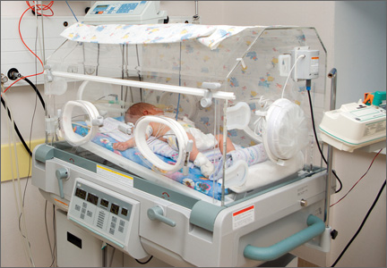
AN OBGYN ENCOUNTERED SHOULDER DYSTOCIA. He used fundal pressure and downward lateral traction to free the baby’s shoulder. The child has a brachial plexus injury of the right shoulder, including nerve avulsion, a fractured clavicle, and permanent disfigurement. She underwent surgery; physical and occupational therapy will continue.
PARENTS' CLAIM The standard sequence of maneuvers should have been attempted before fundal pressure and lateral traction were used—the baby was sufficiently oxygenated to allow time for these maneuvers. Excessive lateral traction caused the injury.
DEFENDANTS' DEFENSE The injuries occurred in utero before or while the fetus progressed down the birth canal, and were due to the maternal forces of labor.
VERDICT A $3,070,000 Michigan verdict was returned against the hospital, ObGyn, and ObGyn group.
WHAT IS THE STANDARD SEQUENCE OF MANEUVERS FOR SHOULDER DYSTOCIA?
Read Dr. Robert L. Barbieri’s May Editorial, You are the second responder to a shoulder dystocia emergency. What do you do first? and Dr. Ronald T. Burkman’s March Stop/Start article, Stop all activities that may lead to further shoulder impaction when you suspect possible shoulder dystocia Meconium aspiration leads to brain injury
LATE IN HER PREGNANCY, a woman went to the emergency department (ED) with hypertension; she was discharged the same day. She saw her ObGyns, Dr. A and Dr. B, three times in the next 2 weeks. A day after her last visit, she returned to the ED in active labor. Dr. B assumed her care. Fetal monitoring indicated a nonreassuring heart rate with decelerations. Dr. B administered oxytocin and labor continued.
The baby was born by cesarean delivery after 25 minutes of fetal bradycardia. She was covered in meconium, with a low heart rate and irregular, labored respirations. The baby was transferred to another hospital, where she was treated for pulmonary hypertension, meconium aspiration, and seizures. The child is totally disabled, and will require constant care for life.
PARENTS' CLAIM The mother’s hypertension was not properly treated. Dr. B and the nurse waited too long to perform a cesarean delivery.
DEFENDANTS' DEFENSE Proper prenatal care was provided. There was no reason for additional testing; fetal heart tones at the mother’s last office visit were reactive. There were no clinical signs of a hematoma or cord varix during office visits. An unpredictable, unpreventable umbilical cord hematoma caused ischemia and hypoxia, and the subsequent brain injury. Meconium had been in the amniotic fluid for at least 10 hours due to the ischemic/hypoxic episode. The hematoma formed between her last office visit and when the mother came to the hospital the next day.
VERDICT Settlements were reached with Dr. A and the hospital. An Arkansas defense verdict was returned for Dr. B and the nurse.
14 months' recovery after mass removed
A GYNECOLOGIC ONCOLOGIST operated on a woman in her 50s to remove a large, noncancerous pelvic mass. The patient, discharged on postoperative day 2, was readmitted the next day with a fever (temperature, 103ºF), nausea, vomiting, and abdominal pain. Four days later, the oncologist repaired a perforated bowel and created an ileostomy. Other procedures were needed to drain abscesses and repair fistulas, and resect a large portion of colon due to continuing infection. Treatment lasted 14 months.
PATIENT'S CLAIM The gynecologic oncologist was negligent in failing to timely diagnose and treat the bowel perforation. Earlier repair would have curtailed development of the abscesses and fistulae.
PHYSICIAN'S DEFENSE Any complications the patient experienced were unrelated to any delay in treatment.
VERDICT A $612,237 Michigan verdict was returned.
Colon perforated during abdominal access
WHEN A MORBIDLY OBESE 37-YEAR-OLD WOMAN reported chronic pelvic pain, her gynecologist suspected endometriosis. Conservative treatment failed and the gynecologist offered laparoscopic hysterectomy.
After abdominal insufflation was unsuccessfully attempted twice using a Veress needle, the gynecologist entered the abdomen with a Visiport optical trocar, and continued the procedure. The gynecologist inspected the abdomen before closing but found no injuries.
The patient did not do well after surgery. CT scan detected a bowel perforation on postoperative day 6. During exploratory laparotomy, a through-and-through “bayonet” colon perforation was repaired. Because of the extensive infection, the patient’s surgical wound was left open and several “washouts” were performed; the wound was closed several weeks later. The patient also underwent two adhesiolysis procedures.
PATIENT'S CLAIM Access to the abdomen was not properly performed and caused colon perforation. The injury should have been found and treated earlier.
PHYSICIAN'S DEFENSE The case was settled before trial.
VERDICT A $750,000 Virginia settlement was reached.
READ How to avoid intestinal and urinary tract injuries during gynecologic laparoscopy, by Michael Baggish, MD (Surgical Techniques, October 2012) What caused this C. diff infection after hysterectomy?
AFTER A HYSTERECTOMY, a 42-year-old woman developed a persistent fever and increased white blood cell count. The gynecologist prescribed ciprofloxacin for a urinary tract infection, and discharged the patient from the hospital on postoperative day 4. She returned to the gynecologist’s office with severe abdominal pain and vomiting 4 days after discharge. The gynecologist prescribed an antacid and told her to continue taking ciprofloxacin.
The patient was taken to the ED by ambulance 3 days later. Testing revealed a Clostridium dificule (C. diff) infection. During emergency surgery, a large portion of her colon was resected, and a colostomy was performed. The colostomy was reversed 6 months later. The patient developed an incisional hernia and has abdominal scarring.
PATIENT'S CLAIM Prophylactic antibiotics should have been prescribed before surgery.
Two possible scenarios were presented: 1) A bowel injury occurred during surgery, and ciprofloxacin likely worsened the infection caused by the bowel injury; or 2) ciprofloxacin triggered the C. diff infection that caused leaking colon perforations and subsequent peritonitis.
The colon perforations could have been avoided if the gynecologist had diagnosed and treated the C. diff infection in a timely manner.
PHYSICIAN'S DEFENSE The patient’s symptoms did not suggest a C. diff infection; testing was not necessary. Ciprofloxacin might have allowed the proliferation of the C. diff infection, but the use of the drug was not negligent. The infection was not preventable and could not have been diagnosed earlier.
VERDICT A $776,000 New York verdict was returned.
Brain injury and cerebral palsy: When did this occur?
DURING LABOR AND DELIVERY, there were periods when the fetal heart-rate tracings were nonreassuring with variable decelerations and fetal tachycardia; some variables were severe. The child suffered anoxic encephalopathy that caused neurologic injury and cerebral palsy.
PARENTS' CLAIM The infant suffered numerous hypoxic incidents before cesarean delivery was performed. An earlier cesarean delivery could have prevented the injury.
PHYSICIAN'S DEFENSE The newborn had a normal blood cord gas level of 7.2 pH and Apgar scores of 9 and 10, at 1 and 5 minutes, respectively. Fetal heart-rate tracings did not show evidence of fetal hypoxia. The brain injury likely occurred prior to the onset of labor and was possibly related to a viral encephalopathy.
VERDICT A Virginia defense verdict was returned. These cases were selected by the editors of OBG Management from Medical Malpractice Verdicts, Settlements & Experts, with permission of the editor, Lewis Laska (www.verdictslaska.com). The information available to the editors about the cases presented here is sometimes incomplete. Moreover, the cases may or may not have merit. Nevertheless, these cases represent the types of clinical situations that typically result in litigation and are meant to illustrate nationwide variation in jury verdicts and awards.

No safety issues detected in HPV vaccine pregnancy registry
The analysis of the Gardasil pregnancy registry data has provided "reassuring" results regarding the potential risks of exposure to the vaccine during pregnancy, Dr. Fabio Lievano reported at a meetingof the Centers for Disease Control and Prevention’s Advisory Committee on Immunization Practices.
Based on more than 2,500 pregnancies reported to Merck and enrolled in the registry over a 6-year period, the overall rates of spontaneous abortion, fetal deaths, and congenital anomalies were "at or below background rates," and no pattern in the type of birth defects has been identified, said Dr. Lievano, executive director of clinical risk management at Merck Research Laboratories. Merck is the manufacturer of the quadrivalent HPV vaccine, which provides coverage against HPV types 6, 11, 16, and 18. The vaccine was approved in the United States in 2006, is marketed as Gardasil, and is administered in a three-dose series.
Gardasil is not recommended for use in pregnancy, and as part of the company’s postmarketing safety commitments for the Food and Drug Administration and European and Canadian regulatory authorities, Merck established a pregnancy registry to monitor outcomes associated with inadvertent pregnancy exposures. Women were enrolled in the pregnancy registry if they were living in the United States, Canada, or France and had received a dose of Gardasil within 1 month of their last menstrual period or at any time during pregnancy.
Between June 1, 2006, and May 31, 2012, 2,802 pregnancies exposed to the vaccine were enrolled in the registry, reported from health care providers and vaccinees. (Enrollment closed on Dec. 31, 2012.) The main outcomes the company monitored were infant outcomes (congenital anomalies) and pregnancy outcomes, which included elective abortions, spontaneous abortions (before 20 weeks’ gestation), fetal deaths (at 20 weeks or later), and live births.
Of the 2,440 prospective pregnancy reports (received before the pregnancy outcome was known), about 91% indicated exposure to the vaccine occurred before the end of the first trimester. Of the total exposed pregnancies, there were 102 (3.6%) elective abortions (including 1 associated with anencephaly and hypoplastic heart) and 105 (3.7%) spontaneous abortions (including 1 triplet pregnancy and 1 with a chromosomal anomaly).
Among the 1,460 newborns, 1,381 (95%) had normal outcomes. Of the remainder, 34 had major congenital anomalies and 45 had minor congenital anomalies.
The rate of spontaneous abortions was 6.7/100 outcomes, Dr. Lievano said, noting that the background rate among clinically recognized pregnancies is 15%. The rate of fetal deaths was 0.8/100 outcomes (live births plus fetal deaths) compared with the background rate of 0.62-1/100.
The overall rate of major congenital anomalies was 2.5/100 live born infants, compared with the background rate of 2.67/100 live born infants. The rate of congenital anomalies appears to be similar to the background rates, and the anomalies reported in the registry have varied in type and etiology, Dr. Lievano said, concluding that the analysis "does not support a causal relationship" between exposure to the vaccine in pregnancy and the birth defects reported in the registry,
Of the 362 retrospective reports of pregnancy exposures in the registry (those with outcomes that were known at the time of enrollment), there were 319 known pregnancy outcomes, which included 61 spontaneous abortions and 8 fetal deaths. Among the retrospective reports, 25 infants had major congenital anomalies, including 13 with an isolated anomaly, 4 with two anomalies each, 3 with multiple anomalies, and 2 with multiple anomalies as part of a chromosomal abnormality, he said.
As of April, the FDA, as well as the European Medicines Agency and Health Canada, consider that the company has met the postmarketing pregnancy registry commitment. The results will be published and will be added to the drug’s label. Merck will continue to update the registry with reports of exposures and will provide periodic safety updates to the regulatory agencies, Dr. Lievano said.
The updated ACIP statement on HPV vaccine, which is being planned, will advise that pregnancy exposures continue to be reported to Merck and to the FDA and CDC’s Vaccine Adverse Event Reporting System (VAERS).
The updated Gardasil pregnancy registry website says that cases of exposure to Gardasil during pregnancy should be reported to Merck at 877-888-4231.
FROM A CDC ADVISORY COMMITTEE MEETING
Does myo-inositol supplementation reduce the rate of gestational diabetes in pregnant women with a family history of type 2 diabetes?
Inositol has generated increasing attention as a treatment for conditions related to pregnancy, including polycystic ovary syndrome1 and neural tube defects.2,3 In this study, pregnant women with a family history of type 2 diabetes were randomly allocated to:
- folic acid alone (400 µg daily)—placebo group
- folic acid plus myo-inositol (400 µg and 4 g daily, respectively)—treatment group.
The goal was to determine whether the addition of myo-inositol could prevent GDM and macrosomia. Because the study was conducted in Italy, GDM was diagnosed using recommendations from the International Association of Diabetes and Pregnancy Study Groups (IADPSG).4
Of the 197 women who completed the study, those given myo-inositol had a lower incidence of GDM than those given placebo: 6 of 99 women in the treatment group developed GDM, compared with 15 of 98 in the placebo group (P = .04). None of the women in the treatment group gave birth to a macrosomic infant (>4,000 g), compared with seven women in the placebo group (P = .007). However, other adverse outcomes, such as cesarean delivery, gestational hypertension, shoulder dystocia, preterm delivery, and fetal respiratory distress syndrome occurred at similar frequencies in the two groups.
RELATED ARTICLE Weight changes between pregnancies tied to risk of gestational diabetes mellitus (Web NEWS, June 2011)
How to improve outcomes in gestational diabetes—for mother and baby
E. Albert Reece, MD, PhD, MBA (March 2011)
Strengths and limitations of the study
Investigators conducted a prospective, randomized, placebo-controlled trial, one of the first moderately sized trials to examine the therapeutic effects of myo-inositol in pregnant women who had a family history of type 2 diabetes (in one or both parents). This study builds upon a previous report from the same investigators of the positive effects of myo-inositol in reducing insulin resistance in a smaller group of women (n = 69) with GDM.5
Another strength is that the investigators focused on a compound, inositol, readily found in foods, which suggests that women could derive benefits from a diet fortified with this supplement.
However, there are several concerns, also touched upon in a previous commentary about this study,6 which include:
- Only white women were included in the study. (As an ethnic group, white women have a lower risk of GDM.)
- All of the women enrolled in this trial had a prepregnancy body mass index within the normal range. Women who have a family history of type 2 diabetes who are also obese are a significant population to study.
- More work is needed to understand the mechanism of action of myo-inositol to help determine the optimal concentration to administer and the route of administration (ie, in pill form or as part of a diet plan).
Another important point: Investigators used IADPSG criteria to diagnose GDM; these criteria tend to identify significantly more cases of diabetes than the Carpenter and Coustan criteria, which are currently used in the United States.7,8 Indeed, a National Institutes of Health expert panel recommended continuing use of the Carpenter and Coustan criteria to diagnose GDM in the United States until more data are collected to show correlation between changing the diagnostic threshold and improved fetal and maternal outcomes.9
WHAT THIS EVIDENCE MEANS FOR PRACTICE
Current standards for identifying GDM include appropriate screening. Methods for prevention of GDM including dietary counseling and advice on an exercise program to reduce fetal macrosomia, but no interventions, to date, have effectively controlled GDM.
The concept of using a dietary supplement to prevent GDM is intriguing, and the results of this trial, coupled with a second recent report on the beneficial effects of myo-inositol to prevent GDM,10 are promising. However, the many concerns raised here—especially the use of IADPSG criteria—make it difficult to apply these findings to traditional diagnostic criteria for US practitioners. Follow-up, large-scale trials are needed to determine how effective this supplement is in preventing GDM and whether its effects translate into benefits for women at increased risk of developing diabetes. The routine use of myo-inositol in patients with a family history of type 2 diabetes should await further studies, including the aforementioned trials.
E. Albert Reece, MD, PhD, MBA
- Morgante G, Orvieto R, Di Sabatino A, Musacchio MC, De Leo V. The role of inositol supplementation in patients with polycystic ovary syndrome, with insulin resistance, undergoing the low-dose gonadotropin ovulation induction regimen. Fertil Steril. 2011;95(8):2642–2644.
- Khandelwal M, Reece EA, Wu YK, Borenstein M. Dietary myo-inositol therapy in hyperglycemia-induced embryopathy. Teratology. 1998;57(2):79–84.
- Cavalli P, Tonni G, Grosso E, Poggiani C. Effects of inositol supplementation in a cohort of mothers at risk of producing an NTD pregnancy. Birth Defects Res A Clin Mol Teratol. 2011;91(11):962–965.
- International Association of Diabetes and Pregnancy Study Groups Consensus Panel. International Association of Diabetes and Pregnancy Study Groups recommendations on the diagnosis and classification of hyperglycemia in pregnancy. Diabetes Care. 2010;33(3):676–682.
- Corrado F, D’Anna R, Di Vieste G, et al. The effect of myoinositol supplementation on insulin resistance in patients with gestational diabetes. Diabet Med. 2011;28(8):972–975.
- Coustan DR. Can a dietary supplement prevent gestational diabetes mellitus? Diabetes Care. 2013;36(4):777–779.
- Reece EA, Moore T. The diagnostic criteria for gestational diabetes: To change or not to change? Am J Obstet Gynecol. 2013;208(4):255–259.
- Langer O, Umans JG, Miodovnik M. Perspectives on the proposed gestational diabetes mellitus diagnostic criteria. Obstet Gynecol. 2013;121(1):177–182.
- National Institutes of Health Draft Consensus Development Panel. Draft Statement: National Institutes of Health Consensus Development Conference Statement: Diagnosing Gestational Diabetes Mellius; March 4–6, 2013; Bethesda, Maryland. http://prevention.nih.gov/cdp/conferences/2013/gdm/files/DraftStatement.pdf. Published March 6, 2013. Accessed May 17, 2013.
- Matarrelli B, Vitacolonna E, D’Angelo M, et al. Effect of dietary myo-inositol supplementation in pregnancy on the incidence of maternal gestational diabetes mellitus and fetal outcomes: a randomized controlled trial [published online ahead of print March 1, 2013]. J Matern Fetal Neonatal Med. doi:10.3109/14767058.2013.766691.
Inositol has generated increasing attention as a treatment for conditions related to pregnancy, including polycystic ovary syndrome1 and neural tube defects.2,3 In this study, pregnant women with a family history of type 2 diabetes were randomly allocated to:
- folic acid alone (400 µg daily)—placebo group
- folic acid plus myo-inositol (400 µg and 4 g daily, respectively)—treatment group.
The goal was to determine whether the addition of myo-inositol could prevent GDM and macrosomia. Because the study was conducted in Italy, GDM was diagnosed using recommendations from the International Association of Diabetes and Pregnancy Study Groups (IADPSG).4
Of the 197 women who completed the study, those given myo-inositol had a lower incidence of GDM than those given placebo: 6 of 99 women in the treatment group developed GDM, compared with 15 of 98 in the placebo group (P = .04). None of the women in the treatment group gave birth to a macrosomic infant (>4,000 g), compared with seven women in the placebo group (P = .007). However, other adverse outcomes, such as cesarean delivery, gestational hypertension, shoulder dystocia, preterm delivery, and fetal respiratory distress syndrome occurred at similar frequencies in the two groups.
RELATED ARTICLE Weight changes between pregnancies tied to risk of gestational diabetes mellitus (Web NEWS, June 2011)
How to improve outcomes in gestational diabetes—for mother and baby
E. Albert Reece, MD, PhD, MBA (March 2011)
Strengths and limitations of the study
Investigators conducted a prospective, randomized, placebo-controlled trial, one of the first moderately sized trials to examine the therapeutic effects of myo-inositol in pregnant women who had a family history of type 2 diabetes (in one or both parents). This study builds upon a previous report from the same investigators of the positive effects of myo-inositol in reducing insulin resistance in a smaller group of women (n = 69) with GDM.5
Another strength is that the investigators focused on a compound, inositol, readily found in foods, which suggests that women could derive benefits from a diet fortified with this supplement.
However, there are several concerns, also touched upon in a previous commentary about this study,6 which include:
- Only white women were included in the study. (As an ethnic group, white women have a lower risk of GDM.)
- All of the women enrolled in this trial had a prepregnancy body mass index within the normal range. Women who have a family history of type 2 diabetes who are also obese are a significant population to study.
- More work is needed to understand the mechanism of action of myo-inositol to help determine the optimal concentration to administer and the route of administration (ie, in pill form or as part of a diet plan).
Another important point: Investigators used IADPSG criteria to diagnose GDM; these criteria tend to identify significantly more cases of diabetes than the Carpenter and Coustan criteria, which are currently used in the United States.7,8 Indeed, a National Institutes of Health expert panel recommended continuing use of the Carpenter and Coustan criteria to diagnose GDM in the United States until more data are collected to show correlation between changing the diagnostic threshold and improved fetal and maternal outcomes.9
WHAT THIS EVIDENCE MEANS FOR PRACTICE
Current standards for identifying GDM include appropriate screening. Methods for prevention of GDM including dietary counseling and advice on an exercise program to reduce fetal macrosomia, but no interventions, to date, have effectively controlled GDM.
The concept of using a dietary supplement to prevent GDM is intriguing, and the results of this trial, coupled with a second recent report on the beneficial effects of myo-inositol to prevent GDM,10 are promising. However, the many concerns raised here—especially the use of IADPSG criteria—make it difficult to apply these findings to traditional diagnostic criteria for US practitioners. Follow-up, large-scale trials are needed to determine how effective this supplement is in preventing GDM and whether its effects translate into benefits for women at increased risk of developing diabetes. The routine use of myo-inositol in patients with a family history of type 2 diabetes should await further studies, including the aforementioned trials.
E. Albert Reece, MD, PhD, MBA
- Morgante G, Orvieto R, Di Sabatino A, Musacchio MC, De Leo V. The role of inositol supplementation in patients with polycystic ovary syndrome, with insulin resistance, undergoing the low-dose gonadotropin ovulation induction regimen. Fertil Steril. 2011;95(8):2642–2644.
- Khandelwal M, Reece EA, Wu YK, Borenstein M. Dietary myo-inositol therapy in hyperglycemia-induced embryopathy. Teratology. 1998;57(2):79–84.
- Cavalli P, Tonni G, Grosso E, Poggiani C. Effects of inositol supplementation in a cohort of mothers at risk of producing an NTD pregnancy. Birth Defects Res A Clin Mol Teratol. 2011;91(11):962–965.
- International Association of Diabetes and Pregnancy Study Groups Consensus Panel. International Association of Diabetes and Pregnancy Study Groups recommendations on the diagnosis and classification of hyperglycemia in pregnancy. Diabetes Care. 2010;33(3):676–682.
- Corrado F, D’Anna R, Di Vieste G, et al. The effect of myoinositol supplementation on insulin resistance in patients with gestational diabetes. Diabet Med. 2011;28(8):972–975.
- Coustan DR. Can a dietary supplement prevent gestational diabetes mellitus? Diabetes Care. 2013;36(4):777–779.
- Reece EA, Moore T. The diagnostic criteria for gestational diabetes: To change or not to change? Am J Obstet Gynecol. 2013;208(4):255–259.
- Langer O, Umans JG, Miodovnik M. Perspectives on the proposed gestational diabetes mellitus diagnostic criteria. Obstet Gynecol. 2013;121(1):177–182.
- National Institutes of Health Draft Consensus Development Panel. Draft Statement: National Institutes of Health Consensus Development Conference Statement: Diagnosing Gestational Diabetes Mellius; March 4–6, 2013; Bethesda, Maryland. http://prevention.nih.gov/cdp/conferences/2013/gdm/files/DraftStatement.pdf. Published March 6, 2013. Accessed May 17, 2013.
- Matarrelli B, Vitacolonna E, D’Angelo M, et al. Effect of dietary myo-inositol supplementation in pregnancy on the incidence of maternal gestational diabetes mellitus and fetal outcomes: a randomized controlled trial [published online ahead of print March 1, 2013]. J Matern Fetal Neonatal Med. doi:10.3109/14767058.2013.766691.

Asthma medications
One of the most common conditions that complicate pregnancy is maternal asthma. Evidence continues to mount that adequate control of asthma, including appropriate use of medications, is the best approach to optimizing outcomes. Yet questions persist about the effect of asthma itself, as well as specific medications and the risk for major congenital malformations. As with any exposure during pregnancy, answering these questions is challenging because of the rarity of specific birth defects and the various and increasing number of medications that might be used to treat or prevent asthma symptoms.
Previously in this column ("Beta2-agonists for asthma, December 2011), we reviewed two studies that suggested short-acting beta-agonists used for the treatment of asthma were associated with an increased risk of oral clefts and that long-acting beta-agonists might be associated with an increased risk of cardiac anomalies (Hum. Reprod. 2011;26:3147-54; Birth Defects Res. A. Clin. Mol. Teratol. 2011;91:937-47).
What have we learned since then? Two studies published in 2013 add to the body of knowledge. The first, a database analysis using the United Kingdom’s General Practice Research Database, assessed pregnancy outcomes between 1991 and 2002 in 7,911 women exposed to asthma medications in the first trimester of pregnancy and 15,840 women who were not exposed (Pharmacotherapy 2013;33:363-8). Major anomalies were identified up to 1 year of age. Minor anomalies, chromosomal anomalies, and those associated with prematurity were excluded.
The overall risk for any exposure, compared with no exposure, for any congenital anomaly was 1.1 (95% confidence interval [CI], 1.0-1.3). No significant differences were found by class of asthma medication. Specific categories of defects also were evaluated, including musculoskeletal anomalies, oral clefts, cardiovascular defects, and multiple anomalies. Some estimates were elevated for specific medication classes.
For example, the relative risk (RR) of cleft lip or palate associated with exposure to long-acting beta-agonists was 2.4, but the confidence interval included 1 (0.3-21.8) based on 424 exposed pregnancies. The authors concluded that they found no significant increased risk of congenital anomalies associated with exposure to asthma, asthma medications, or any specific asthma medication classes in the first trimester. Limitations of the study included the inability to verify that exposure took place and insufficient data to adjust for confounding by vitamin supplementation, alcohol use, socioeconomic status, or markers of disease severity (other than number of medications prescribed).
The second study addressed the issue of maternal asthma itself and the risk for congenital anomalies (BJOG 2013;120:812-22). Using a meta-analysis approach, 21 cohort studies published between 1975 and 2012 met the criteria for inclusion. Combining major and minor congenital anomalies, the authors found a slight but statistically significant increased risk for any defect (RR, 1.11; 95% CI, 1.01-1.68), but when the analysis was restricted to major defects alone, the summary estimate was elevated but no longer significant (RR, 1.31; 95% CI, 0.57-3.02). When the specific defect grouping of oral clefts was examined, however, there was a significantly elevated overall relative risk of 1.30 (95% CI, 1.01-1.68). Limitations of this study include differing quality of studies and the appropriateness of combining data to derive a summary estimate.
Although both studies provide reassurance about the overall risk of major defects in the offspring of women with asthma, both suggest that more work needs to be done to follow up on the risk for oral clefts and whether this is linked to underlying disease severity and/or use of specific medications. Furthermore, safety of specific long-acting beta-agonist medications in pregnancy should be further examined.
The take-home message, however, continues to be that the risk for major defects in the offspring of pregnant women with asthma appears to be low, which supports the recommendation to follow guidelines for appropriate treatment of women with asthma both during and outside of pregnancy to control symptoms.
Dr. Chambers is professor of pediatrics and family and preventive medicine at the University of California, San Diego. She is director of the California Teratogen Information Service and Clinical Research Program. Dr. Chambers is a past president of the Organization of Teratology Information Specialists and past president of the Teratology Society. She said that she had no relevant financial disclosures.

Low-carb, high-fat diet may not be best for gestational diabetes
Earn 0.25 hours AMA PRA Category 1 credit: Read this article, and click the link at the end to take the post-test.
CHICAGO – Women with gestational diabetes on a conventional low-carbohydrate, high-fat diet were more insulin resistant, and their infants had slightly higher rates of adiposity, than did with women who consumed a diet high in complex carbohydrates and low in fat, according to a randomized pilot study of 11 women.
Both diets controlled maternal glucose and weight. "So, they’re both okay, except that tissue data and fasting levels imply that higher fat content is exacerbating insulin resistance during pregnancy," Teri L. Hernandez, Ph.D., said in an interview after presenting her findings at the annual scientific sessions of the American Diabetes Association.
It’s too soon to tell if and when the findings will have implications on practice. "It’s a good study," said Dr. Assiamira Ferrara, senior research scientist at Kaiser Permanente, Oakland, Calif., who was a moderator and not involved in the study. "But we need a bigger sample size and more feasibility studies on whether women will adhere to the diet" before we try this outside of a research environment.
The conventional diet recommended to women who have gestational diabetes (GDM) has mainly focused on carbohydrate (CHO) restriction. But Dr. Hernandez and her colleagues said that the restrictions result in greater fat intake, which in turn could promote insulin resistance and increase fetal adiposity. Meanwhile, owing to patient noncompliance and a lack of controlled designs, evidence remains confounded, she reported.
In a recent unpublished systematic review of prospective, randomized, controlled trials of diet interventions in women with GDM, Dr. Hernandez and her colleagues found that women tolerated higher complex carb/low glycemic index diets and that diets higher in unrefined carbs effectively blunted postprandial glycemia, reduced the need for insulin therapy, and improved insulin sensitivity, hemoglobin A1c, and systolic blood pressure.
Dr. Hernandez of the departments of medicine and nursing at University of Colorado at Denver in Aurora, said that she has a larger number women in her ongoing study, but the findings so far "lend evidence to the idea that women can tolerate more carb than we thought.
"These women are worried about their baby’s outcome, and they’re afraid their babies are going to be born too big, so they become very fearful of carbohydrates. What this says is that they can actually have toast and other carbs in their diet and still have a great outcome, and it could even help improve their insulin resistance," she said in an interview.
Researchers randomized five women to the conventional low-carb/high-fat diet, and 6 women to the high-carb/low-fat diet.
The low-carb/high fat diet comprised 40% CHO, 45% fat, and 15% protein; the high-carb/low fat diet contained 60% CHO, 25% fat, and 15% protein. Simple sugars made up about 18% or less of total daily calories.
The subjects were rather healthy women with mild gestational diabetes, were highly compliant, and were closely matched, with a mean body mass index of 33.5 kg/m2, and were 29-30 years old. They were provided with all the meals.
During the study period, weight gain was similar in both groups, and their glycemic profiles were below target.
However, the high-carb/low-fat diet group had lower fasting glucose and fasting insulin at 6 and 7 weeks, compared with the low-carb/high-fat diet group (P = .007 and .06, respectively)
Results also showed that the postprandial free fatty acids were significantly higher in the low-carb/high-fat diet group (P = .037). And, at week 37, fasting glucose, insulin, and maternal insulin resistance (HOMA-IR) were significantly higher in the low-carb/high-fat diet, compared with the low-fat/high-carb diet (P = .007, .06, and .02, respectively).
Meanwhile, infant adiposity was slightly higher in the infants of the low-carb/high-fat groups, compared with the high-carb/low-fat group (14% v. 11%). And, regardless of diet, higher fasting insulin and HOMA-IR at 37 weeks were associated with greater infant adiposity (P less than .05).
Dr. Hernandez said that both diets are doing a good job, and thus far, there haven’t been any adverse outcomes because of either diet.
Dr. Hernandez had no disclosures. Dr. Ferrara is an employee of Takeda Global Research and Development and has received research support from Takeda Pharmaceutical.
To earn 0.25 hours AMA PRA Category 1 credit after reading this article, take the post-test here.
On Twitter @NaseemSMiller
AT THE ADA ANNUAL SCIENTIFIC SESSIONS

Low-carb, high-fat diet may not be best for gestational diabetes
CHICAGO – Women with gestational diabetes on a conventional low-carbohydrate, high-fat diet were more insulin resistant, and their infants had slightly higher rates of adiposity, than did with women who consumed a diet high in complex carbohydrates and low in fat, according to a randomized pilot study of 11 women.
Both diets controlled maternal glucose and weight. "So, they’re both okay, except that tissue data and fasting levels imply that higher fat content is exacerbating insulin resistance during pregnancy," Teri L. Hernandez, Ph.D., said in an interview after presenting her findings at the annual scientific sessions of the American Diabetes Association.

It’s too soon to tell if and when the findings will have implications on practice. "It’s a good study," said Dr. Assiamira Ferrara, senior research scientist at Kaiser Permanente, Oakland, Calif., who was a moderator and not involved in the study. "But we need a bigger sample size and more feasibility studies on whether women will adhere to the diet" before we try this outside of a research environment.
The conventional diet recommended to women who have gestational diabetes (GDM) has mainly focused on carbohydrate (CHO) restriction. But Dr. Hernandez and her colleagues said that the restrictions result in greater fat intake, which in turn could promote insulin resistance and increase fetal adiposity. Meanwhile, owing to patient noncompliance and a lack of controlled designs, evidence remains confounded, she reported.
In a recent unpublished systematic review of prospective, randomized, controlled trials of diet interventions in women with GDM, Dr. Hernandez and her colleagues found that women tolerated higher complex carb/low glycemic index diets and that diets higher in unrefined carbs effectively blunted postprandial glycemia, reduced the need for insulin therapy, and improved insulin sensitivity, hemoglobin A1c, and systolic blood pressure.
Dr. Hernandez of the departments of medicine and nursing at University of Colorado at Denver in Aurora, said that she has a larger number women in her ongoing study, but the findings so far "lend evidence to the idea that women can tolerate more carb than we thought.
"These women are worried about their baby’s outcome, and they’re afraid their babies are going to be born too big, so they become very fearful of carbohydrates. What this says is that they can actually have toast and other carbs in their diet and still have a great outcome, and it could even help improve their insulin resistance," she said in an interview.
Researchers randomized five women to the conventional low-carb/high-fat diet, and 6 women to the high-carb/low-fat diet.
The low-carb/high fat diet comprised 40% CHO, 45% fat, and 15% protein; the high-carb/low fat diet contained 60% CHO, 25% fat, and 15% protein. Simple sugars made up about 18% or less of total daily calories.
The subjects were rather healthy women with mild gestational diabetes, were highly compliant, and were closely matched, with a mean body mass index of 33.5 kg/m2, and were 29-30 years old. They were provided with all the meals.
During the study period, weight gain was similar in both groups, and their glycemic profiles were below target.
However, the high-carb/low-fat diet group had lower fasting glucose and fasting insulin at 6 and 7 weeks, compared with the low-carb/high-fat diet group (P = .007 and .06, respectively)
Results also showed that the postprandial free fatty acids were significantly higher in the low-carb/high-fat diet group (P = .037). And, at week 37, fasting glucose, insulin, and maternal insulin resistance (HOMA-IR) were significantly higher in the low-carb/high-fat diet, compared with the low-fat/high-carb diet (P = .007, .06, and .02, respectively).
Meanwhile, infant adiposity was slightly higher in the infants of the low-carb/high-fat groups, compared with the high-carb/low-fat group (14% v. 11%). And, regardless of diet, higher fasting insulin and HOMA-IR at 37 weeks were associated with greater infant adiposity (P less than .05).
Dr. Hernandez said that both diets are doing a good job, and thus far, there haven’t been any adverse outcomes because of either diet.
Dr. Hernandez had no disclosures. Dr. Ferrara is an employee of Takeda Global Research and Development and has received research support from Takeda Pharmaceutical.
On Twitter @NaseemSMiller
CHICAGO – Women with gestational diabetes on a conventional low-carbohydrate, high-fat diet were more insulin resistant, and their infants had slightly higher rates of adiposity, than did with women who consumed a diet high in complex carbohydrates and low in fat, according to a randomized pilot study of 11 women.
Both diets controlled maternal glucose and weight. "So, they’re both okay, except that tissue data and fasting levels imply that higher fat content is exacerbating insulin resistance during pregnancy," Teri L. Hernandez, Ph.D., said in an interview after presenting her findings at the annual scientific sessions of the American Diabetes Association.
It’s too soon to tell if and when the findings will have implications on practice. "It’s a good study," said Dr. Assiamira Ferrara, senior research scientist at Kaiser Permanente, Oakland, Calif., who was a moderator and not involved in the study. "But we need a bigger sample size and more feasibility studies on whether women will adhere to the diet" before we try this outside of a research environment.
The conventional diet recommended to women who have gestational diabetes (GDM) has mainly focused on carbohydrate (CHO) restriction. But Dr. Hernandez and her colleagues said that the restrictions result in greater fat intake, which in turn could promote insulin resistance and increase fetal adiposity. Meanwhile, owing to patient noncompliance and a lack of controlled designs, evidence remains confounded, she reported.
In a recent unpublished systematic review of prospective, randomized, controlled trials of diet interventions in women with GDM, Dr. Hernandez and her colleagues found that women tolerated higher complex carb/low glycemic index diets and that diets higher in unrefined carbs effectively blunted postprandial glycemia, reduced the need for insulin therapy, and improved insulin sensitivity, hemoglobin A1c, and systolic blood pressure.
Dr. Hernandez of the departments of medicine and nursing at University of Colorado at Denver in Aurora, said that she has a larger number women in her ongoing study, but the findings so far "lend evidence to the idea that women can tolerate more carb than we thought.
"These women are worried about their baby’s outcome, and they’re afraid their babies are going to be born too big, so they become very fearful of carbohydrates. What this says is that they can actually have toast and other carbs in their diet and still have a great outcome, and it could even help improve their insulin resistance," she said in an interview.
Researchers randomized five women to the conventional low-carb/high-fat diet, and 6 women to the high-carb/low-fat diet.
The low-carb/high fat diet comprised 40% CHO, 45% fat, and 15% protein; the high-carb/low fat diet contained 60% CHO, 25% fat, and 15% protein. Simple sugars made up about 18% or less of total daily calories.
The subjects were rather healthy women with mild gestational diabetes, were highly compliant, and were closely matched, with a mean body mass index of 33.5 kg/m2, and were 29-30 years old. They were provided with all the meals.
During the study period, weight gain was similar in both groups, and their glycemic profiles were below target.
However, the high-carb/low-fat diet group had lower fasting glucose and fasting insulin at 6 and 7 weeks, compared with the low-carb/high-fat diet group (P = .007 and .06, respectively)
Results also showed that the postprandial free fatty acids were significantly higher in the low-carb/high-fat diet group (P = .037). And, at week 37, fasting glucose, insulin, and maternal insulin resistance (HOMA-IR) were significantly higher in the low-carb/high-fat diet, compared with the low-fat/high-carb diet (P = .007, .06, and .02, respectively).
Meanwhile, infant adiposity was slightly higher in the infants of the low-carb/high-fat groups, compared with the high-carb/low-fat group (14% v. 11%). And, regardless of diet, higher fasting insulin and HOMA-IR at 37 weeks were associated with greater infant adiposity (P less than .05).
Dr. Hernandez said that both diets are doing a good job, and thus far, there haven’t been any adverse outcomes because of either diet.
Dr. Hernandez had no disclosures. Dr. Ferrara is an employee of Takeda Global Research and Development and has received research support from Takeda Pharmaceutical.
On Twitter @NaseemSMiller
AT THE ADA ANNUAL SCIENTIFIC SESSIONS
Major finding: At week 37, fasting glucose, insulin, and maternal insulin resistance (HOMA-IR) were significantly higher in the low carb/high fat diet, compared with the low fat/high carb diet.
Data source: Randomized pilot study of 11 women with mild gestational diabetes.
Disclosures: Dr. Hernandez had no disclosures. Dr. Ferrara is an employee of Takeda Global Research and Development and has received research support from Takeda Pharmaceutical.

Use 6-cm dilation to judge labor progress
SAN FRANCISCO – A threshold of 6-cm cervical dilation is more accurate than the conventional 4 cm to determine when a woman enters the active phase of labor, a reevaluation of evidence suggests.
The historical evidence behind the commonly used assumption that 4-cm dilation signals the start of active labor contains methodological flaws, doesn’t match today’s population of pregnant women, and is contradicted by more recent studies supporting the 6-cm threshold, Tekoa King, C.N.M, Ph.D., said at a meeting on antepartum and intrapartum management sponsored by the University of California, San Francisco.

Using the 4-cm threshold, a woman "may just be in the normal latent phase of labor," she said.
Switching to the 6-cm threshold should delay or reduce the use of epidural anesthesia and might lower the high rate of cesarean sections in the United States. "Six centimeters is the new four centimeters," said Dr. King, a certified nurse-midwife and clinical professor of nursing at the university.
The 4-cm threshold for active labor arose out of two studies in the 1950s by Friedman et al. One study plotted the course of labor in centimeters of dilation over time in 500 nulliparous pregnant women. The same investigators profiled 200 women who were considered in the 1950s to have "ideal labor" – meaning term, vertex, singleton pregnancies – but the women commonly received considerable amounts of morphine and may or may not have had instrumented deliveries using outlet forceps. "They wouldn’t be what we would consider ideal today," Dr. King said.
Comparing the two cohorts, investigators in the 1950s found that the "ideal" group had shorter labors. They concluded that women who had been contracting more than 24 hours had a prolonged latent phase, and that the slowest rate of dilation in the active phase of labor was 1.2 cm/hr. Thus was born the "Friedman curve" that underpinned the decades-long dogma that women need to dilate about 1 cm/hr in the active phase of labor.
More recently, other data show that "the Friedman curve was really codified by the way we examined women every 2 hours. If you examine them more frequently, you’re going to get a different curve. Probably what we should be doing is examining them a lot less frequently until they’re 6 cm, and perhaps a little more frequently from 6 cm to complete" dilation, depending on whether or not there are other indications for examination, she said.
A 2002 study by Zhang et al. of 1,329 nulliparous, term, singleton, vertex pregnancies with normal-weight babies, spontaneous onset of labor, and vaginal delivery used a different approach from the Friedman studies. It measured the time between each centimeter change in dilation instead of the time it took to go from 4-cm to 10-cm dilation and then calculating an interval average. The newer study found that it was common to have very slow progress before 7 cm, there was no deceleration phase, and the slowest but still normal rate of cervical dilation was less than 1 cm/hr, with a wide range of variability (Am. J. Obstet. Gynecol. 2002;187:824-8).
To go from 6-cm to 7-cm dilation, for example, took little more than half an hour on average, but it ranged from 0.2 hours to more than 2 hours. "This makes sense," Dr. King said. "What’s really happening between 6 and 7 or 7 and 8 cm? Internal rotation. We often forget that internal rotation and descent station are progress. We just get ourselves fixated on cervical dilation" and end up performing a cesarean section in women for "arrested labor" at a time when they’re having normal progress.
The same investigators followed that with a 2010 study analyzing data on more than 50,000 singleton, vertex pregnancies with spontaneous onset of labor, vaginal delivery and "normal outcome." Again, they used the "repeated measures" approach to estimate the labor curves and to "redefine normal," Dr. King said. They found that the median rate of change was about 2 cm/hr and the slowest rate of normal change was 0.4 cm/hr (Obstet. Gynecol. 2010;116:1281-7).
On Friedman’s curve, what he called the point of inflection (or the beginning of the active phase of labor) was at 4-cm cervical dilation. But Zhang’s curve suggests that the point of inflection when labor starts to progress faster is at 6-cm dilation. "That’s our real world today, and here we are, using 4 cm. Basically, we’re treating women who are in the latent phase of labor as though they were active," she said.
The other insight from the work of Zhang et al. is that dilation progresses faster as the cervix becomes more dilated, not at a steady rate of 1 cm/hr.
Zhang et al. also reported that 40% of women who undergo induction of labor get a cesarean section when they are 4 cm dilated. "That’s probably where we really need to start paying some attention to what we’re doing," Dr. King said. Too many cesarean sections are being done at cervical dilations of 6 cm or less, she said.
Other recent data show that women with induced labor need significantly more time to reach 6-cm dilation compared with women with spontaneous labor, but after 6 cm the rate of progression is similar (Obstet. Gynecol. 2012;119:1113-8).
Greater patience with induced labor could reduce the rate of cesarean sections. "These are the women who are getting sectioned," she said.
A separate study suggests that women trying for a vaginal delivery after a prior cesarean section should be assessed by the same progression curves as women without a prior cesarean section (Obstet. Gynecol. 2012;119:732-6).
Clinical variables also have changed since the 1950s in ways that affect the progress of labor. Pregnant women in the United States today are more likely to be obese. A high body mass index prolongs the time it takes for cervical dilation in labor. "We may need a whole new labor curve for these women," Dr. King said.
She reported having no financial disclosures.
On Twitter @sherryboschert
SAN FRANCISCO – A threshold of 6-cm cervical dilation is more accurate than the conventional 4 cm to determine when a woman enters the active phase of labor, a reevaluation of evidence suggests.
The historical evidence behind the commonly used assumption that 4-cm dilation signals the start of active labor contains methodological flaws, doesn’t match today’s population of pregnant women, and is contradicted by more recent studies supporting the 6-cm threshold, Tekoa King, C.N.M, Ph.D., said at a meeting on antepartum and intrapartum management sponsored by the University of California, San Francisco.
Using the 4-cm threshold, a woman "may just be in the normal latent phase of labor," she said.
Switching to the 6-cm threshold should delay or reduce the use of epidural anesthesia and might lower the high rate of cesarean sections in the United States. "Six centimeters is the new four centimeters," said Dr. King, a certified nurse-midwife and clinical professor of nursing at the university.
The 4-cm threshold for active labor arose out of two studies in the 1950s by Friedman et al. One study plotted the course of labor in centimeters of dilation over time in 500 nulliparous pregnant women. The same investigators profiled 200 women who were considered in the 1950s to have "ideal labor" – meaning term, vertex, singleton pregnancies – but the women commonly received considerable amounts of morphine and may or may not have had instrumented deliveries using outlet forceps. "They wouldn’t be what we would consider ideal today," Dr. King said.
Comparing the two cohorts, investigators in the 1950s found that the "ideal" group had shorter labors. They concluded that women who had been contracting more than 24 hours had a prolonged latent phase, and that the slowest rate of dilation in the active phase of labor was 1.2 cm/hr. Thus was born the "Friedman curve" that underpinned the decades-long dogma that women need to dilate about 1 cm/hr in the active phase of labor.
More recently, other data show that "the Friedman curve was really codified by the way we examined women every 2 hours. If you examine them more frequently, you’re going to get a different curve. Probably what we should be doing is examining them a lot less frequently until they’re 6 cm, and perhaps a little more frequently from 6 cm to complete" dilation, depending on whether or not there are other indications for examination, she said.
A 2002 study by Zhang et al. of 1,329 nulliparous, term, singleton, vertex pregnancies with normal-weight babies, spontaneous onset of labor, and vaginal delivery used a different approach from the Friedman studies. It measured the time between each centimeter change in dilation instead of the time it took to go from 4-cm to 10-cm dilation and then calculating an interval average. The newer study found that it was common to have very slow progress before 7 cm, there was no deceleration phase, and the slowest but still normal rate of cervical dilation was less than 1 cm/hr, with a wide range of variability (Am. J. Obstet. Gynecol. 2002;187:824-8).
To go from 6-cm to 7-cm dilation, for example, took little more than half an hour on average, but it ranged from 0.2 hours to more than 2 hours. "This makes sense," Dr. King said. "What’s really happening between 6 and 7 or 7 and 8 cm? Internal rotation. We often forget that internal rotation and descent station are progress. We just get ourselves fixated on cervical dilation" and end up performing a cesarean section in women for "arrested labor" at a time when they’re having normal progress.
The same investigators followed that with a 2010 study analyzing data on more than 50,000 singleton, vertex pregnancies with spontaneous onset of labor, vaginal delivery and "normal outcome." Again, they used the "repeated measures" approach to estimate the labor curves and to "redefine normal," Dr. King said. They found that the median rate of change was about 2 cm/hr and the slowest rate of normal change was 0.4 cm/hr (Obstet. Gynecol. 2010;116:1281-7).
On Friedman’s curve, what he called the point of inflection (or the beginning of the active phase of labor) was at 4-cm cervical dilation. But Zhang’s curve suggests that the point of inflection when labor starts to progress faster is at 6-cm dilation. "That’s our real world today, and here we are, using 4 cm. Basically, we’re treating women who are in the latent phase of labor as though they were active," she said.
The other insight from the work of Zhang et al. is that dilation progresses faster as the cervix becomes more dilated, not at a steady rate of 1 cm/hr.
Zhang et al. also reported that 40% of women who undergo induction of labor get a cesarean section when they are 4 cm dilated. "That’s probably where we really need to start paying some attention to what we’re doing," Dr. King said. Too many cesarean sections are being done at cervical dilations of 6 cm or less, she said.
Other recent data show that women with induced labor need significantly more time to reach 6-cm dilation compared with women with spontaneous labor, but after 6 cm the rate of progression is similar (Obstet. Gynecol. 2012;119:1113-8).
Greater patience with induced labor could reduce the rate of cesarean sections. "These are the women who are getting sectioned," she said.
A separate study suggests that women trying for a vaginal delivery after a prior cesarean section should be assessed by the same progression curves as women without a prior cesarean section (Obstet. Gynecol. 2012;119:732-6).
Clinical variables also have changed since the 1950s in ways that affect the progress of labor. Pregnant women in the United States today are more likely to be obese. A high body mass index prolongs the time it takes for cervical dilation in labor. "We may need a whole new labor curve for these women," Dr. King said.
She reported having no financial disclosures.
On Twitter @sherryboschert
EXPERT ANALYSIS FROM A MEETING ON ANTEPARTUM AND INTRAPARTUM MANAGEMENT
