User login
Vertebral fractures in COVID-19 linked to mortality
Vertebral fractures appear to be common in people with severe COVID-19, and also raise the mortality risk, findings from a retrospective cohort suggest.
Among 114 patients with COVID-19 who underwent lateral chest x-rays at the San Raffaele Hospital ED in Milan, more than a third were found to have thoracic vertebral fractures. And, those individuals were more than twice as likely to die as were those without vertebral fractures.
“Morphometric vertebral fractures are one of the most common comorbidities among adults hospitalized with COVID-19, and the presence of such fractures may predict the severity of disease outcomes,” lead investigator Andrea Giustina, MD, said in an interview.
This is the first study to examine vertebral fracture prevalence in any coronavirus disease, but such fractures have been linked to an increased risk of pneumonia and impaired respiratory function, including restrictive pulmonary dysfunction. One possible mechanism may be that they cause anatomical changes, such as kyphosis, which negatively impact respiratory function by decreasing vital capacity, forced expiratory volume in 1 second, and inspiratory time, explained Dr. Giustina, professor of endocrinology, San Raffaele Vita Salute University, Milan, and president of the European Society of Endocrinology. The results were published in the Journal of Clinical Endocrinology and Metabolism.
Clinically, the findings suggest that all patients with COVID-19 who are undergoing chest x-rays should have morphometric vertebral x-ray evaluation, said Dr. Giustina.
“One interesting aspect of the study is that without morphometry, approximatively two thirds of vertebral fractures [would have been] missed. Therefore, they are largely underestimated in clinical practice,” he noted.
Thoracic vertebral fractures assessed via lateral chest x-rays
The 114 study subjects included were those whose lateral chest x-rays allowed for a high-quality assessment and in which all the thoracic tract of T4-T12 were viewable and assessable. None had been using glucocorticoids and only 3% had a prior diagnosis of osteoporosis.
The majority (75%) were male, and median age was 57 years. Most (79%) were hospitalized after evaluation in the ED. Of those, 12% (13) were admitted to the ICU and 15% (16) died.
Thoracic vertebral fractures were detected on the lateral chest x-rays in 36% (41) of the patients. In contrast, in studies of women aged 50 years and older from the general European population, morphometric vertebral fracture prevalence ranged from 18% to 26%, the investigators noted.
Of the total 65 vertebral fractures detected, 60% were classified as mild (height ratio decrease <25%), 33.3% as moderate (25%-40% decrease) and 7.7% as severe (>40%). Patients with more than one vertebral fracture were classified by their most severe one.
Those with versus without vertebral fractures didn’t differ by sex, body mass index, or clinical or biological parameters evaluated in the ED. But, compared with those without vertebral fractures, those with them were significantly older (68 vs. 54 years) and were more likely to have arterial hypertension (56% vs. 30%) and coronary artery disease (22% vs. 7%).
In multivariate analysis, age was the only statistically significant predictor of vertebral fractures (odds ratio, 1.04; P < .001).
Mortality doubled, though not significantly
Those with vertebral fractures were more likely to be hospitalized, although not significantly (88% vs. 74%). There was no significant difference in ICU admission (11% vs. 12.5%).
However, those with vertebral fractures required noninvasive mechanical ventilation significantly more often (48.8% vs. 27.4%; P = .02), and were more than twice as likely to die (22% vs. 10%; P = .07). While the difference in overall mortality wasn’t quite statistically significant, those with severe vertebral fractures were significantly more likely to die, compared with those with mild or moderate fractures (60%, 7%, 24%, respectively, for severe, moderate, and mild; P = .04), despite no significant differences in clinical or laboratory parameters.
“Our data from the field reinforce the need of implementing previously published recommendations concerning the importance of bone fragility care during the COVID pandemic with at least those patients already treated with antiosteoporotic drugs maintaining their adherence to treatments including vitamin D, which have also been suggested very recently to have no relevant predisposing effect on COVID-19,” Dr. Giustina and colleagues wrote.
Moreover, they added, “continuity of care should also include bone density monitoring despite very restricted access to clinical facilities, during the COVID-19 pandemic. Finally, all patients with fractures should start antiresorptive treatment right away, even during hospital stay.”
The authors reported having no disclosures.
SOURCE: Giustina A et al. J Clin Endocrinol Metab. 2020 Oct 21. doi: 10.1210/clinem/dgaa738.
FROM THE JOURNAL OF CLINICAL ENDOCRINOLOGY AND METABOLISM
Combine calculators and medications to manage risk in osteoporosis patients
Updated assessment and treatment options provide more tools to help clinicians manage osteoporosis and reduce fracture risk, according to Rick Pope, MPAS, PA-C.
Criteria from the National Osteoporosis Foundation for the diagnosis of osteoporosis expanded in 2020 to include a T score measure of –2.5 or less at the wrist in postmenopausal women or in men aged 50 years and older (in addition to existing criteria of –2.5 or lower T scores at the lumbar spine, femoral neck, or total hip), he said in a presentation at the virtual annual Metabolic and Endocrine Disease Summit by Global Academy for Medical Education.
Other updated diagnostic criteria for osteoporosis include a low-trauma hip fracture regardless of bone mineral density, and a history of fracture of the pelvis or wrist in the context of osteopenia (in addition to the existing criteria of fracture of the vertebrae or proximal humerus).
When a diagnosis of osteoporosis is established, the Fracture Risk Assessment Tool calculator continues to serve as useful tool that allows clinicians to easily input patient data and obtain a projection of fracture risk, Mr. Pope said.
During a clinical visit, be sure to measure patients’ height, and look for kyphosis to help evaluate fall risk. Progressive kyphosis is important because the head weight can increase to 40 pounds if the kyphosis progresses to 30 degrees, and puts further stress on the vertebrae, he emphasized. In addition, looking at gait is important, especially for older patients, said Mr. Pope. “I want to get an assessment of how steady they are on their feet.”
Vertebral fracture assessment (VFA) is a useful strategy to evaluate the spine for silent compression fractures, especially in someone who has lost 1.5 inches in height or is on chronic steroids, Mr. Pope said. VFA has several advantages, including lower cost and lower radiation exposure than plain radiographs of the spine.
In addition, trabecular bone score (TBS) allows clinicians to evaluate bone microarchitecture, and this score can serve as an important indicator of fracture risk, Mr. Pope said.
As for treatment options, managing skeletal health in osteoporosis patients includes advising patients on healthy lifestyle practices that include not only adequate calcium and vitamin D, but also smoking cessation and a combination of weight-bearing, dynamic balance, and resistive exercises, he noted.
When considering medications, patient factors determine the most appropriate drug to use, Mr. Pope emphasized.
Bisphosphonates remain an option for treatment and have shown effectiveness at reducing fracture risk in postmenopausal women with osteoporosis, but concerns persist about side effects such as osteonecrosis of the jaw and atypical femoral fractures (AFF), he noted.
Reassure patients that AFF is more of an issue with long-term bisphosphonate use, Mr. Pope said, citing a 2012 study in which the risk of atypical femoral fracture was 1.78 per 100,000 person-years among individuals with 0.1-1.9 years of bisphosphonate exposure, but this jumped to 113 per 100,000 person-years among those with 8-9.9 years of bisphosphonate exposure.
“Eight years seems to be the sweet spot,” before a significant increase, he said. In his clinic, clinicians stop patients at about 8 years of bisphosphonate treatment, and then consider restarting.
However, nonbisphosphonate treatments are also available, including the monoclonal antibody denosumab. “It is different than bisphosphonates, and the effect wears off rapidly,” said Mr. Pope. Also, creatinine clearance is not an issue with denosumab. However, when patients have gone past the 10-year mark, should be switched to an alternative treatment because of an increased fracture risk at that point.
One relatively new treatment, abaloparatide, is currently indicated only for postmenopausal women with osteoporosis. Data have shown an 86% reduction in vertebral fracture risk, but the drug carries a black-box warning for osteosarcoma, said Mr. Pope.
Romosozumab, another newcomer drug, is indicated only for postmenopausal osteoporotic women at high risk for fracture with multiple risk factors who have failed other therapies. Romosozumab carries a black-box warning for cardiovascular risk for those with a history of MI or stroke. “This is a completely different mechanism of action” from other drugs, Mr. Pope said. The drug is given twice a month for a total of 12 months, and must be administered by a health professional in an office setting.
Mr. Pope had no financial conflicts to disclose. Global Academy for Medical Education and this news organization are owned by the same parent company.
FROM MEDS 2020
Burosumab is a ‘game changer,’ effective in all subgroups of XLH
A recently approved agent, burosumab (Crysvita), was better than placebo across a range of efficacy outcomes for 14 predefined subgroups of adults with X-linked hypophosphatemia (XLH), new research shows.
The authors analyzed data from the initial 24-week randomized blinded phase of the pivotal phase 3 trial that led to regulatory approval of this drug in the United States in 2018 for XLH, a rare form of rickets characterized by low serum phosphorus levels, skeletal defects, pain, and stiffness.
As in the main analysis, in the subgroups, among patients who received burosumab, serum phosphorus levels were improved, and outcomes were better on the following measures: Western Ontario and McMaster Universities Arthritis Index (WOMAC) stiffness scale, the WOMAC physical function measure, and the Brief Pain Inventory (BPI), which were the main efficacy outcomes. Improvements were seen for many other outcomes as well.
Maria-Luisa Brandi, MD, Careggi University Hospital, Florence, Italy, presented the new subanalysis during the virtual American Society of Bone and Mineral Research (ASBMR) 2020 annual meeting.
The subgroup results were consistent with the overall trial findings, “showing a favorable direction of effect of burosumab relative to placebo” except for results in patients recruited in Asia and non-White patients; those results were considered inconclusive because there were too few participants in those categories, she told Medscape Medical News,.
Lorenz Hofbauer, MD, scientific chair of the ASBMR meeting, said that the take-away message is that the drug “works to reduce pain and disability” in adults with XLH with more severe/less severe symptoms, and “it provides new hope for many patients suffering from this disease,” he told Medscape Medical News.
Burosemab also appears superior to what has previously been considered standard therapy for XLH, phosphate/calcitriol, the experts say.
‘Rare is relative,’ burosumab is a ‘transformative therapy’
“The disease prevalence is 1 to 9 in a million,” Brandi said. “Undiagnosed adults are treated by the doctor that makes the diagnosis, usually a nephrologist or a rheumatologist or a bone doctor; this depends on the prevalent complications in a given patient. The endocrinologist who treats this patient is the one expert in bone disorders.”
Hofbauer noted, however, that “[r]are is relative. If you run a bone clinic, you will see four to five patients with XLH; if you are a regional center, 20 to 30 patients. People with rare disease travel more than 1000 miles to see experts.”
The US Food and Drug Administration approved burosumab for use in children and adults with XLH 2 years ago. The European Medicines Agency (EMA) approved it for use in children.
The drug is expected to be approved by the EMA for adults with XLH some time this year, said Hofbauer, who is from Dresden Technical University, Dresden, Germany.
Burosumab is a “game changer” with respect to previous treatments, he stressed.
This study is one of the top five clinical abstracts of the ASBMR meeting, which are selected on the basis of “scientific content/novelty, making a difference in clinical practice,” Hofbauer explained. He noted that “new drugs that work are always in the top ranks.”
Craig Munns, PhD, who was senior author of a recent review about burosumab, agrees.
“Burosumab is transformative, as it is a paradigm shift in the way we manage XLH,” he told Medscape Medical News.
“Standard therapy for children is with oral phosphate and calcitriol, and many adults do not receive any therapy,” said Munns, from the University of Sydney, Sydney, Australia.
“Phosphate and calcitriol need to be taken multiple times per day, is an incomplete therapy, and has many complications. Burosumab offers a 2-weekly (children) or 4-weekly (adult) dosing regime with superior outcomes compared to no treatment or phosphate/calcitriol,” he emphasized.
Efficacy in 14 predefined subgroups
“Burosumab is an anti-FGF-23 [anti–fibroblast growth factor-23] antibody for a rare genetic disease, XLH, in which the gene for PHEX is defective,” Hofbauer explained.
“PHEX is an enzyme that clears FGF-23; if it does not work, then FGF-23 accumulates in the body and causes phosphate wasting with wide consequences for bone, muscle, and joints. Burosumab is a smart approach, since it blocks these excessive FGF-23 effects.”
Children with XLH have rickets, deformities in the lower skeleton, and short stature, Brandi noted, whereas adults have fractures, pseudofractures, enthesopathy (calcification of joint capsule, tendon insertions, and ligaments), pain, stiffness, and impaired physical function.
However, “treatment with oral phosphate and vitamin D is associated with nephrocalcinosis and hyperparathyroidism,” she said.
In the phase 3 trial, 134 adults (aged 18 to 65 years) with XLH were randomly assigned in a double-blind manner to receive either burosumab or placebo for 24 weeks, followed by 24 weeks of open-label burosumab. The patients’ serum phosphorus levels were <2.5 mg/dL, and they were experiencing measurable bone/joint pain.
Baseline characteristics were similar for the patients who received placebo (66) and those who received burosumab (68). The mean age of the patients was 40 years; 65% were women; and 81% were White.
The current exploratory analysis examined efficacy outcomes in patients grouped according to the following factors and characteristics: sex; age (≤41 years or >41 years); race (non-White, White); region (Asia, North America/Europe); baseline WOMAC pain score; WOMAC total pain; WOMAC stiffness; WOMAC physical function; BPI worst pain; BPI average pain; opioid use; pain medication use; active fractures and pseudofractures; and 6-minute walking test distance.
The efficacy outcomes were as follows: serum phosphorus level (primary outcome), BPI worst pain, WOMAC stiffness, and WOMAC physical function (key secondary outcomes); and WOMAC pain, WOMAC total score, BPI average pain, BPI pain interference, BPI worst fatigue, BPI global score, patient global impression (PGI), and 6-minute walking distance.
In the overall cohort, at 24 weeks, in comparison with patients who received placebo, patients who received burosumab had favorable responses with respect to serum phosphorus level, WOMAC stiffness (P =. 012),WOMAC physical function (P = .048), and BPI worst pain (P = .092, not significant), as well as significant improvements in WOMAC total score and the 6-minute walk test. There were nonsignificant improvements in WOMAC pain and BPI average pain.
In the subgroup analysis, burosumab was superior to placebo for the primary outcome (serum phosphorus) in all subgroups. It was also superior to placebo for the key secondary outcomes (worst pain, stiffness, and physical function) across all subgroups except for patients from Asia (18 patients) and non-White patients (26).
The study was funded by Kyowa Kirin in partnership with Ultragenyx. Brandi receives consultancy and speaker fees as well as research grants from Kyowa Kirin and other pharmaceutical companies. Munns has received research funding from Kyowa Kirin.
This article first appeared on Medscape.com.
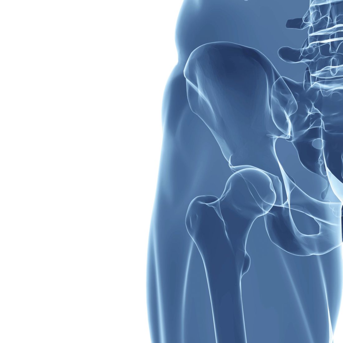
Low DHT linked to hip fracture in men
In older men, circulating levels of dihydrotestosterone (DHT) and sex hormone–binding globulin (SHBG) independently predict risk of hip fracture, but testosterone does not, according to a study involving more than 1,000 men.
These findings could influence clinical measurement of male hormone levels and possibly intervention for low DHT, reported lead author Emily A. Rosenberg, MD, of Brigham and Women’s Hospital in Boston and colleagues.
“Male aging is associated with a decrease in serum sex hormones, and this decline has been shown to influence bone health, although the links between androgen levels in men and bone mineral density and fracture risk remain an ongoing source of debate,” the investigators wrote in Metabolism.
According to Dr. Rosenberg and colleagues, most previous studies in this area have focused on total or bioavailable testosterone; however, DHT demonstrates greater affinity with and slower dissociation from the androgen receptor, which could translate to a more significant role in bone metabolism. With the advent of mass spectrometry–based DHT assays, it is now possible to accurately measure small concentrations of DHT in blood, they added.
Their prospective, multicenter, cohort study involved 1,128 men who were 65 years or older and without history of cardiovascular disease. Beginning in 1989-1990, participants underwent a baseline examination that included standardized medical history questionnaires, physical exam, and laboratory testing. Additional participants joined the study in 1992-1993, and in 1994-1995, a subset of participants (n = 439) underwent dual-energy x-ray absorptiometry (DXA) scanning.
Hormone assays were conducted in 2010 using frozen serum samples from 1994-1995. Testosterone and DHT were measured by liquid chromatography–tandem mass spectrometry assay, while SHBG was measured by fluoroimmunoassay.
The primary outcome, incident hip fracture, was identified from medical records through 2013. Secondary outcomes included lean body mass and bone mineral density of the hip. A variety of covariates were also recorded, including age, sex, weight, alcohol consumption, smoking status, and others.
After a median follow-up of 10.2 years (interquartile range, 5.9-15.5 years), 106 cases of hip fracture occurred, which translated to an incidence rate of 0.89 per 100 person-years. Cox regression models mutually adjusted for covariates, and the other analyses showed that each standard deviation increase in DHT correlated with a 26% decreased risk of hip fracture (adjusted hazard ratio, 0.74; 95% confidence interval, 0.55-1.00; P = .049). Conversely, each standard deviation increase in SBHG was associated with a 26% increased risk of hip fracture (aHR, 1.26; 95% CI, 1.01-1.58; P = .045). In contrast with both DHT and SBHG, testosterone was not significantly associated with the primary outcome (aHR, 1.16; 95% CI, 0.86-1.56; P = .324).
Further analysis showed that testosterone, DHT, and SBHG were not significantly associated with bone mineral density of the hip. In adjusted models, testosterone and DHT were independently associated with higher lean body mass; however, in mutually adjusted models, these associations were not statistically significant, although they remained similar and positive.
“More research is needed to determine the mechanism(s) by which DHT may affect bone health and whether interventions that regulate DHT might be used to reduce risk of hip fracture,” the investigators concluded. “While our results require confirmation, there may be a role for measurement of DHT along with testosterone when the clinical scenario requires measurement of male hormone levels.”
The study was funded by the National Heart, Lung and Blood Institute and the National Institute on Aging. The investigators reported no conflicts of interest.
SOURCE: Rosenberg EA et al. Metabolism. 2020 Oct 12. doi: 10.1016/j.metabol.2020.154399.
FROM METABOLISM
Osteoporosis Journal Scans: October 2020

Some young women can present with very low bone density and multiple fragility fractures. While multiple FDA-approved therapies exist for post-menopausal osteoporosis, pre-menopausal osteoporosis remains an “orphan disease.” Previous evidence suggests that bone anabolic agents such as teriparatide may be useful for women with idiopathic pre-menopausal osteoporosis (IOP). In a phase 2 randomized clinical trial from New York and Nebraska, 41 women with IOP and multiple fractures were randomized to receive placebo or teriparatide in a cross-over study design. The primary endpoint was bone density in the spine and hip after 6 months of treatment, which was significantly increased by teriparatide versus placebo. In addition, bone biopsies were obtained for measurement of bone formation rate using quadruple labeling with tetracycline and demeclocycline three months into treatment. As expected, biopsies showed that teriparatide treatment increased bone formation rates using this ‘gold standard’ method. This prospective randomized study adds to a growing body of evidence that teriparatide may be a safe method to boost bone density in women with IOP. Future studies are needed to assess the impact of this therapy on fracture risk in this specific patient population.
Type 2 diabetes and osteoporosis are both major problems in our aging population. While some diabetes medications such as thiazolidinediones clearly increase fracture risk, the effects of newer diabetes medications on bone biology and fracture risk remain incompletely understood. In addition to beneficial effects on glycemic control, SGLT2 inhibitors show promise with improving cardiovascular and renal outcomes, even in patients without diabetes. Some studies have suggested adverse effects of SGLT2 inhibitor monotherapy on bone density and fracture risk. However, the impact of combined therapy with SGLT2 inhibitors and metformin on fracture risk remains to be established. In this meta-analysis of 25 randomized controlled trials involving 19,500 patients, fracture risk was assessed, based on available information, for individuals who received metformin monotherapy or metformin plus SGLT2 inhibitor treatment. In general, combination therapy did not increase fracture risk compared to metformin alone. Only 6 of the 25 RCTs included in the meta-analysis investigated bone density or bone turnover markers. In these 6 studies, no obvious changes in skeletal outcomes were noted when comparing metformin alone versus metformin plus SGLT2 inhibitor therapy. Although these data are somewhat reassuring for the skeletal safety of combination metformin/SGLT2 inhibitor therapy, confidence is limited by relatively short follow-up time and lack of detailed information about fractures in these studies which focused primarily on diabetes-related outcomes. Future prospective studies are needed to specifically address the skeletal impact of this commonly-used combination of diabetes medications.
Marc Wein, M.D., Ph.D
Assistant Professor of Medicine
Massachusetts General Hospital Endocrine Unit, Harvard Medical School
SGLT2 inhibitors with metformin do not influence fracture risk
Key clinical point: Sodium-glucose transporter-2 inhibitors (SGLT2is) combined with metformin therapy did not influence fracture risk in patients with type 2 diabetes mellitus (T2DM).
Major finding: SGLT2is and metformin combination therapy did not increase the risk of fracture vs. metformin monotherapy or other comparators in patients with T2DM (odds ratio, 0.97; 95% confidence interval, 0.71-1.32).
Study details: A meta-analysis of 25 randomized controlled trials including 19,500 participants with T2DM.
Disclosures: No study sponsor was identified. The authors declared no conflicts of interest.
Source: Qian BB et al. Osteoporos Int. 2020 Aug 11. doi: 10.1007/s00198-020-05590-y.
Sepsis and osteoporosis: What’s the link?
Key clinical point: Adults younger than 65 years with sepsis are at an increased risk of developing osteoporosis.
Major finding: The risk for osteoporosis was significantly higher in the sepsis vs. nonsepsis group (adjusted hazard ratio [aHR], 1.17; 95% confidence interval [CI], 1.04-1.31). The risk for osteoporosis in the sepsis vs. nonsepsis group was significantly higher for young patients aged 20-49 years (aHR, 1.93; 95% CI, 1.08-3.44) and patients aged 50-64 years (aHR, 2.01; 95% CI, 1.52-2.65).
Study details: This Taiwanese population-based study included 13,178 patients diagnosed with sepsis and 13,178 propensity-score matched individuals without sepsis using data from the insurance claims database.
Disclosures: No study sponsor was identified. The authors declared no conflicts of interest.
Source: Lee YF et al. Osteoporos Int. 2020 Aug 22. doi: 10.1007/s00198-020-05599-3.
Osteoporosis increases likelihood of revision surgery after long spinal fusion for adult spinal deformity
Key clinical point: The presence of osteoporosis correlates with a higher likelihood for revision surgery within 2 years following a long spinal fusion for adult spinal deformity (ASD).
Major finding: The rate of revision surgery was significantly higher in ASD patients with osteoporosis vs. those without osteoporosis (40.5% vs. 28.0%; P = .01). The incidence of multiple revision surgeries was similar in both groups (8.4% vs. 8.6%; P = .95). Age and sex were not statistically correlated with the incidence of revision surgery.
Study details: A retrospective comparative study of 399 patients with ASD (40 years or older) who underwent long spinal fusion surgery (osteoporotic group, n=131; nonosteoporotic group, n=268).
Disclosures: The study did not receive any funding.
Source: Gupta A et al. Spine J. 2020 Aug 10. doi: 10.1016/j.spinee.2020.08.002.
Osteoporosis: Predictors of vertebral fracture risk in patients receiving denosumab
Key clinical point: Both total hip bone mineral density (BMD) change and T-score can serve as surrogates for predicting vertebral fracture risk in patients with osteoporosis receiving denosumab treatment.
Major finding: Total hip BMD change and T-score were strong predictors of subsequent vertebral fracture risk, with total hip BMD change accounting for 73% and T-score accounting for 23% of the treatment effect.
Study details: Post hoc analysis of the DIRECT trial involving 1,011 Japanese postmenopausal women and men who were randomly assigned (2:2:1) to receive denosumab, placebo, or alendronate.
Disclosures: The study was funded by Daiichi Sankyo Co., Ltd., Tokyo, Japan. N Okubo, T Osakabe, K Watanabe, and H Takami were Daiichi Sankyo employees. Other authors reported relationships with various pharmaceutical companies, including Daiichi Sankyo.
Source: Okubo N et al. Calcif Tissue Int. 2020 Aug 25. doi: 10.1007/s00223-020-00750-y.
Metformin may attenuate osteoporosis risk in diabetic patients with carcinoma in situ
Key clinical point: Diabetic patients with carcinoma in situ (CIS) under metformin therapy presented a lower osteoporosis rate vs. those who were not receiving metformin.
Major finding: The risk of osteoporosis was lower in diabetic patients with CIS who were receiving metformin therapy vs. those who were not receiving metformin (adjusted hazard ratio, 0.82; P = .022).
Study details: This retrospective matched-cohort study included 7,827 diabetic patients with CIS treated with metformin and 23,481 matched controls with no metformin therapy.
Disclosures: The study was funded by the Tri-Service General Hospital Research Foundation, the National Defense Medical Center, Ministry of National Defense-Medical Affairs Bureau, and the Teh-Tzer Study Group for Human Medical Research Foundation. The authors declared no conflicts of interest.
Source: Lu CH et al. J Clin Med. 2020 Sep 2. doi: 10.3390/jcm9092839.