User login
Approach could overcome drug resistance

Photo courtesy of the CDC
When treating patients with malaria and other infectious diseases, doctors should consider using drug combinations that reach similar parts of the body, according to researchers.
The group found that imperfect drug penetration—when drugs don’t reach all parts of the body—accelerates treatment resistance.
When there is a “pocket” of the body where only one drug is present, such as the brain or the digestive system, a pathogen can quickly develop resistance to one drug at a time.
“If there is a space where there is only one drug, that’s the place where the pathogen can start its escape,” said Pleuni Pennings, PhD, of San Francisco State University in California.
“Once it no longer has the first drug to deal with, it’s very easy for it to quickly become resistant to a second drug.”
Dr Pennings and her colleagues reported these findings in PNAS.
The team believes this research could have major implications for how treatment plans are designed and prescribed to patients with malaria, HIV, tuberculosis, and other ailments.
Because pathogens can quickly develop resistance to a single drug, providers often prescribe multiple drugs to increase their effectiveness.
The results of the study suggest that, when doing so, doctors should carefully consider which parts of the body each drug will reach and whether selecting medications with imperfect but similar penetrations might be the best treatment option.
“It may be better, in some cases, to leave a pocket of the body without any drugs instead of leaving a pocket with just one drug,” Dr Pennings said.
The study is the first to look at the connection between drug penetration and multidrug resistance. Dr Pennings and her colleagues ran computer simulations to observe the behavior of pathogens in response to changes in drugs and their levels of penetration.
The team found that, in instances where even small parts of the body could only be reached by one drug, the pathogen’s ability to build resistance to both drugs was accelerated compared to situations where no such pockets existed.
“This requires a new way of thinking about drug combinations that is a bit counterintuitive,” Dr Pennings said. “Suppose that drug A does not reach the brain, but drug B does. You’ll see the pathogen evolving resistance to drug B and assume that’s where the problem lies. But, in fact, it is drug A that is not doing its job because it’s not reaching the brain, and that’s the drug you may have to actually fix.”
With future research, Dr Pennings and her colleagues hope to outline the most effective drug combinations by exploring which parts of the body cannot be reached by specific drugs and where and how quickly specific pathogens are able to develop resistance. ![]()
Readmissions for severe sepsis fairly common

Photo courtesy of the CDC
DENVER—Severe sepsis is a significant cause of rehospitalization, along the lines of more commonly discussed conditions such as heart failure and pneumonia, according to research presented at the 2015 American Thoracic Society International Conference.
“Severe sepsis continues to be a common cause of hospitalization and has the associated high costs,” said Darya Rudym, MD, of New York University School of Medicine in New York.
She presented this finding at the meeting as publication number A3699.
Some quality measures associated with sepsis, such as length of hospital stay, have been studied previously. But Dr Rudym and her colleagues wanted to look at the rate of readmission within 30 days after discharge among patients with a diagnosis of severe sepsis.
The researchers also wanted to compare this rate to the readmission rates for outcomes reported by the Centers for Medicare and Medicaid Services, such as acute myocardial infarction, heart failure, and pneumonia.
The team examined inpatient discharges from Bellevue Hospital in New York City between July 2011 and July 2014 and identified subsequent readmissions to the hospital within 30 days.
During the 3-year study period, there were 22,712 discharges and an overall readmission rate of 15.31%, or 3477 patients.
The researchers used 3 different methods to identify patients with severe sepsis: Angus implementation, Martin implementation, and explicit ICD-9-CM sepsis codes.
There were 1801 cases of severe sepsis according to Angus implementation, 798 cases according to Martin implementation, and 579 cases according to ICD-9-CM sepsis codes.
In all, 266, 119, and 71 of these patients, respectively, were readmitted to the hospital within 30 days. This accounted for 14.77%, 14.91%, and 12.26% of cases, respectively.
The readmission rate for myocardial infarction was 8.67%. It was 14.46% for pneumonia, and 15% for heart failure.
“Readmission rates in severe sepsis are shown not to be significantly different from readmission rates in heart failure and pneumonia,” Dr Rudym noted. ![]()
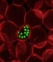
Team discovers target for antimalarial drugs

a red blood cell
Image courtesy of St. Jude
Children’s Research Hospital
New research indicates that manipulating the permeability of parasitophorous vacuoles could defeat malaria parasites.
The researchers unearthed this finding while studying the way in which the Toxoplasma gondii parasite, which causes toxoplasmosis, and Plasmodium parasites,
which cause malaria, access vital nutrients from their host cells.
The team described this work in Cell Host & Microbe.
Roughly a third of the world’s deadly infectious diseases are caused by pathogens that spend a large portion of their life inside parasitophorous vacuoles. This type of vacuole separates the host cytoplasm and the parasite by a membrane, protecting the parasite from the host cell’s defenses and providing an environment tailored to the parasite’s needs.
However, the membrane of the vacuole also acts as a barrier between the parasite and the host cell. This makes it more difficult for the parasite to release proteins involved in the transformation of the host cell beyond the membrane to spread the disease and for the pathogen to gain access to vital nutrients.
“Ultimately, what defines a parasite is that they require certain key nutrients from their host,” said study author Dan Gold, PhD, of the Massachusetts Institute of Technology in Cambridge.
“So they have had to evolve ways to get around their own barriers to gain access to these nutrients.”
Previous research suggested the vacuoles are selectively permeable to small molecules, allowing certain nutrients to pass through pores in the membrane. But until now, no one has been able to determine the molecular makeup of these pores or how they are formed.
When studying Toxoplasma, Dr Gold and his colleagues discovered 2 proteins secreted by the parasite, known as GRA17 and GRA23, which are responsible for forming these pores in the vacuole. The researchers discovered the proteins’ roles by accident while investigating how the parasites are able to release their own proteins out into the host cell beyond the vacuole membrane.
Similar research into how the related Plasmodium pathogens perform this trick revealed a protein export complex that transports encoded proteins from a parasite into its host red blood cell, which transforms the cell in a way that is vital to the spread of malaria.
“The clinical symptoms of malaria are dependent on this process and this remodeling of the red blood cell that occurs,” Dr Gold said.
The researchers identified proteins secreted by Toxoplasma that appeared to be homologues of this protein export complex in Plasmodium. But when they stopped these proteins from functioning, the team found it made no difference to the export of proteins from the parasite beyond the vacuole.
“We were left wondering what GRA17 and GRA23 actually do if they are not involved in protein export, and so we went back to look at this longstanding phenomenon of nutrient transport,” Dr Gold said.
When they added dyes to the host cell and knocked out the 2 proteins, the researchers found that it prevented the dyes flowing into the vacuole.
“That was our first indication that these proteins actually have a role in small-molecule transfer,” Dr Gold said.
When the researchers expressed a Plasmodium export complex gene in the modified Toxoplasma, they found the dyes were able to flow into the vacuole once again, suggesting this small-molecule transport function had been restored.
Since these proteins are only found in the parasite phylum Apicomplexa, to which both Toxoplasma and Plasmodium belong, they could be used as a drug target against the diseases they cause, the researchers said.
“This very strongly suggests that you could find small-molecule drugs to target these pores, which would be very damaging to these parasites but likely wouldn’t have any interaction with any human molecules,” Dr Gold said. “So I think this is a really strong potential drug target for restricting the access of these parasites to a set of nutrients.”![]()
Equation provides new insight into blood flow

Engineers have devised an equation that yields simple predictions as to how quickly blood cells will migrate away from blood-vessel walls, how they will behave when they collide with each other, and how they will segregate during flow.
In the long run, these insights could help practitioners manipulate the mechanics of blood to design better blood transfusions, new techniques for drug delivery, and new processes for isolating blood-borne tumor cells.
Mike Graham, PhD, of the University of Wisconsin-Madison, and his colleagues described this work in Physical Review Letters.
“I’m really excited about this paper because it’s the first analytical theory for this phenomenon,” Dr Graham said. “It’s not very common that theory is ahead of experiments, but we’re in that position now.”
Dr Graham and his colleagues created complex computer simulations that showed how relatively stiff white blood cells and platelets interact with more flexible red blood cells.
As the different cells collide during blood flow, white cells tend to be pushed toward the walls of a blood vessel. This segregation process, called margination, creates some advantages; for example, letting white blood cells quickly exit the blood vessel to head to the site of an injury or infection.
However, the mechanical details of blood could spell both good news and bad in areas ranging from drug delivery to blood disorders to the spread of disease.
“I view my role as providing a fundamental basis of understanding for practitioners and for other engineers who are more directly connected with applications,” Dr Graham said.
Now, he is aiming to draw a firmer connection between mechanical insights and the biological functions they might impact. His group is working to refine the new equation to suit more complex flow situations and pursuing an experimental collaboration with Wilbur Lam, MD, PhD, a hematologist at Georgia Tech and Emory University in Atlanta.
Building on Dr Graham’s theoretical and simulation work, Dr Lam’s research group is creating microfluidic devices to study the behavior of blood cells. Dr Lam has developed a way to grow endothelial cells inside the artificial channels of the microfluidic devices.
“I think, together, our labs have really stumbled on how fluid mechanics may be able to explain a lot of the biological phenomena we see in blood,” Dr Lam said. “This can be related to a new way of understanding inflammation, infections, even transfusion medicine. It really pervades many different problems we see in hematology.”
Dr Graham said that capturing the physical nuances of blood vessels’ shape, size, and relative stiffness has tremendous value, even given the myriad other forces at work in the human body.
“We’d like to be able to convince practitioners that you don’t have to worry about all the details to capture the fundamental understanding of what’s going on,” Dr Graham said. “It’s extremely challenging to incorporate all the phenomena that might be important into a simulation. You have to make your case convincingly—if you want somebody to apply this research—that you’ve kept the important parts.”
Both researchers pointed out that sickle cell anemia has long been understood as both a mechanical and a biological problem. The defective red blood cells the disease causes are not only misshapen, but also stiffer than healthy red blood cells, meaning they block blood flow.
Yet, on a more detailed mechanical level, Drs Graham and Lam believe that sickle cells may literally poke and irritate the inner walls of blood vessels. If so, that would make sickle cell anemia not just a blood disorder but a disorder of the entire circulatory system. Their combined research strengths now create an opportunity to test that hypothesis.
“Biologists and hematologists have known for decades that these cells can get stuck, but what is less understood is that the blood vessel walls in the entire patient are really inflamed, and we don’t really know why,” Dr Lam said.
The researchers noted that a better understanding of blood-flow mechanics could help to make blood transfusions safer as well. Transfusions can sometimes set off heart attacks or lung damage, and the medical community isn’t entirely sure why. Dr Lam wants to find out if certain cells in stored, donated blood have mechanical properties that put patients at greater risk.
Though the collaboration between Drs Graham and Lam is still in an early stage, both researchers see the possibility of opening a new frontier in blood research.
“This would be a whole new category of things we could be looking at, and that’s why it’s so exciting,” Dr Lam said. “Suddenly, we have applications where the mechanics can be just as important.” ![]()
New method to assess cancer risk from pollutants

Photo by Tiffany Dawn Nicholson
Scientists say they have developed a faster, more accurate method to assess cancer risk from certain common environmental pollutants.
The group found they could analyze the immediate genetic responses of the skin cells of exposed mice and apply statistical approaches to determine whether or not those cells would eventually become cancerous.
The study focused on a class of pollutants known as polycyclic aromatic hydrocarbons (PAHs) that commonly occur in the environment as mixtures such as diesel exhaust and cigarette smoke.
“After only 12 hours, we could predict the ability of certain PAH mixtures to cause cancer, rather than waiting 25 weeks for tumors to develop,” said study author Susan Tilton, PhD, of Oregon State University in Corvallis.
For at least some PAH mixtures, the new method is not only quicker but produces more accurate cancer-risk assessments than are currently possible, she added.
Dr Tilton and her colleagues described the method in Toxicological Sciences.
“Our work was intended as a proof of concept,” Dr Tilton noted. “The method needs to be tested with a larger group of chemicals and mixtures. But we now have a model that we can use to develop larger-scale screening tests with human cells in a laboratory dish.”
The researchers believe the model will be particularly useful for screening PAHs, a large class of pollutants that result from combustion of organic matter and fossil fuels. PAHs are widespread contaminants of air, water, and soil. There are hundreds of different kinds, and some are known carcinogens, but many have not been tested.
Humans are primarily exposed to PAHs in the environment as mixtures, which makes it harder to assess their cancer risk. The standard calculation, Dr Tilton said, is to identify the risk of each element in the mix—if it’s known—and add them together.
But this method doesn’t work with most PAH mixes. It assumes the risk for each component is known, as well as which components are in a given mix. Often, that information is not available.
For this study, Dr Tilton and her colleagues examined 3 PAH mixtures that are common in the environment—coal tar, diesel exhaust, and cigarette smoke—and various mixtures of them.
The group found that each substance touched off a rapid and distinctive cascade of biological and metabolic changes in the skin cells of a mouse. The response amounted to a unique “fingerprint” of the genetic changes that occur as cells reacted to exposure to each chemical.
By matching patterns of genetic changes known to occur as cells become cancerous, the researchers found that some of the cellular responses were early indicators of developing cancers.
They also found that the standard method to calculate carcinogenic material underestimated the cancer risk of some mixtures and overestimated the combined risk of others.
“Our study is a first step in moving away from risk assessments based on individual components of these PAH mixtures and developing more accurate methods that look at the mixture as a whole,” Dr Tilton said. “We’re hoping to bring the methodology to the point where we no longer need to use tumors as our endpoint.” ![]()
Explaining obesity in cancer survivors

Researchers have identified several factors that may influence the risk of obesity in childhood cancer survivors.
Previous research showed that obesity rates are elevated in childhood cancer survivors who were exposed to cranial radiation.
But the new study has shown that other types of treatment, a patient’s age, and certain genetic variants are associated with obesity in this population.
Carmen Wilson, PhD, of St. Jude Children’s Research Hospital in Memphis, Tennessee, and her colleagues reported these findings in Cancer.
The researchers evaluated 1,996 childhood cancer survivors treated at St. Jude. The patients’ median age at diagnosis was 7.2 years (range, 0.1-24.8), and their median age at follow-up was 32.4 years (range, 18.9-63.8).
At the time of evaluation, 645 patients (32.3%) were of normal weight, 71 (3.6%) were underweight, 556 (27.9%) were overweight, and 723 (36.2%) were obese.
The prevalence of obesity was highest in male leukemia survivors (42.5%) and females who survived neuroblastoma (43.6%), followed closely by those who survived leukemia (43.1%).
Multivariable analyses showed that 3 factors were independently associated with an increased risk of obesity: older age at the time of evaluation (≥30 years vs <30 years; P<0.001), undergoing cranial radiation (P<0.001), and receiving glucocorticoids (P=0.004).
On the other hand, receiving chest, abdominal, or pelvic radiation was associated with a decreased risk of obesity (P<0.001).
The researchers also identified 166 single nucleotide polymorphisms that were associated with obesity among cancer survivors who had received cranial radiation. The strongest association was in variants of genes involved in neuron growth, repair, and connectivity.
Among survivors who did not receive cranial radiation, only 1 single nucleotide polymorphism—rs12073359, located on chromosome 1—was associated with an increased risk of obesity.
Dr Wilson said these findings might help us identify the childhood cancer survivors who are most likely to become obese. The results may also provide a foundation for future research efforts aimed at characterizing molecular pathways involved in the link between childhood cancer treatment and obesity. ![]()
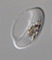
Antimalarial drug unavailable, CDC says

Photo courtesy of the FDA
The antimalarial drug chloroquine is not currently available from US suppliers, according to the Centers for Disease Control and Prevention (CDC).
The agency said it will provide updates as more information becomes available from the Food and Drug Administration.
Chloroquine is used as malaria treatment and prophylaxis, but hydroxychloroquine sulfate can be prescribed in place of chloroquine when indicated.
Healthcare providers who need assistance diagnosing or managing suspected or confirmed cases of malaria can call the CDC Malaria Hotline at 1-855-856-4713 (Monday through Friday, 9 am to 5pm, Eastern time).
For emergency consultation after hours, providers can call 1-770-488-7100 and ask to speak with a CDC Malaria Branch clinician. ![]()
Photo courtesy of the FDA
The antimalarial drug chloroquine is not currently available from US suppliers, according to the Centers for Disease Control and Prevention (CDC).
The agency said it will provide updates as more information becomes available from the Food and Drug Administration.
Chloroquine is used as malaria treatment and prophylaxis, but hydroxychloroquine sulfate can be prescribed in place of chloroquine when indicated.
Healthcare providers who need assistance diagnosing or managing suspected or confirmed cases of malaria can call the CDC Malaria Hotline at 1-855-856-4713 (Monday through Friday, 9 am to 5pm, Eastern time).
For emergency consultation after hours, providers can call 1-770-488-7100 and ask to speak with a CDC Malaria Branch clinician.
Impotence drug could prevent malaria transmission
blood cell that has stiffened
after treatment
© 2015 Ramdani et al.
The erectile dysfunction drug sildenafil (Viagra) could prevent transmission of the malaria parasite, according to research published in PLOS Pathogens.
Investigators found that sildenafil increases the stiffness of erythrocytes infected by the parasite Plasmodium falciparum.
This allows the cells to be eliminated from the bloodstream and may therefore reduce transmission of the malaria parasite from humans to mosquitoes.
The investigators noted that P falciparum has a complex developmental cycle that is completed partly in humans and partly in mosquitoes. Treatments for malaria target the asexual forms of this parasite that cause symptoms, but not the sexual forms transmitted from a human to a mosquito.
Malaria eradication therefore necessitates new types of treatments against sexual forms of the parasite in order to block transmission and prevent dissemination of the disease.
The sexual forms of P falciparum develop in human erythrocytes sequestered in the bone marrow before they are released into the blood. They are then accessible to mosquitoes, which can ingest them when they bite.
Circulating erythrocytes are deformable, thus preventing their clearance via the spleen. And gametocyte-infected erythrocytes can easily pass through the spleen and persist for several days in the blood circulation.
With this in mind, Ghania Ramdani, of Université Paris Descartes in France, and colleagues sought to stiffen the infected erythrocytes so they would be removed from circulation.
The team found that the deformability of gametocyte-infected erythrocytes is regulated by a signaling pathway that involves cAMP. When cAMP molecules accumulate, an erythrocyte becomes stiffer. And cAMP is degraded by the enzyme phosphodiesterase, which promotes erythrocyte deformability.
Using an in vitro model reproducing filtration by the spleen, the investigators were able to identify several pharmacological agents that inhibit phosophodiesterases and can therefore increase the stiffness of infected erythrocytes.
One of these agents is sildenafil. The team showed that a standard dose of the drug had the potential to increase the stiffness of sexual forms of the parasite and therefore favor the elimination of infected erythrocytes from the circulation.
This discovery could lead to new ways to stop the spread of malaria, the investigators said. They believe that modifying the active substance in sildenafil to block its erectile effect, or testing similar agents devoid of this effect, could indeed result in a treatment to prevent transmission of the parasite from humans to mosquitoes.
Malaria vaccine proves partially protective

Photo by James Gathany
A new malaria vaccine candidate proved partially effective in adult males living in an area of low malaria transmission.
The T-cell vaccine consists of the recombinant viral vectors chimpanzee adenovirus 63 (ChAd63) and modified vaccinia Ankara (MVA), both encoding the multiple epitope string and thrombospondin-related adhesion protein (ME-TRAP), a fusion of protein fragments found on the surface of the malaria parasite Plasmodium falciparum.
In what’s known as a prime-boost strategy, an initial dose of the vaccine “primes” the immune system by exposing it to the malaria antigen and is followed by a vaccine booster, which re-stimulates the immune system to further solidify immunity.
Caroline Ogwang, of the Kenya Medical Research Institute–Wellcome Trust Research Programme in Kilifi, Kenya, and her colleagues described their trial of the vaccine in Science Translational Medicine.
The researchers tested the vaccine in an area of low malaria transmission in Kenya. They enrolled 121 healthy adult men and randomized them to receive the malaria vaccine or a control rabies vaccine.
The subjects were also treated with antimalarial drugs to clear any previous infection and were closely monitored for 8 weeks for P falciparum infection.
The malaria vaccine appeared to be safe, prompting no serious adverse events. The most common local adverse event associated with both ChAd63 ME-TRAP and MVA ME-TRAP was mild to moderate pain that lasted from a few hours to 3 days.
Subjects also experienced various systemic adverse events of mild to moderate intensity that lasted from a few hours to 5 days. Other events were reported within 30 days of vaccination as well, but these were not considered vaccine-related.
As for efficacy, blood tests revealed that the vaccine activated a strong immune response from T cells. For at least 2 weeks after vaccination, the vaccine showed partial efficacy in protecting against malaria infection.
The vaccine reduced subjects’ risk of infection by 67% (P=0.002) during the 8-week monitoring period. And the researchers said T-cell responses to TRAP peptides 21 to 30 were significantly associated with protection (hazard ratio=0.24, P=0.016).
The team is continuing clinical testing of the vaccine, including possible combinations with other vaccination strategies to increase efficacy.
Study reveals ‘doorway’ into RBCs
infecting an RBC
Photo courtesy of St. Jude
Children’s Research Hospital
A protein on the surface of red blood cells (RBCs) serves as an essential entry point for malaria parasite invasion, according to researchers.
They found the presence of this protein, CD55, was critical to the Plasmodium falciparum parasite’s ability to attach itself to the RBC surface.
The team believes this discovery, published in Science, opens up a promising new avenue for developing therapies to treat and prevent malaria.
“Plasmodium falciparum malaria parasites have evolved several key-like molecules to enter into human red blood cells through different door-like host receptors,” said study author Manoj Duraisingh, PhD, of the Harvard T. H. Chan School of Public Health in Boston, Massachusetts.
“Hence, if one red blood cell door is blocked, the parasite finds another way to enter. We have now identified an essential host factor which, when removed, prevents all parasite strains from entering red blood cells.”
The researchers accomplished this by developing a new technique to tap into a relatively unexplored area: identifying characteristics of a host RBC that make it susceptible to parasites. RBCs are difficult targets for such efforts as they lack a nucleus, which makes genetic manipulation impossible.
So the team transformed stem cells into RBCs, which allowed them to conduct a genetic screen for host determinants of P falciparum infection. They found that malaria parasites failed to attach properly to the surface of RBCs that lacked CD55.
The protein was required for invasion in all tested strains of the parasite, including those developed in a lab and those isolated from patients. This makes CD55 a primary candidate for intervention, the researchers said.
“The discovery of CD55 as an essential host factor for P falciparum raises the intriguing possibility of host-directed therapeutics for malaria, as is used in HIV,” said study author Elizabeth Egan, MD, PhD, also of the Harvard T. H. Chan School of Public Health.
“CD55 also gives us a hook with which to search for new parasite proteins important for invasion, which could serve as vaccine targets.”